
A 50 years old lady came to the surgeon with the complaints of a firm, painless mass in the upper lateral quadrant of her right breast. On examination the nipple was found to be retracted. Axillary lymph nodes were also palpable and firm. She was diagnosed as a case of Breast cancer. 3 7 3 4 a. What are the structures lying deep to the breast? b. Describe the lymphatic dralnage of the breast. c. What causes the retractlon of nipple? d. What is the anatomical basis of peau d' orange? e. Explain the anatomical basis of spread of cancer to vertebrae
lymphatic drainage of breast axillary nodes diagram
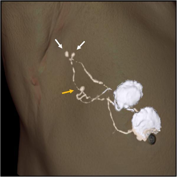
A three-dimensional (3D) volume-rendered CT lymphography image of the axillary region in a patient with breast cancer, demonstrating lymphatic drainage pathways and sentinel nodes. The image displays a skin-rendered surface with overlaid hyper-dense (white) structures representing contrast-enhanced lymphatic vessels and nodes. Two proximal sentinel lymph nodes are indicated by white arrows, appearing as small, ovoid, high-density structures connected by delicate, branching lymphatic channels. A third, more distal sentinel node is highlighted by a yellow arrow, located along a separate lymphatic tributary. The lymphatic flow originates from large, irregular high-density areas representing the intradermal contrast injection sites near the nipple-areolar complex. A metallic localizing marker is visible on the skin surface to facilitate surgical planning. This diagnostic imaging highlights the utility of CT lymphography in identifying multiple or widely distributed sentinel nodes that might be overlooked during conventional radioisotope or dye-only mapping due to spatial separation or signal interference from the primary injection site.
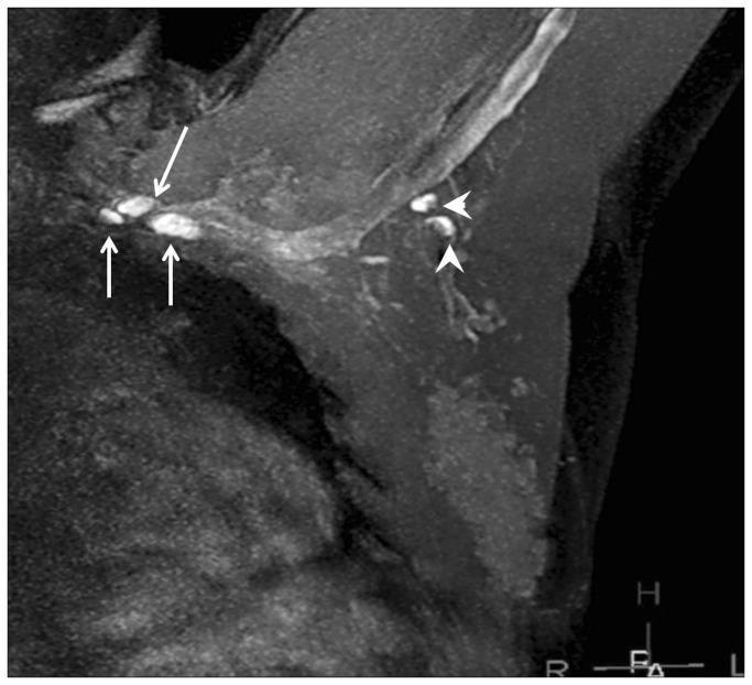
This diagnostic image is a Magnetic Resonance Lymphangiography (MRL) scan demonstrating the axillary lymphatic system in a breast cancer context. The grayscale image shows anatomical structures of the breast and axilla with gadolinium-based contrast enhancement highlighting the lymphatic pathway. Two distinct levels of axillary lymph nodes are identified by markers: Level 1 lymph nodes are indicated by arrowheads, appearing as bright, rounded hyperintense structures clustered in the lower axilla. Level 3 lymph nodes, indicated by arrows, are situated more superiorly and medially (infraclavicular region) and also exhibit high signal intensity, though they appear slightly smaller in this view. The image illustrates the use of MRL for visualizing nodal anatomy and mapping the lymphatic drainage pathway. High signal intensity (bright white) indicates successful contrast uptake, while the surrounding darker gray areas represent soft tissue and fat with lower signal intensity. This modality is used in oncology to assess lymph node morphology and potential metastatic involvement by identifying enhancement defects or vessel dilation.
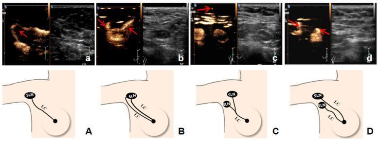
This composite educational graphic illustrates four distinct patterns of axillary lymphatic drainage from the breast to sentinel lymph nodes (SLNs), using Contrast-Enhanced Ultrasound (CEUS) imaging and corresponding anatomical diagrams. The visual material is divided into four sets (A–D), each featuring a split-screen ultrasound image (CEUS and B-mode) alongside a schematic illustration. The primary components described are Lymphatic Channels (LC) and Sentinel Lymph Nodes (SLN). The four patterns shown are: (A) a single LC draining to a single SLN; (B) multiple LCs converging into a single SLN; (C) a single LC branching to multiple SLNs; and (D) multiple LCs draining to multiple separate SLNs. In the CEUS images, the LCs and SLNs are identified by high echogenicity (bright contrast enhancement) indicated by red arrows, facilitating real-time visualization of the drainage pathway. This content is intended for surgical oncology and radiology education, specifically regarding preoperative sentinel lymph node biopsy (SLNB) mapping in breast cancer staging. It demonstrates how CEUS can be used to identify complex lymphatic anatomy that may impact surgical planning.
Breast Cancer - Anatomy-Based Clinical Answers
a. Structures Lying Deep to the Breast
- Deep (pectoral) fascia - the investing fascia overlying the pectoralis major
- Pectoralis major muscle - the primary muscle deep to the breast, spanning from ribs II to VI laterally
- Pectoralis minor muscle - enclosed in the clavipectoral fascia, which extends laterally to fuse with the axillary fascia
- Serratus anterior (inferolaterally)
- External oblique (inferiorly, overlying ribs 6-7)
- Ribs and intercostal muscles (underlying the inferior extent)
- Sabiston Textbook of Surgery, 21st ed.
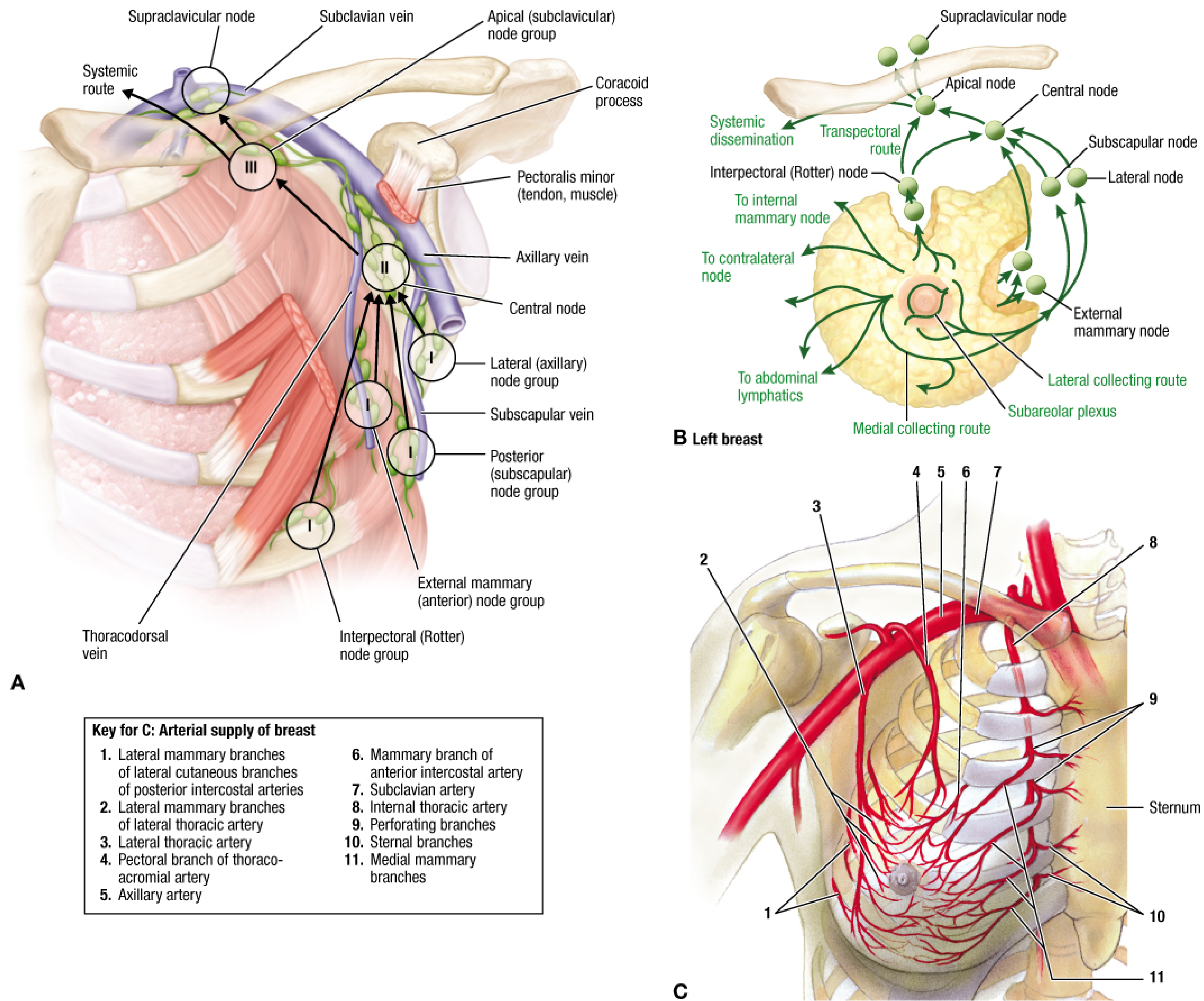
b. Lymphatic Drainage of the Breast
Primary Route (75%) - Axillary Nodes
- Level I (inferior to pectoralis minor): external mammary, lateral axillary, subscapular nodes
- Level II (posterior to pectoralis minor): central, interpectoral (Rotter's) nodes
- Level III (medial/superior to pectoralis minor): apical (subclavian) nodes --> subclavian trunk --> venous angle
Secondary Routes
- Internal mammary (parasternal) nodes - particularly for medial quadrant tumors; via medial collecting route
- Supraclavicular nodes - especially in advanced disease
- Interpectoral (Rotter's) nodes - between pectoralis major and minor
- Deltopectoral nodes - along the cephalic vein
- Contralateral breast - cross-midline drainage
- Abdominal lymphatics - via inferior phrenic nodes (rare; relevant for subdiaphragmatic spread)
- Fischer's Mastery of Surgery, 8th ed.
c. What Causes Retraction of the Nipple?
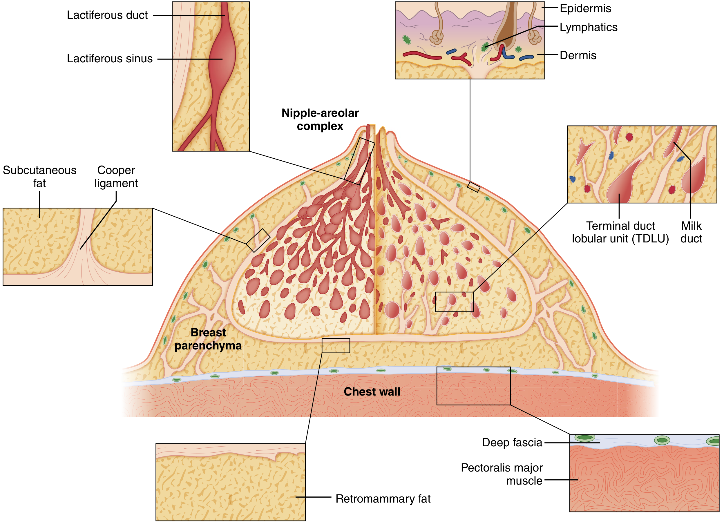
- The breast is supported by fibrous bands called the suspensory ligaments of Cooper (Cooper's ligaments). These run between the deep fascia of the breast (over pectoralis major) and the dermis of the overlying skin, providing shape and structural support.
- These ligaments also attach to and support the nipple-areolar complex.
- When a carcinoma grows in the breast, it elicits a fibrotic (desmoplastic) reaction in the surrounding stroma.
- The cancer infiltrates and shortens the Cooper's ligaments. Since these ligaments are anchored to both the deep fascia and the skin/nipple, their fibrotic shortening pulls the nipple inward, causing retraction.
- In cancers of the retroareolar region, direct involvement of the lactiferous ducts by the tumor also contributes - ductal fibrosis shortens the ductal system and draws the nipple inward.
"Carcinoma of the breast creates tension on these ligaments, causing pitting of the skin." - Gray's Anatomy for Students
- Sabiston Textbook of Surgery; Gray's Anatomy for Students
d. Anatomical Basis of Peau d'Orange
Anatomical Explanation:
- Tumor cells block the subdermal lymphatics directly (lymphatic permeation) OR block the axillary lymph nodes, causing back-pressure in the cutaneous lymphatics.
- This results in dermal lymphoedema - fluid accumulation in the dermis causes diffuse skin thickening and swelling.
- The skin is tethered at multiple points to the underlying dermis by the hair follicles and sweat gland ducts, which pass through the oedematous skin. These tethering points cannot expand - they remain fixed while the surrounding skin swells.
- The result is a skin surface that looks like an orange peel - generalised swelling with multiple small pits corresponding to the hair follicle orifices.
"Peau d'orange is a sign of locally advanced disease due to obstruction of cutaneous lymphatic drainage of the breast, by infiltration of either subdermal lymphatics or axillary lymph nodes by tumour cells." - Bailey and Love's Short Practice of Surgery, 28th ed.
- Robbins & Kumar Basic Pathology; Bailey and Love's, 28th ed.; Gray's Anatomy for Students
e. Anatomical Basis of Spread of Cancer to Vertebrae
1. Haematogenous Spread via Batson's Vertebral Venous Plexus (Primary Mechanism)
- Batson's plexus is a network of valveless epidural veins running along the anterior epidural space of the vertebral column.
- These veins communicate freely with the intercostal veins, internal thoracic veins, and the azygos system - all of which drain the breast region.
- Because these veins have no valves, blood can flow in either direction depending on intrathoracic and intra-abdominal pressure changes (e.g. during coughing, straining, Valsalva).
- Tumor emboli from the breast can therefore enter these veins and travel retrograde into the vertebral venous sinusoids, seeding the bone marrow of vertebral bodies.
- This route bypasses the lungs entirely, explaining why some patients develop vertebral metastases without pulmonary metastases.
2. Arterial (Systemic) Route
- Tumor cells enter the systemic circulation via the axillary or internal thoracic veins --> subclavian vein --> right heart --> pulmonary circulation.
- After passing through the lungs, tumor emboli in the arterial circulation can reach the nutrient arteries of the vertebrae, particularly targeting the red bone marrow of the vertebral bodies.
Pattern of Vertebral Involvement
- The thoracic vertebrae are most commonly involved in breast cancer (reflecting direct haematogenous spread via Batson's plexus from the thoracic intercostal veins).
- The order of frequency of skeletal metastases is: lumbar vertebrae > neck of femur > thoracic vertebrae > ribs > skull.
- Bone metastases from breast cancer are generally osteolytic (though osteosclerotic and mixed types also occur).
- Extensive bone marrow replacement may cause a leukoerythroblastic anaemia.
"The propensity of solid tumors to metastasize to the vertebral column probably reflects the high proportion of bone marrow located in the axial skeleton... spread through Batson's plexus, a network of veins along the anterior epidural space." - Harrison's Principles of Internal Medicine, 22nd ed.
- Bailey and Love's Short Practice of Surgery, 28th ed.; Harrison's Principles of Internal Medicine, 22nd ed.
Summary Table
| Feature | Anatomical Basis |
|---|---|
| Nipple retraction | Fibrotic shortening of Cooper's ligaments by tumor desmoplasia |
| Peau d'orange | Dermal lymphoedema from blockage of subdermal lymphatics or axillary nodes; skin tethered at hair follicle orifices |
| Axillary node involvement | 75% of breast lymph drains to axillary nodes via subareolar plexus |
| Vertebral spread | Batson's valveless vertebral venous plexus allows retrograde hematogenous seeding of vertebral bone marrow |