
Down syndrome
Reading File
Now let me search the medical library for Down syndrome content in parallel.
Finding Sources
Searching PubMed
"Down syndrome"[MeSH Terms] AND management
Good results. Now let me read the key sections in parallel - the genetics textbook (most comprehensive) and search for clinical features/management.
Reading File
Finding Sources
Excellent. Now let me read the Robbins pathology content and more of the Thompson genetics content for comprehensive clinical features, cytogenetics, and management.
Reading File
Reading File
Let me now read the Robbins content on clinical features and also fetch the FISH image.
Reading File
Loading Image
I now have comprehensive information from multiple authoritative textbooks. Let me compile the full answer.
Down Syndrome (Trisomy 21)
Overview
Down syndrome is the most common chromosomal disorder and the single most common genetic cause of moderate intellectual disability. The incidence is approximately 1 in 700 live births in the general population. Despite the well-known association with advanced maternal age, more than half of all affected newborns are born to mothers under 35, simply because younger women have higher overall birth rates.
- Thompson & Thompson Genetics, 9th ed., p. 103
- Robbins Pathologic Basis of Disease
Cytogenetics: Three Karyotypic Forms
| Type | Frequency | Karyotype | Key Feature |
|---|---|---|---|
| Trisomy 21 | ~95% | 47,XX or XY,+21 | Meiotic nondisjunction; sporadic |
| Robertsonian Translocation | ~4% | 46 chromosomes (21q fused to 14 or 22) | May be familial; maternal carrier risk |
| Mosaic | ~1-2% | Mix of 46 and 47 chromosome cells | Variable/milder phenotype |
Trisomy 21 results from meiotic nondisjunction - the extra chromosome 21 is of maternal origin in ~95% of cases, predominantly from an error in maternal meiosis I. Paternal nondisjunction accounts for about 10% of standard trisomies, often in meiosis II.
Robertsonian translocation Down syndrome involves fusion of 21q to another acrocentric chromosome (usually chromosome 14). Despite only 46 chromosomes, the patient is trisomic for all genes on 21q. Critically, it shows no relation to maternal age but carries a high recurrence risk when a parent is a carrier (45 chromosomes, with one chromosome 14 and 21 replaced by the translocation chromosome).
Mosaic Down syndrome arises from mitotic nondisjunction early in embryogenesis. Phenotype is variably milder depending on the proportion of trisomy 21 cells.
Below: FISH analysis of an interphase nucleus using chromosome 13 probes (green) and chromosome 21 probes (red), showing three red signals confirming trisomy 21.
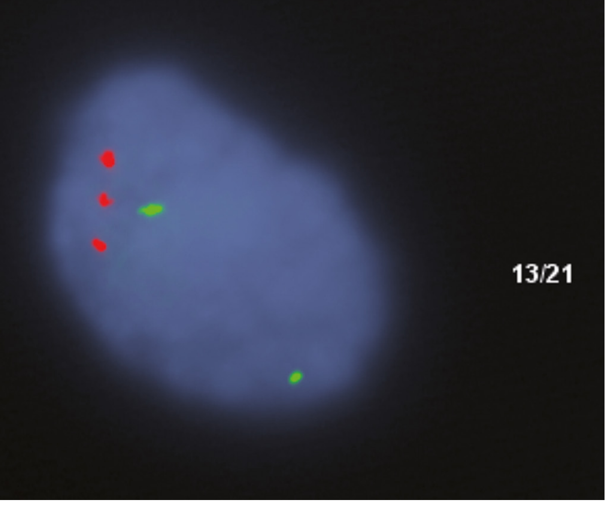
Fig. FISH 13/21 probe - 3 red signals (chromosome 21) confirm trisomy 21. Robbins Pathologic Basis of Disease.
Maternal Age Effect
| Maternal Age | Risk of Trisomy 21 |
|---|---|
| Under 20 | ~1 in 1,550 |
| 30 years | Risk begins to rise sharply |
| Over 45 | ~1 in 25 |
The strong maternal age correlation reflects the prolonged arrest of oocytes in meiosis I, making them more susceptible to nondisjunction over time.
Clinical Features
Physical features at birth:
- Hypotonia (often the first sign noticed in the newborn)
- Flat facial profile with oblique palpebral fissures and epicanthic folds
- Brachycephaly with flat occiput
- Short, broad neck with loose skin at the nape
- Short, broad hands - often with a single transverse palmar crease and fifth finger clinodactyly (incurved fifth digit)
- Short stature
Intellectual disability:
- Most common major concern; ranges from mild to moderate
- Development delay typically becomes obvious by end of the first year
- Many individuals with Down syndrome attend school and develop into interactive, semi-independent persons
Congenital heart disease (~40-50%):
- Atrioventricular septal defects (43% of cardiac cases)
- Ventricular septal defects (32%)
- Atrial septal defects (19%)
- Tetralogy of Fallot (6%)
- Cardiac disease accounts for most deaths in infancy and early childhood
GI malformations: Duodenal atresia, tracheoesophageal fistula, esophageal atresia - significantly more common than in other disorders
Leukemia risk:
- 20-fold increased risk for precursor B-cell acute lymphoblastic leukemia (ALL)
- 500-fold increased risk for acute myeloid leukemia (AML)
- Overall ~15-fold increased leukemia risk compared to general population
Alzheimer disease: Virtually all patients older than 40 develop neuropathologic changes of Alzheimer disease (cortical atrophy, ventricular dilatation, neurofibrillary tangles). This is attributed to overexpression of the amyloid precursor protein (APP) gene on chromosome 21. Onset is several decades earlier than in the general population.
Immune dysfunction:
- Mainly T-cell function abnormalities
- Increased susceptibility to serious infections (especially pulmonary)
- Predisposition to thyroid autoimmunity
Molecular / Pathogenetic Basis
The phenotype results from gene dosage effects across multiple loci on chromosome 21 - not a single gene mutation. Key mechanisms include:
- APP overexpression - drives early-onset Alzheimer disease (amyloid-beta aggregation)
- Mitochondrial dysfunction - ~10% of overexpressed genes regulate mitochondrial function; broken/swollen cristae are observed, with reactive oxygen species generation and apoptosis activation
- lncRNAs - chromosome 21 has the highest density of long non-coding RNAs; their contributions to the phenotype are still being studied
Prenatal Screening & Diagnosis
Non-invasive prenatal testing (NIPT): ~5-10% of cell-free DNA in maternal blood is fetal. Next-generation sequencing can determine gene dosage of chromosome 21 with high precision - a powerful non-invasive screen for trisomy 21 and other trisomies. Positive screens require confirmation by karyotyping.
Invasive confirmation:
- Chorionic villus sampling (CVS) - first trimester
- Amniocentesis - second trimester
Serum markers (second trimester "quadruple test"): AFP, hCG, unconjugated estriol, and inhibin A
Ultrasound markers: Nuchal translucency (first trimester), "double-bubble sign" (duodenal atresia), short femur, echogenic bowel
Recurrence Risk
| Karyotype | Recurrence Risk |
|---|---|
| Standard trisomy 21 | Low (~1% above baseline age-related risk) |
| Translocation (carrier mother) | ~10-15% risk per pregnancy |
| Translocation (carrier father) | ~2-5% risk per pregnancy |
| Mosaic parent | Variable |
Prognosis & Lifespan
Improved medical care has dramatically extended life expectancy. The median age at death is now 47 years (up from 25 years in 1983). Major causes of morbidity and mortality include congenital heart disease (early life), recurrent infections, leukemia, and Alzheimer disease (later life).
Sources: Robbins & Cotran Pathologic Basis of Disease | Thompson & Thompson Genetics and Genomics in Medicine, 9th ed. | Emery's Elements of Medical Genetics and Genomics
This is a shared conversation. Sign in to Orris to start your own chat.