
Cld
Chronic liver disease
What aspect of Chronic Liver Disease are you looking for? · What's your context?
Chronic Liver Disease (CLD) - Comprehensive Review
1. DEFINITION & OVERVIEW
2. ETIOLOGY (Causes)
| Category | Examples |
|---|---|
| Viral | Hepatitis B (HBV), Hepatitis C (HCV) |
| Alcohol | Alcoholic liver disease / Laennec's cirrhosis (10-20% of chronic alcoholics) |
| Metabolic | NAFLD/NASH (increasingly common), Wilson's disease, Hemochromatosis |
| Autoimmune | Autoimmune hepatitis, Primary biliary cholangitis (PBC), Primary sclerosing cholangitis (PSC) |
| Biliary | Chronic biliary obstruction, biliary cirrhosis |
| Drugs/Toxins | Methotrexate, amiodarone, isoniazid |
| Cryptogenic | Unknown etiology (~10-15%) |
| Vascular | Budd-Chiari syndrome, cardiac cirrhosis |
Key exam point: NASH has become the leading cause of cryptogenic cirrhosis; the process typically spans >20 years.
- Rosen's Emergency Medicine, p. 1247
3. PATHOPHYSIOLOGY
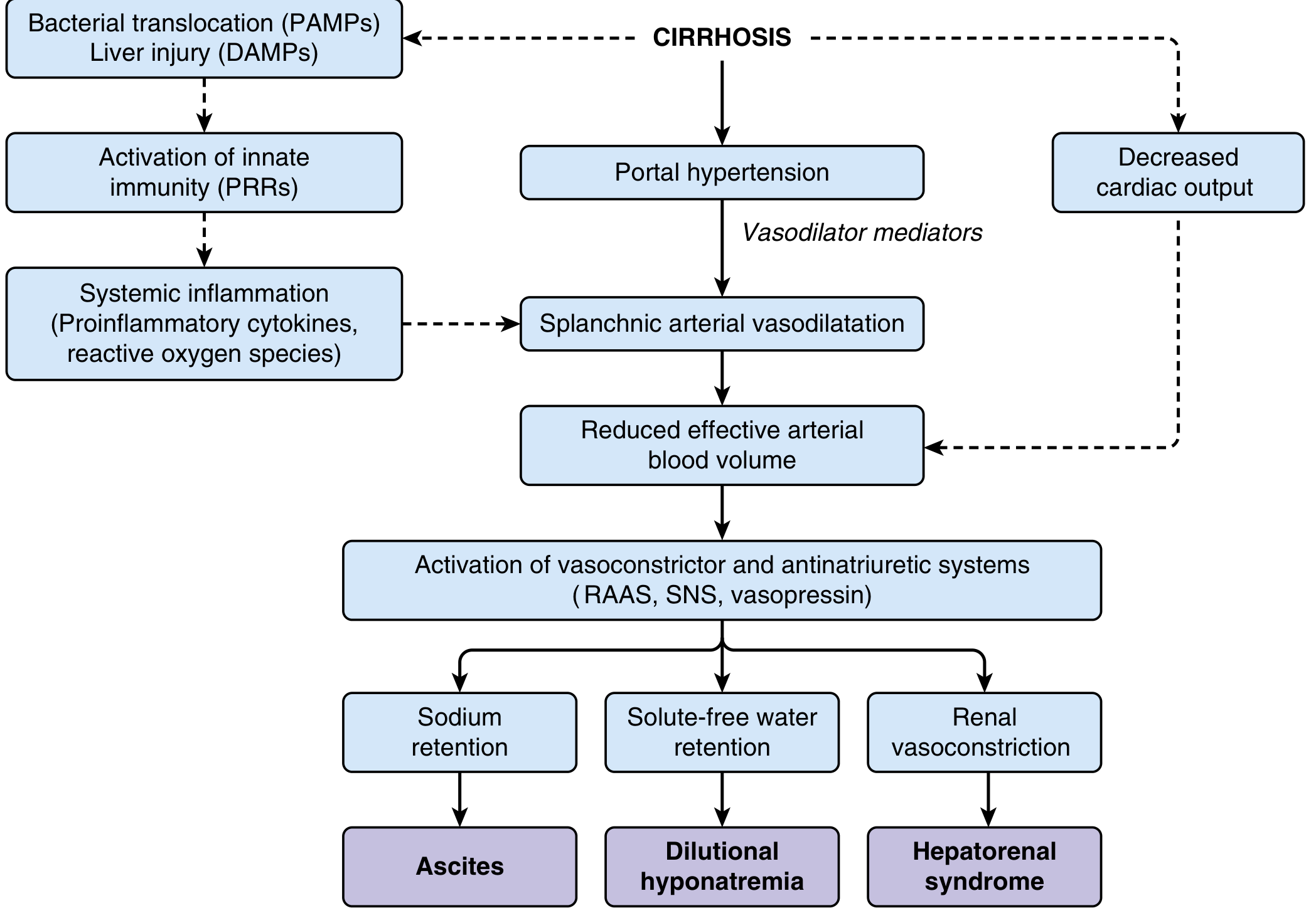
- Loss of hepatocytes → impaired synthetic function (coagulopathy, hypoalbuminemia, jaundice)
- Fibrosis + nodules → distorted architecture → increased intrahepatic resistance → portal hypertension
- Portal hypertension → splanchnic vasodilation (mediated by nitric oxide and vasodilator substances)
- Splanchnic vasodilation → reduced effective arterial blood volume
- Compensatory activation of RAAS + SNS + vasopressin
- Consequences: Na retention → Ascites; Water retention → Dilutional hyponatremia; Renal vasoconstriction → HRS
4. CLINICAL FEATURES
Compensated vs. Decompensated
| Feature | Compensated | Decompensated |
|---|---|---|
| Symptoms | Often asymptomatic; fatigue, anorexia | Jaundice, ascites, encephalopathy, bleeding |
| 5-yr survival | ~80% | ~30% |
| Child-Pugh | Class A (5-6) | Class B/C (≥7) |
Physical Examination Signs
- Skin: Spider angiomata, palmar erythema, jaundice, ecchymosis, Dupuytren contracture
- Abdomen: Hepatomegaly or shrunken liver, splenomegaly, ascites, caput medusae (dilated abdominal veins)
- Endocrine/Metabolic: Gynecomastia, testicular atrophy, loss of body hair (in men)
- General: Muscle wasting, peripheral edema, leukonychia (white nails), clubbing
Biliary cirrhosis is distinctive: patients present with pruritus and jaundice before end-stage disease.
- Rosen's Emergency Medicine, p. 1247
5. INVESTIGATIONS
Blood Tests
| Test | Finding in CLD |
|---|---|
| AST/ALT | Mildly elevated (rarely >5x in cirrhosis) |
| Bilirubin | Elevated (late finding) |
| Albumin | Low (impaired synthesis) |
| PT/INR | Prolonged (coagulopathy) |
| Platelets | Low (hypersplenism) |
| BUN/Creatinine | Elevated (dehydration or HRS) |
| Alkaline phosphatase | Disproportionately elevated in biliary disease |
Imaging
- Ultrasound: Nodular liver surface, splenomegaly, ascites - first-line
- Transient elastography (FibroScan): Non-invasive staging of fibrosis; also predicts varices and liver-related death
- CT/MRI: Better characterization, HCC surveillance
Liver Biopsy
- Gold standard for confirming cirrhosis and grading fibrosis
- Limitations: sampling error, interobserver variability, procedure risk
- Increasingly replaced by non-invasive markers
6. SEVERITY SCORING SYSTEMS (HIGH-YIELD EXAM TOPIC)
Child-Pugh Score (5 parameters)
| Parameter | 1 point | 2 points | 3 points |
|---|---|---|---|
| Bilirubin (mg/dL) | <2 | 2-3 | >3 |
| Albumin (g/dL) | >3.5 | 2.8-3.5 | <2.8 |
| PT prolongation (sec) / INR | <4 / <1.7 | 4-6 / 1.7-2.3 | >6 / >2.3 |
| Ascites | None | Mild | Moderate-severe |
| Encephalopathy | None | Grade 1-2 | Grade 3-4 |
- Class A (5-6): Compensated; well-tolerated surgery
- Class B (7-9): Moderate risk
- Class C (≥10): Decompensated; surgery contraindicated; listed for transplant
MELD Score
- Formula: 3.78 × ln[Bilirubin] + 11.2 × ln[INR] + 9.57 × ln[Creatinine] + 6.43
- Now includes serum sodium (MELD-Na / MELD 3.0) for improved accuracy
- Used for organ allocation in liver transplant listing in the United States
- Higher score = higher short-term mortality
- Harrison's Principles, p. 814-821
7. COMPLICATIONS (The "Big 5")
7a. Ascites
- Most common complication; develops in ~60% within 10 years of compensated cirrhosis
- Pathogenesis: Na retention (RAAS activation) → extracellular fluid expansion → peritoneal accumulation
- Diagnosis: SAAG (Serum-Ascites Albumin Gradient)
- SAAG ≥1.1 g/dL = portal hypertension (cirrhosis, cardiac, Budd-Chiari)
- SAAG <1.1 = non-portal (TB, malignancy, pancreatitis)
| Grade | Management |
|---|---|
| Grade 1 (mild) | Sodium restriction (<2g/day), monitor |
| Grade 2 (moderate) | Spironolactone ± furosemide (100:40 ratio) |
| Grade 3 (tense) | Large-volume paracentesis (LVP) + albumin 8g/L drained |
| Refractory | Repeat LVP, TIPS (transjugular intrahepatic portosystemic shunt), transplant |
Key: Give albumin 8 g per litre when removing >5 L by paracentesis. ACE inhibitors and ARBs are contraindicated in decompensated cirrhosis with ascites.
- Rosen's Emergency Medicine, p. 2500-2506
7b. Spontaneous Bacterial Peritonitis (SBP)
- Acute infection of ascitic fluid WITHOUT a surgical source
- Common organisms: E. coli > Klebsiella > Streptococcus pneumoniae
- Diagnosis: Ascitic fluid PMN (neutrophil) count >250 cells/mm³
- Treatment: Cefotaxime 2 g IV q8h for 5 days + albumin 1.5 g/kg on day 1, 1 g/kg on day 3
- Prophylaxis: Norfloxacin or ciprofloxacin in high-risk patients
- Rosen's Emergency Medicine, p. 2524-2535
7c. Variceal Bleeding
- From gastroesophageal varices due to portal hypertension (portal pressure >12 mmHg)
- Mortality: 10-15% per episode of upper GI bleeding from varices
- Acute management:
- Resuscitate (platelets >50,000 for procedures)
- Vasoactive drugs: Octreotide/somatostatin (reduce splanchnic flow)
- Antibiotic prophylaxis: Ceftriaxone (reduces SBP risk post-bleed)
- Endoscopic band ligation (EBL) - treatment of choice
- Balloon tamponade (Sengstaken-Blakemore tube) - bridge only
- TIPS - for refractory bleeding
- Secondary prophylaxis: Non-selective beta-blockers (propranolol/nadolol) + EBL
- Coagulopathy note: Prefer cryoprecipitate (1 unit/10 kg) over FFP for active bleeding in liver disease
7d. Hepatic Encephalopathy (HE)
- Neuropsychiatric dysfunction from hyperammonemia + altered neurotransmission (GABA-BZ receptor upregulation)
- Pathogenesis: Reduced hepatocyte function + portosystemic shunting → ammonia bypasses liver → blood-brain barrier disruption
- Note: Ammonia levels do NOT directly correlate with severity
| Grade | Features |
|---|---|
| 0 (Minimal/Covert) | Subclinical; detected only on psychometric tests |
| 1 | Trivial lack of awareness, euphoria/anxiety, impaired attention |
| 2 | Lethargy, disorientation, inappropriate behavior, asterixis |
| 3 | Somnolence, confusion, asterixis, bizarre behavior |
| 4 | Coma |
- GI bleeding, hypokalemia, infection, dehydration, constipation, sedatives
- Correct precipitants
- Lactulose 30-60 g/day (acidifies colon, traps NH4+, promotes evacuation)
- Rifaximin 400 mg PO q8h (non-absorbed antibiotic, reduces ammonia production)
- Branched-chain amino acids (BCAA) as adjunct
- Liver transplant is the definitive cure
HE develops in 50-70% of cirrhotic patients; 1-year survival without transplant is ~42%, 3-year ~23%
- Sleisenger & Fordtran's, p. 1815
7e. Hepatorenal Syndrome (HRS)
- Functional renal failure (no intrinsic renal disease) due to severe renal vasoconstriction in advanced cirrhosis
- Diagnostic criteria: Rising creatinine in liver failure + oliguria (<400 mL/day) + urine Na <10 mEq/L + no improvement after 2 days of diuretic withdrawal and albumin loading
| Type | Feature |
|---|---|
| HRS-AKI (formerly Type 1) | Rapid doubling of creatinine in <2 weeks; poor prognosis |
| HRS-CKD (formerly Type 2) | Gradual decline; associated with refractory ascites |
- Bridge therapy: Vasoconstrictors + albumin
- Terlipressin (vasopressin analogue) - most evidence
- Norepinephrine - equivalent efficacy
- Octreotide + midodrine combination
- TIPS
- Extracorporeal albumin dialysis (MARS)
- Definitive: Liver transplantation (reverses HRS)
- Goldman-Cecil Medicine; Harrison's Principles, p. 2221-2224
8. OTHER COMPLICATIONS
| Complication | Key Point |
|---|---|
| Hepatocellular carcinoma (HCC) | 6-monthly US ± AFP screening in all cirrhotics |
| Hepatopulmonary syndrome | Intrapulmonary shunting → hypoxia; platypnoea-orthodeoxia |
| Portopulmonary hypertension | Pulmonary arterial HTN; rules out transplant if severe |
| Hepatic hydrothorax | Right-sided pleural effusion (portal hypertension); fluid from peritoneum via diaphragmatic defect |
| Coagulopathy | Low platelets (hypersplenism) + low clotting factors + low fibrinogen |
| Hypersplenism | Thrombocytopenia, leukopenia, anemia |
| Hyponatremia | Dilutional; water restriction + V2 receptor antagonists (tolvaptan) |
| Osteoporosis | Especially in PBC/PSC; calcium + vitamin D supplementation |
9. MANAGEMENT OVERVIEW
General Principles
- Treat the underlying cause (antivirals for HBV/HCV, abstinence for alcohol, immunosuppression for AIH)
- Alcohol: strict abstinence for all patients
- Vaccinations: Hep A, Hep B, pneumococcal, annual influenza
- Avoid hepatotoxic drugs and NSAIDs (worsen renal function in cirrhosis)
- Regular surveillance: 6-monthly liver ultrasound for HCC; upper endoscopy at diagnosis to assess varices
Liver Transplantation
- Indication: Child-Pugh class B (score ≥7) or MELD ≥15, or major complication (HCC within Milan criteria, HRS, refractory ascites)
- Child-Pugh class C - surgery is otherwise contraindicated
- HBV cirrhosis: Long-term TDF/TAF/entecavir → can achieve cirrhosis regression in 75% at 5 years
10. HIGH-YIELD EXAM MNEMONICS
- Alcohol
- Biliary disease (PBC, PSC)
- Chronics - Viral hepatitis C, Cryptogenic
- Drugs/metabolic (NAFLD, Wilson's, Hemochromatosis)
- Etc. - Autoimmune hepatitis, Budd-Chiari
- Bilirubin
- Encephalopathy
- Albumin
- PT/INR
- Tension ascites
- GI bleeding
- Infection (SBP)
- Bowel obstruction/constipation
- Alcohol binge
- Drugs (sedatives, opioids, diuretics)
- Sodium and electrolyte disturbance (hypokalemia)
Causes of hepatomegaly
Causes of Hepatomegaly
Exam tip: The liver can feel hard and irregular (cirrhosis), tender and smooth (acute hepatitis, hepatic congestion from RHF), or nodular with bruit (HCC). The texture and tenderness give you the diagnosis.
- Goldman-Cecil Medicine, p. 1543-1544
Classification by Mechanism
1. INFLAMMATORY / INFECTIVE
| Cause | Notes |
|---|---|
| Acute viral hepatitis (A, B, C, D, E) | Tender, smooth, enlarged; "starry sky" on USS |
| Chronic viral hepatitis (B, C) | Coarsened echotexture on USS |
| Alcoholic hepatitis | Tender hepatomegaly; AST:ALT >2:1 |
| Infectious mononucleosis (EBV) | Young patients; splenomegaly + pharyngitis |
| Cytomegalovirus (CMV) | Especially in immunocompromised |
| Amoebic liver abscess | Tender, smooth; right lobe most affected |
| Pyogenic liver abscess | Marked tenderness; swinging fever |
| Hydatid disease (Echinococcus) | Cystic lesion; travel history |
| Malaria (Plasmodium) | Tropical; + splenomegaly |
| Schistosomiasis | Tropical; portal fibrosis, pipestem fibrosis |
| Leptospirosis | Weil's disease; jaundice + renal failure |
| Tuberculosis (miliary/hepatic) | Systemic infiltration; granulomas |
| Visceral leishmaniasis (Kala-azar) | Massive hepatosplenomegaly |
| Brucellosis | Animal contact; granulomatous hepatitis |
2. CONGESTIVE / VASCULAR
| Cause | Notes |
|---|---|
| Right heart failure (RHF) | Tender, pulsatile; elevated JVP; hepatojugular reflux |
| Constrictive pericarditis | Firm liver; Kussmaul sign; "nutmeg liver" on histology |
| Tricuspid regurgitation | Pulsatile hepatomegaly - characteristic finding |
| Budd-Chiari syndrome | Hepatic vein obstruction; ascites + tender hepatomegaly; caudate lobe hypertrophy |
| Inferior vena cava obstruction | Venous congestion |
| Sinusoidal obstruction syndrome (SOS) | Post-bone marrow transplant; tender hepatomegaly + jaundice + fluid retention |
Pulsatile hepatomegaly = tricuspid regurgitation until proven otherwise.
3. INFILTRATIVE
| Cause | Notes |
|---|---|
| Fatty liver (NAFLD/NASH) | Most common; diffuse increased echogenicity on USS |
| Alcoholic fatty liver | Reversible; large, smooth, tender liver |
| Amyloidosis | Firm, non-tender; "waxy" consistency |
| Sarcoidosis | Granulomas; + lymphadenopathy, lung involvement |
| Haemochromatosis | Iron overload; "bronze diabetes"; hepatomegaly + cirrhosis |
| Wilson's disease | Copper deposition; K-F rings; young patients |
| Glycogen storage diseases (GSD I - Von Gierke's) | Enlarged hepatocytes with pale cytoplasm; presents in infancy with hypoglycemia |
| Gaucher's disease | Lipid storage; glucocerebrosidase deficiency |
| Niemann-Pick disease | Sphingomyelin storage |
| α-1 antitrypsin deficiency | PAS+ inclusions in hepatocytes |
4. NEOPLASTIC / SPACE-OCCUPYING
| Cause | Notes |
|---|---|
| Hepatocellular carcinoma (HCC) | Arterial bruit; ↑ AFP; background cirrhosis |
| Metastatic carcinoma | Most common malignant cause; hard, nodular, knobbly; from colorectal, breast, lung, stomach, pancreas |
| Cholangiocarcinoma | Jaundice + hepatomegaly |
| Hepatic lymphoma | Primary or secondary; homogeneous enlargement |
| Hepatic haemangioma | Benign; most common benign liver tumour; no bruit |
| Hepatic adenoma | OCP use; risk of rupture |
| Focal nodular hyperplasia (FNH) | Central stellate scar on CT/MRI |
| Hepatic cysts | Simple cysts - incidental; polycystic liver disease |
| Leukaemia / lymphoma | Infiltration of liver; + lymphadenopathy, splenomegaly |
5. CHOLESTATIC / BILIARY
| Cause | Notes |
|---|---|
| Biliary tract obstruction (extrahepatic) | Dilated ducts on USS; ↑ ALP, bilirubin; pruritus |
| Primary biliary cholangitis (PBC) | Middle-aged women; anti-mitochondrial antibodies (AMA) |
| Primary sclerosing cholangitis (PSC) | Associated with IBD; MRCP: "beading" of bile ducts |
6. CIRRHOSIS
- Early/active cirrhosis → enlarged liver
- End-stage cirrhosis → shrunken, fibrosed liver (right lobe atrophies, caudate/left lobe hypertrophy)
- Nodular surface on ultrasound
Clinical Approach: Texture of the Liver (Exam Favourite)
| Texture | Likely Cause |
|---|---|
| Soft, tender, smooth | Acute hepatitis, RHF/congestion, abscess |
| Smooth, non-tender | Fatty liver, early cirrhosis, leukaemia |
| Firm, irregular, nodular | Cirrhosis, metastases |
| Hard, rock-hard nodular | Metastatic malignancy |
| Pulsatile | Tricuspid regurgitation |
Diagnostic Approach - Algorithm
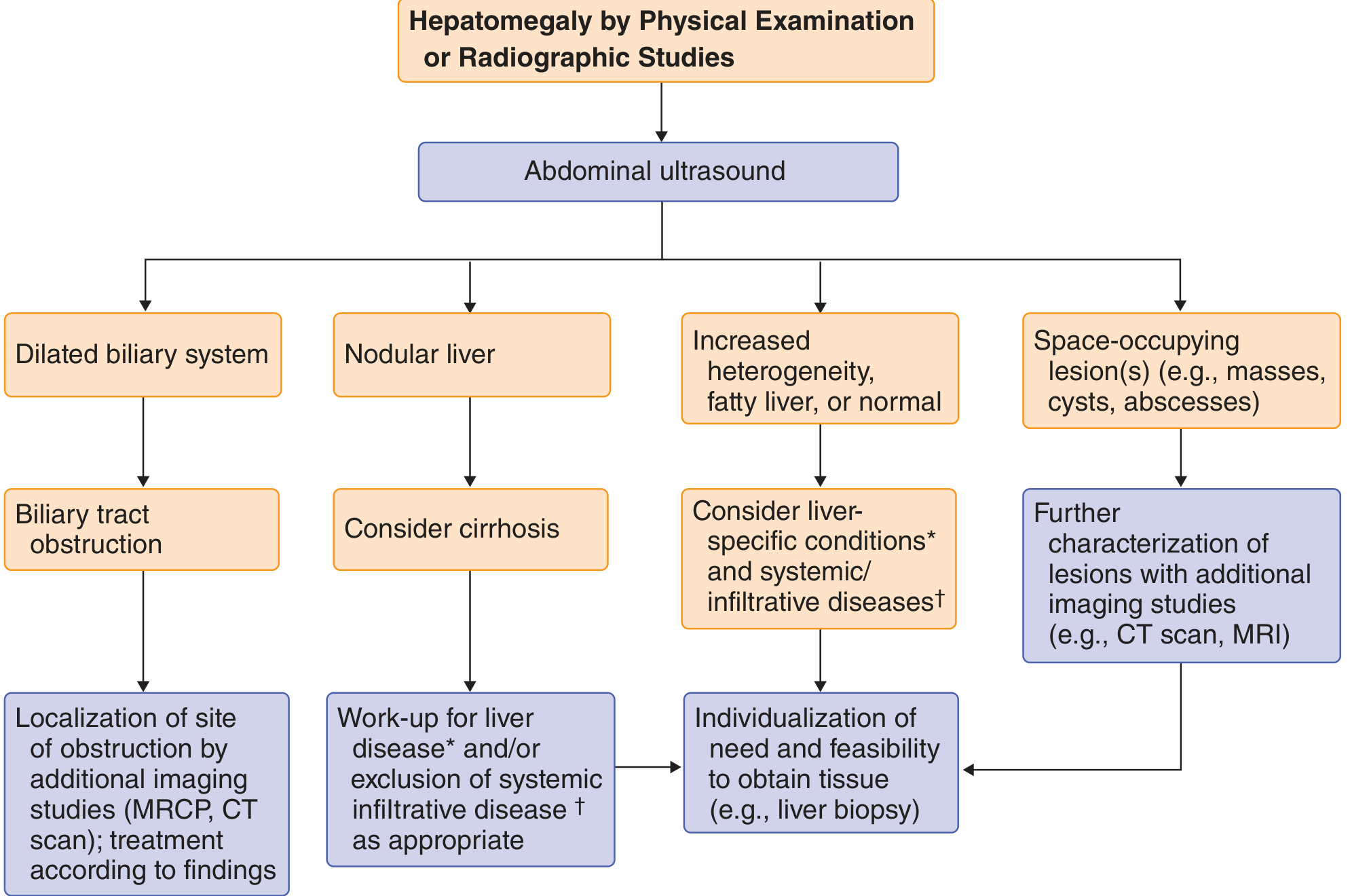
- Ultrasound abdomen - first-line
- Dilated biliary system → biliary obstruction → MRCP/CT
- Nodular liver → consider cirrhosis → liver-specific workup
- Increased heterogenicity / fatty liver → liver-specific conditions (hepatitis, NAFLD, NASH, autoimmune, metabolic) ± systemic/infiltrative diseases (amyloid, lymphoma, sarcoidosis, TB)
- Space-occupying lesions (masses, cysts, abscesses) → CT/MRI for characterisation
- Blood tests: LFTs, viral hepatitis serology, AFP, autoimmune markers, ferritin, ceruloplasmin
- Liver biopsy - if diagnosis unclear after imaging
High-Yield Exam Mnemonics
- Infections (hepatitis, abscess, malaria, amoeba)
- Neoplasia (HCC, mets, lymphoma)
- Vascular (RHF, Budd-Chiari, TR)
- Infiltrative (fatty liver, amyloid, sarcoid, GSD)
- Toxic/alcohol
- Endocrine/metabolic (DM, haemochromatosis, Wilson's)
- Drugs
- Malignancy (primary + secondary)
- Ducts (biliary obstruction)
- Storage diseases (Gaucher, Niemann-Pick)
Causes of hepatomegaly
Causes of Hepatomegaly - Quick Reference
By Mechanism (6 Groups)
| Category | Key Examples |
|---|---|
| 1. Inflammatory / Infective | Viral hepatitis (A-E), amoebic/pyogenic abscess, malaria, EBV, TB, kala-azar, schistosomiasis |
| 2. Congestive / Vascular | Right heart failure, tricuspid regurgitation (pulsatile!), Budd-Chiari syndrome, constrictive pericarditis |
| 3. Infiltrative | NAFLD/fatty liver, amyloidosis, sarcoidosis, haemochromatosis, Wilson's, glycogen storage diseases |
| 4. Neoplastic | HCC, metastases (most common malignant cause), lymphoma, haemangioma, hepatic adenoma |
| 5. Cholestatic / Biliary | Biliary obstruction, PBC, PSC |
| 6. Cirrhosis | Early/active cirrhosis (enlarged) - late cirrhosis gives a shrunken liver |
Texture Clue (Exam Favourite)
| Feel | Think |
|---|---|
| Soft, smooth, tender | Acute hepatitis, RHF, abscess |
| Smooth, non-tender | Fatty liver, leukaemia, amyloid |
| Firm, nodular | Cirrhosis, metastases |
| Rock-hard nodular | Metastatic malignancy |
| Pulsatile | Tricuspid regurgitation |
Mnemonic - "INVITED MDs"
Diagnostic Approach
- Ultrasound first - guides everything
- Dilated ducts → biliary obstruction → MRCP/CT
- Nodular → cirrhosis workup
- Fatty/heterogeneous → hepatitis panel, metabolic screen
- Mass/cyst/abscess → CT/MRI for characterisation
- Labs: LFTs, viral serology, AFP, AMA, ANA, ferritin, ceruloplasmin
- Liver biopsy if diagnosis remains unclear
Splenomegaly
Splenomegaly - Comprehensive Review
Functions of the Spleen (Context for Causes)
- Work hypertrophy - increased filtering/phagocytic demand (infection, haemolysis)
- Congestion - impaired venous drainage (portal hypertension, cardiac failure)
- Infiltration - abnormal cells/deposits (malignancy, storage diseases)
- Immunological hyperactivity - antigen stimulation (autoimmune, infection)
Classification of Causes
1. INFECTIVE / INFLAMMATORY
| Sub-category | Examples |
|---|---|
| Viral | Infectious mononucleosis (EBV) - most common viral cause; CMV; HIV; viral hepatitis |
| Bacterial | Infective endocarditis, typhoid fever, brucellosis, syphilis, TB, rickettsial (Rocky Mountain spotted fever), leptospirosis |
| Parasitic | Malaria (nearly always causes splenomegaly), kala-azar (visceral leishmaniasis) - causes massive splenomegaly, schistosomiasis, trypanosomiasis, toxoplasmosis, echinococcosis |
| Fungal | Histoplasmosis, disseminated cryptococcosis |
| Splenic abscess | From endocarditis emboli, typhoid, osteomyelitis, pancreatic necrosis |
Geography and travel history are key clues - malaria and kala-azar dominate in the tropics.
- Goldman-Cecil Medicine, p. 1435
2. CONGESTIVE / VASCULAR
| Cause | Notes |
|---|---|
| Cirrhosis of the liver | Main cause of massive congestive splenomegaly; portal hypertension |
| Portal vein thrombosis | Direct compression/obstruction of portal flow |
| Splenic vein thrombosis | Left-sided (sinistral) portal hypertension |
| Right heart failure | Systemic venous congestion; moderately enlarged spleen (rarely >500g) |
| Constrictive pericarditis | Venous backpressure |
| Tricuspid valve disease / cor pulmonale | Right-sided congestion |
| Budd-Chiari syndrome | Hepatic vein obstruction → portal hypertension |
| Schistosomiasis | "Pipestem" hepatic fibrosis → portal hypertension |
Cirrhosis is the most common cause of congestive splenomegaly. A palpable spleen tip in chronic liver disease implies portal hypertension.
- Robbins & Cotran Pathologic Basis of Disease
3. HAEMATOLOGICAL
Haemolytic Anaemias
| Cause | Notes |
|---|---|
| Hereditary spherocytosis | Autosomal dominant; spleen destroys spherocytes |
| Autoimmune haemolytic anaemia (AIHA) | Antibody-coated RBCs phagocytosed by splenic macrophages |
| Haemoglobinopathies | Sickle cell (early - later autosplenectomy), thalassaemia major |
| G6PD deficiency | Episodic haemolysis |
Myeloproliferative Neoplasms (causes massive splenomegaly)
| Cause | Notes |
|---|---|
| Primary myelofibrosis | Massive splenomegaly from extramedullary haematopoiesis; leukoerythroblastic blood film |
| Chronic myeloid leukaemia (CML) | Hallmark is massive splenomegaly; Philadelphia chromosome |
| Polycythaemia vera | Splenomegaly from extramedullary haematopoiesis |
| Essential thrombocythaemia | Moderate splenomegaly |
Lymphoproliferative Disorders
| Cause | Notes |
|---|---|
| Lymphoma (Hodgkin & Non-Hodgkin) | ± lymphadenopathy |
| Hairy cell leukaemia | Isolated splenomegaly WITHOUT lymphadenopathy - characteristic |
| Chronic lymphocytic leukaemia (CLL) | + lymphadenopathy |
| Acute leukaemias | Variable |
4. AUTOIMMUNE / IMMUNE-MEDIATED
| Cause | Notes |
|---|---|
| Rheumatoid arthritis | Mild-moderate; Felty syndrome = RA + splenomegaly + neutropenia |
| SLE | Autoimmune activation |
| ITP | Spleen seldom palpable despite destroying platelets |
| Drug reaction | Phenytoin, antibiotics (serum sickness) |
5. INFILTRATIVE / STORAGE DISEASES
| Cause | Notes |
|---|---|
| Gaucher's disease | Most common storage disease; glucocerebrosidase deficiency; bone crises |
| Niemann-Pick disease | Sphingomyelinase deficiency |
| Amyloidosis | Firm, non-tender splenomegaly |
| Sarcoidosis | Granulomatous infiltration; + hilar lymphadenopathy |
| Haemochromatosis | Iron deposition (usually secondary to cirrhosis) |
| Extramedullary haematopoiesis | Bone marrow failure/fibrosis |
6. OTHER
| Cause | Notes |
|---|---|
| Thyrotoxicosis | Mild, uncommon |
| Haemophagocytic lymphohistiocytosis (HLH) | Severe systemic inflammatory syndrome; fever + cytopenias + splenomegaly |
| Splenic cysts/pseudocysts | Benign; incidental |
| Metastatic malignancy | Rare; melanoma is the most likely solid tumour to metastasise to spleen |
MASSIVE Splenomegaly - Causes to Know (High-Yield)
| Mnemonic: "MMCH KG" |
|---|
| Myelofibrosis (primary) |
| Malaria (chronic) |
| CML (chronic myeloid leukaemia) |
| Hairy cell leukaemia |
| Kala-azar (visceral leishmaniasis) |
| Gaucher's disease |
Hypersplenism
- Splenomegaly
- One or more peripheral cytopenias (anaemia, leukopenia, thrombocytopenia)
- Normal or hypercellular bone marrow (compensating)
- Improvement after splenectomy
- 90% of peripheral platelets
- 65% of granulocytes
- 30% of red blood cells
When hypersplenism is the sole driver of cytopenias, splenectomy is often curative.
- Frameworks for Internal Medicine, p. 336
Clinical Assessment
Symptoms
- Usually asymptomatic (incidental finding on imaging)
- Left upper quadrant dragging discomfort / pain radiating to left shoulder (infarct)
- Early satiety (gastric compression)
- Cytopenias: fatigue, bleeding, infections
- Rarely: catastrophic splenic rupture (can be first sign with EBV!)
Examination Technique
- Position patient in right lateral decubitus with knees flexed
- Begin palpation from the right iliac fossa (to avoid missing massive spleen)
- Spleen descends on deep inspiration toward right iliac fossa
- Notch on medial border is pathognomonic
- Record in cm below left costal margin in midclavicular line
Key Differentiating Features of Spleen vs. Kidney
| Feature | Spleen | Kidney |
|---|---|---|
| Moves with respiration | Yes - downward & medially | Minimally |
| Ballottable | No | Yes |
| Can get above it | No | Sometimes |
| Notch on medial border | Yes | No |
| Band of colonic resonance | No (dull to percussion) | Yes |
Diagnostic Approach
- History: Age, travel history (tropical), drug history, family history
- Examination: Size of spleen, lymphadenopathy, hepatomegaly, jaundice, fever
- FBC + blood film: Cytopenias, abnormal cells, parasites (malaria film)
- LFTs, viral serology: EBV (Monospot/Paul-Bunnell), hepatitis screen
- Ultrasound: Confirms size, portal vein Doppler (portal HTN), lesions
- CT abdomen: Better anatomical detail, lymphadenopathy
- Bone marrow biopsy: If haematological malignancy suspected
- Splenic biopsy: Rarely needed; risk of haemorrhage
Complications of Splenomegaly
| Complication | Notes |
|---|---|
| Hypersplenism | Pancytopenia from sequestration |
| Splenic infarct | Pain; sickle cell, endocarditis, myeloproliferative |
| Splenic rupture | Spontaneous or traumatic; EBV classic cause |
| Splenic abscess | From septic emboli or haematogenous spread |
Causes of hepatosplenomegaly.
Causes of Hepatosplenomegaly
A palpable spleen tip in chronic liver disease implies portal hypertension. Fever + hepatosplenomegaly suggests systemic infection (EBV, malaria, visceral leishmaniasis), granulomatous disease (TB, histoplasmosis), or lymphoma.
- Goldman-Cecil Medicine, p. 2941
Classification by Cause
1. INFECTIVE (Most Common Group)
| Cause | Key Features |
|---|---|
| Infectious mononucleosis (EBV) | Classic triad: pharyngitis + lymphadenopathy + hepatosplenomegaly; young adults; Monospot +ve; rash with amoxicillin |
| CMV | Heterophil-negative mono syndrome; immunocompromised |
| Malaria | Nearly always causes hepatosplenomegaly; travel history; Plasmodium vivax most commonly; periodic fever |
| Visceral leishmaniasis (Kala-azar) | Massive hepatosplenomegaly; months of fever, weight loss, anaemia; sub-Saharan Africa, Indian subcontinent |
| Typhoid fever | Salmonella typhi; rose spots; relative bradycardia |
| Brucellosis | Animal contact; undulant fever; lymphadenopathy; malodorous perspiration; hallmark feature |
| Tuberculosis (miliary) | Systemic dissemination; granulomas in both organs |
| Schistosomiasis | Portal fibrosis ("pipestem" fibrosis) → portal HTN; tropical |
| Syphilis (secondary) | Spirochaetaemia; rash including palms & soles |
| Leptospirosis | Animal urine exposure; Weil's disease: jaundice + renal failure + hepatosplenomegaly |
| Infective endocarditis | Septic emboli; immunological activation |
| Histoplasmosis | Fungal; immunocompromised; bird/bat droppings |
| Toxoplasmosis | Intracellular parasite; immunocompromised |
| Trypanosomiasis | African sleeping sickness; tsetse fly |
| HIV | Often due to co-infection (hepatitis C, CMV, MAC) |
| Viral hepatitis (A, B, C, E) | Active hepatitis with systemic immune activation |
2. PORTAL HYPERTENSION / CONGESTIVE
| Cause | Key Features |
|---|---|
| Cirrhosis of the liver | Most common overall cause; portal HTN → splenomegaly; early cirrhosis enlarges liver; late = shrunken liver |
| Schistosomiasis | Pipestem fibrosis → portal hypertension; both organs enlarged |
| Right heart failure | Congestive hepatomegaly + passive splenic congestion |
| Constrictive pericarditis | Elevated JVP; Kussmaul sign |
| Budd-Chiari syndrome | Hepatic vein thrombosis; caudate lobe hypertrophy; + ascites |
| Portal / splenic vein thrombosis | Localised portal hypertension |
3. HAEMATOLOGICAL MALIGNANCIES
| Cause | Key Features |
|---|---|
| Lymphoma (Hodgkin & NHL) | Hepatosplenomegaly ± generalised lymphadenopathy; B symptoms (fever, night sweats, weight loss) |
| Leukaemia (CML, CLL, ALL, AML) | CML: massive hepatosplenomegaly; Philadelphia chromosome (t9;22) |
| Primary myelofibrosis | Massive splenomegaly (extramedullary haematopoiesis); leukoerythroblastic film; teardrop cells |
| Polycythaemia vera | Plethora; pruritis after bath; elevated Hb/Hct |
4. INFILTRATIVE / STORAGE DISEASES
| Cause | Key Features |
|---|---|
| Gaucher's disease | Most common lysosomal storage disorder; glucocerebrosidase deficiency; marked hepatosplenomegaly + bone crises; "wrinkled tissue paper" macrophages |
| Niemann-Pick disease | Sphingomyelinase deficiency; Type A: severe neonatal form with hepatosplenomegaly by 3 months |
| Amyloidosis | Firm, smooth, non-tender; AL or AA types; Congo red staining |
| Sarcoidosis | Non-caseating granulomas in liver + spleen; hilar lymphadenopathy; elevated ACE |
| Haemochromatosis | Iron overload; hepatomegaly + cirrhosis; spleen enlarged secondary to portal HTN |
| Glycogen storage diseases (von Gierke - GSD I) | Infants; hypoglycaemia + massive hepatomegaly; splenomegaly present |
| Hurler syndrome (MPS I) | Mucopolysaccharidosis; children; coarse facies + organomegaly |
5. AUTOIMMUNE / INFLAMMATORY
| Cause | Key Features |
|---|---|
| Systemic lupus erythematosus (SLE) | Multi-system; ANA +ve; lupus hepatitis |
| Rheumatoid arthritis / Felty syndrome | RA + splenomegaly + neutropenia; hepatomegaly from Felty's or co-pathology |
| Still's disease (SJIA) | Fever + rash + lymphadenopathy + hepatosplenomegaly; serositis; very high ferritin; macrophage activation syndrome |
| Haemophagocytic lymphohistiocytosis (HLH) | Cytokine storm; fever + hepatosplenomegaly + cytopenias + very high ferritin (>10,000); triggered by EBV or malignancy |
6. MISCELLANEOUS
| Cause | Key Features |
|---|---|
| Thyrotoxicosis | Mild; warm, tender goitre |
| Chronic myeloid conditions (extramedullary haematopoiesis) | Bone marrow failure states |
| Wilson's disease | Copper overload; K-F rings; young patient |
| α-1 antitrypsin deficiency | Liver disease (PAS+ inclusions) + emphysema |
Exam Summary Table: Key Distinguishing Features
| Cause | Key Associated Feature |
|---|---|
| EBV | Young, pharyngitis, posterior cervical lymphadenopathy, rash with amoxicillin |
| Malaria | Travel, cyclical fever, anaemia, thrombocytopenia |
| Kala-azar | Tropical, months of fever, massive splenomegaly > hepatomegaly, anaemia, hypergammaglobulinaemia |
| Cirrhosis | Spider naevi, caput medusae, ascites, jaundice |
| CML | Massive splenomegaly, Philadelphia chromosome, basophilia |
| Lymphoma | B symptoms, lymphadenopathy, elevated LDH |
| Gaucher's | Bone pain/crises, Ashkenazi Jewish heritage, no neurological symptoms (type 1) |
| Sarcoidosis | Bilateral hilar lymphadenopathy on CXR, elevated ACE, erythema nodosum |
| Brucellosis | Animal contact, undulant fever, malodorous sweating |
| HLH | Very high ferritin >10,000, cytopenias, triggered by EBV or haematological malignancy |
| Still's disease | Quotidian salmon-coloured rash, arthritis, serositis, high ferritin |
Mnemonic: "I CLAIM SHE"
- I - Infections (EBV, malaria, kala-azar, typhoid, TB, brucellosis)
- C - Cirrhosis / portal hypertension
- L - Leukaemia & lymphoma
- A - Amyloid / Autoimmune (SLE, RA, Still's, HLH)
- I - Infiltrative (sarcoidosis, Gaucher's, Niemann-Pick)
- M - Myeloproliferative disorders (CML, myelofibrosis, PV)
- S - Storage diseases (glycogen, mucopolysaccharidoses)
- H - Heart failure (right-sided congestive)
- E - Endocrine/metabolic (haemochromatosis, Wilson's, thyrotoxicosis)
Create a diagnostic flowchart or comparison table for hepatosplenomegaly workup
A detailed medical diagnostic flowchart for hepatosplenomegaly workup. The flowchart starts at the top with "HEPATOSPLENOMEGALY CONFIRMED (Liver + Spleen both enlarged)" in a bold header box. It branches into two main paths: "Step 1: History & Examination" and flows down through investigations. The chart has these sequential steps: 1. Top box: "HEPATOSPLENOMEGALY" 2. Second level: Three parallel boxes for key history clues: "Fever + Travel?" | "B symptoms + Lymphadenopathy?" | "Chronic Liver Disease signs?" 3. Each of those branches to initial investigations box: "Step 2: First-line Investigations: FBC + film, LFTs, ESR/CRP, Blood cultures, Monospot/EBV serology, Malaria films x3, Hepatitis serology, LDH, Ferritin" 4. Results branch into 4 diagnostic pathways: - PATH A (Infection): "Fever + Lymphadenopathy → EBV, CMV, HIV, TB, Brucellosis, Malaria, Kala-azar, Typhoid" in green - PATH B (Portal HTN/Liver): "Abnormal LFTs + Cirrhosis signs → Cirrhosis, Portal HTN, Budd-Chiari, Heart Failure" in blue - PATH C (Haematological): "Abnormal blood film + High LDH → Lymphoma, CML, Myelofibrosis, Leukaemia" in orange - PATH D (Infiltrative): "Normal/mild LFTs + No fever → Sarcoidosis, Amyloid, Gaucher's, Storage diseases" in purple 5. Final box at bottom: "Step 3: Second-line Tests based on pathway: Imaging (USS/CT), Bone marrow biopsy, Lymph node biopsy, Liver biopsy, Specialised serology" Clean professional medical illustration, white background, color-coded pathways, clear arrows, sans-serif font, suitable for medical students and clinical exam preparation.
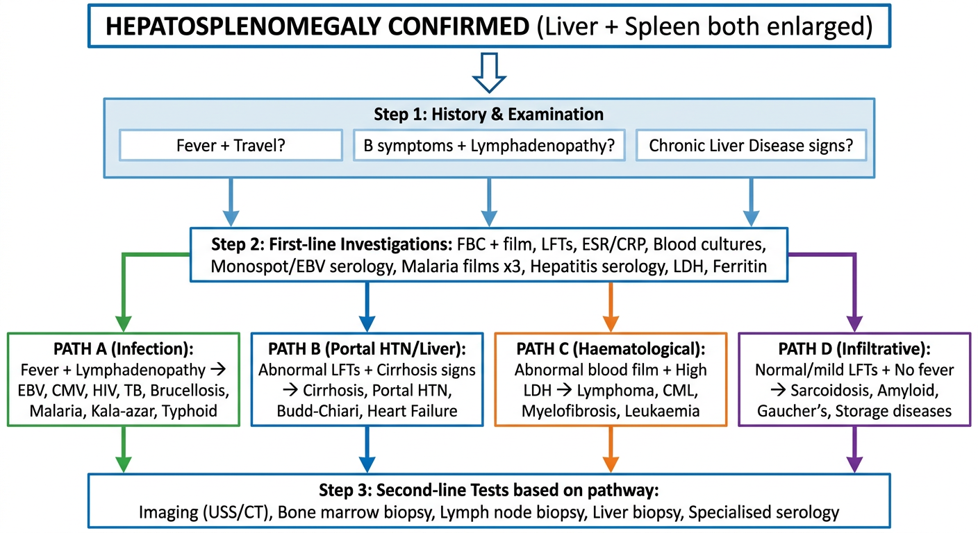
Hepatosplenomegaly Workup - Comparison Table
Step 1: History Clues That Guide the Diagnosis
| Clue | Think |
|---|---|
| Fever + travel to tropics | Malaria, kala-azar, schistosomiasis, typhoid |
| Fever + pharyngitis + young patient | EBV (infectious mononucleosis) |
| Fever + months of weight loss | TB, lymphoma, kala-azar, HIV |
| B symptoms + lymphadenopathy | Lymphoma (Hodgkin / NHL) |
| Jaundice + spider naevi + ascites | Cirrhosis with portal hypertension |
| Animal/farm contact + undulant fever | Brucellosis |
| Bone pain + Ashkenazi Jewish | Gaucher's disease |
| Child + coarse facies + developmental delay | Mucopolysaccharidosis (Hurler) |
| Arthritis + quotidian rash + very high ferritin | Still's disease / HLH |
| Rash on palms and soles | Secondary syphilis |
Step 2: First-Line Investigations (Order in ALL patients)
| Test | What it detects |
|---|---|
| FBC + blood film | Anaemia, cytopenias, blast cells, malaria parasites, atypical lymphocytes (EBV) |
| LFTs (AST/ALT/ALP/bilirubin/albumin) | Hepatocellular vs cholestatic pattern; synthetic function |
| ESR / CRP | Non-specific inflammation |
| Monospot (Paul-Bunnell) + EBV serology | Infectious mononucleosis |
| Malaria thick & thin films x3 | If any travel history |
| Hepatitis A/B/C/E serology | Viral hepatitis |
| LDH | Haemolysis, lymphoma, leukaemia |
| Ferritin | Very high (>10,000) in HLH; elevated in haemochromatosis, inflammation |
| Blood cultures x3 | Bacterial sepsis, brucellosis, typhoid, endocarditis |
| Abdominal ultrasound + Doppler | Confirms organomegaly; portal vein flow; ascites; masses; lymph nodes |
Step 3: Pathway-Based Second-Line Tests
| Pathway | Pattern | Second-Line Tests | Likely Diagnoses |
|---|---|---|---|
| A - Infective | Fever + elevated WBC/CRP + lymphadenopathy | EBV VCA IgM/IgG; CMV PCR; HIV viral load; Blood cultures; Brucella serology; Widal test; Leishmania rK39 antigen; Splenic aspirate for Leishmania | EBV, CMV, HIV, typhoid, brucellosis, kala-azar, malaria, TB |
| B - Portal HTN / Liver | Abnormal LFTs + cirrhosis signs + ascites | Hepatitis serology; INR/PT; Albumin; Liver USS with Doppler; CT abdomen; Liver biopsy; SAAG on ascitic fluid | Cirrhosis, Budd-Chiari, cardiac failure, schistosomiasis |
| C - Haematological | Abnormal blood film + high LDH + lymphadenopathy | CT chest/abdomen/pelvis; Bone marrow biopsy; Lymph node biopsy; BCR-ABL (CML); JAK2 mutation (PV/MF); Flow cytometry | CML, lymphoma, myelofibrosis, leukaemia, hairy cell leukaemia |
| D - Infiltrative / Storage | No/mild fever, minimal LFT derangement, no lymphadenopathy | Serum ACE (sarcoidosis); β-glucocerebrosidase (Gaucher's); Serum protein electrophoresis + Congo red biopsy (amyloid); Ceruloplasmin (Wilson's); Ferritin + HFE gene (haemochromatosis) | Gaucher's, sarcoidosis, amyloid, haemochromatosis, Wilson's |
| E - Autoimmune/Inflammatory | Young patient + arthritis + very high ferritin + rash | ANA, anti-dsDNA (SLE); RF, anti-CCP (RA/Felty); Ferritin >10,000 + triglycerides + fibrinogen (HLH); Bone marrow biopsy (haemophagocytosis) | SLE, Still's disease, HLH, Felty syndrome |
Quick Pattern Recognition (Exam Shortcuts)
| Pattern | Diagnosis |
|---|---|
| Fever + pharyngitis + posterior cervical LN + hepatosplenomegaly + atypical lymphocytes | EBV mononucleosis |
| Months of fever + massive hepatosplenomegaly + anaemia + weight loss (tropics) | Kala-azar |
| Massive splenomegaly > hepatomegaly + basophilia + Philadelphia chromosome | CML |
| B symptoms + mediastinal widening on CXR + hepatosplenomegaly | Hodgkin lymphoma |
| Child + bone crises + hepatosplenomegaly + no neurological features | Gaucher's disease (Type 1) |
| Ferritin >10,000 + cytopenias + fever + hepatosplenomegaly | HLH |
| Ascites + caput medusae + spider naevi + hepatosplenomegaly | Cirrhosis / Portal HTN |
| Bilateral hilar lymphadenopathy + hepatosplenomegaly + elevated ACE | Sarcoidosis |
| Quotidian salmon rash + arthritis + hepatosplenomegaly + serositis | Still's disease |
History I should ask for an per abdomen case
History for a Per Abdomen Case
1. PRESENTING COMPLAINT (PC)
- Abdominal pain / swelling / distension
- Jaundice
- Vomiting / nausea
- Change in bowel habits
- Loss of weight / appetite
- Bleeding (haematemesis, malena, rectal bleeding)
2. HISTORY OF PRESENTING COMPLAINT (HPC)
For Abdominal Pain - Use "SOCRATES"
| Letter | Question |
|---|---|
| S - Site | "Where exactly is the pain?" (point with one finger) |
| O - Onset | "Did it come on suddenly or gradually?" |
| C - Character | "Is it sharp, dull, crampy, burning, colicky?" |
| R - Radiation | "Does it spread anywhere? Back? Shoulder tip? Groin?" |
| A - Associated symptoms | (see below) |
| T - Timing | "Is it constant or does it come and go?" |
| E - Exacerbating/relieving factors | "What makes it better or worse? Food? Posture? Movement?" |
| S - Severity | "On a scale of 1-10, how bad is the pain?" |
For Abdominal Swelling / Distension
- Onset: sudden or gradual?
- Which part of the abdomen?
- Any pain associated with it?
- Any change in urine output or colour?
- Ankle swelling (ascites from cardiac/liver/nephrotic)?
For Jaundice
- Duration and progression
- Dark urine / pale stools / pruritus (obstructive jaundice triad)
- Fever and rigors (Charcot's triad → cholangitis)?
- Contact with jaundiced persons (hepatitis)
- Blood transfusions / IV drug use (hepatitis B/C)
- Alcohol history
- Recent travel
3. ASSOCIATED SYMPTOMS - Ask Specifically
Gastrointestinal
| Symptom | Why it matters |
|---|---|
| Nausea / vomiting | Content? Blood (haematemesis)? |
| Dysphagia | Oesophageal pathology |
| Heartburn / regurgitation | GORD |
| Anorexia / weight loss | Malignancy, chronic liver disease |
| Bowel habits | Constipation vs diarrhoea |
| Blood in stool | Frank red (lower GI) vs melaena (upper GI) |
| Mucus in stool | IBD, colorectal carcinoma |
| Pale/clay stools | Biliary obstruction |
| Dark urine | Haemolysis, hepatitis, biliary obstruction |
| Abdominal distension | Ascites, obstruction, gas |
| Tenesmus | Rectal pathology |
Systemic
| Symptom | Why it matters |
|---|---|
| Fever / chills / rigors | Infection, cholangitis, abscess, malaria |
| Night sweats | TB, lymphoma |
| Fatigue / weakness | Anaemia, CLD, malignancy |
| Pruritus (itching) | Obstructive jaundice, cholestasis, PBC |
| Easy bruising / bleeding | Coagulopathy (liver disease) |
| Peripheral oedema | Hypoalbuminaemia, cardiac failure |
| Shortness of breath | Massive ascites, hepatic hydrothorax, anaemia |
| Joint pains | Haemochromatosis, IBD, SLE |
| Skin rash | SLE, Still's disease, secondary syphilis |
4. PAST MEDICAL HISTORY (PMH)
- Previous episodes of similar complaints
- Known liver disease / hepatitis (B, C)
- Known gallstones / biliary disease
- Diabetes mellitus (NAFLD)
- Hypertension / cardiac disease (congestive hepatomegaly)
- Malignancy (any prior cancer → metastases)
- Previous abdominal surgery
- Blood transfusions
- Autoimmune diseases (SLE, rheumatoid arthritis, IBD)
- Haematological conditions (thalassaemia, sickle cell)
5. DRUG HISTORY (DH)
| Category | Examples |
|---|---|
| Hepatotoxic drugs | Methotrexate, isoniazid, paracetamol (overdose), amiodarone, statins, rifampicin |
| Drugs causing splenomegaly | Phenytoin |
| Anticoagulants | Warfarin, heparin (bleeding risk) |
| OCP | Risk of hepatic adenoma, Budd-Chiari |
| NSAIDs | GI bleeding, renal impairment in liver disease |
| Alcohol | Quantify - units per week (safe limit: ♂ <14 units/week, ♀ <14 units/week) |
| Herbal / traditional medicines | Often hepatotoxic |
6. FAMILY HISTORY (FH)
- Liver disease / jaundice in family
- Haematological disorders (thalassaemia, sickle cell, hereditary spherocytosis)
- Storage diseases (Gaucher's, Wilson's disease - autosomal recessive)
- Haemochromatosis (HFE gene mutation)
- Malignancy (colorectal cancer, HCC)
- Polycystic kidney/liver disease
7. SOCIAL HISTORY (SH)
Alcohol
- How much? How often? For how long?
- Type of alcohol
- CAGE questionnaire: Cut down? Annoyed? Guilty? Eye-opener?
Smoking
- Pack years (risk of pancreatitis, malignancy)
Occupation
- Farmer / abattoir (brucellosis, leptospirosis)
- Healthcare worker (hepatitis B/C risk)
- Chemical / solvent exposure (hepatotoxins)
Travel History
- Travel to tropics → malaria, kala-azar, typhoid, schistosomiasis, hepatitis A/E
- Where? When? Prophylaxis taken?
Sexual History
- Unprotected intercourse → hepatitis B/C, HIV, syphilis
IV Drug Use / Tattoos / Piercings
- Hepatitis B, C, HIV risk
Diet
- Raw shellfish → hepatitis A, E
- Animal contact → brucellosis, leptospirosis
Living Conditions
- Overcrowding → TB, typhoid, hepatitis A
8. REVIEW OF SYSTEMS (ROS)
- Cardiorespiratory: cough, breathlessness, chest pain (right heart failure causing hepatomegaly)
- Urological: decreased urine output (hepatorenal syndrome, nephrotic)
- Neurological: confusion, flapping tremor/asterixis (hepatic encephalopathy)
- Musculoskeletal: arthralgia, bone pain (Gaucher's, haemochromatosis)
- Endocrine: thyroid symptoms (thyrotoxicosis → mild hepatosplenomegaly)
OSCE Memory Aid: "ABCDE Symptoms" for Abdominal History
| Letter | Symptom group |
|---|---|
| A | Appetite/Anorexia/Abdominal pain |
| B | Bowel habit change / Bleeding (haematemesis, melaena, PR bleed) |
| C | Colour change (jaundice, dark urine, pale stools) |
| D | Distension / Dysphagia / Dark urine |
| E | Energy loss / constitutional (fever, weight loss, night sweats) |
Clinical Correlation: History Clues → Likely Diagnosis
| History Clue | Think |
|---|---|
| Alcohol excess + jaundice + RUQ pain | Alcoholic hepatitis / cirrhosis |
| IV drug use + jaundice + fever | Hepatitis B/C, endocarditis |
| Travel to tropics + fever + anaemia | Malaria, kala-azar, typhoid |
| Young patient + pharyngitis + hepatosplenomegaly | EBV mononucleosis |
| OCP use + Budd-Chiari features | Hepatic vein thrombosis |
| Pale stools + dark urine + pruritus + fever + RUQ pain | Cholangitis (Charcot's triad) |
| Weight loss + B symptoms + lymphadenopathy | Lymphoma / TB |
| Family history + bronze skin + DM + arthropathy | Haemochromatosis |
| Child + bone pain + Ashkenazi Jewish | Gaucher's disease |
| Arthritis + butterfly rash | SLE hepatitis |
I need it like symptoms and what all it rules out and what all negative history I should ask to rule out what
Per Abdomen History - Symptom-Based with Rule In / Rule Out Logic
PRESENTING COMPLAINT: Hepatosplenomegaly / Abdominal Mass / Jaundice
A. FEVER
If PRESENT → Suggests:
- Infection: malaria, kala-azar, typhoid, EBV, brucellosis, TB, leptospirosis
- Lymphoma (B symptoms)
- Still's disease / HLH
- SBP (in ascites patient)
- Cholangitis
Negative History to Ask (to rule out specific causes):
| Question | Rules Out if Negative |
|---|---|
| "Did you travel to any tropical country recently?" | Malaria, kala-azar, typhoid, schistosomiasis |
| "Did you have sore throat along with the fever?" | EBV (infectious mononucleosis) |
| "Was the fever coming and going in cycles / every 48-72 hrs?" | Malaria (tertian/quartan pattern) |
| "Did you have rigors (violent shivering) with the fever?" | Malaria, cholangitis, sepsis |
| "Did you have months of low-grade fever with weight loss?" | TB, lymphoma, kala-azar |
| "Any contact with animals / farm / unpasteurised milk?" | Brucellosis, leptospirosis |
| "Any night sweats along with the fever?" | TB, lymphoma |
| "Any skin rash with the fever?" | Still's disease, typhoid (rose spots), syphilis |
| "Any abdominal pain with the fever?" | Cholangitis, liver abscess, SBP |
B. JAUNDICE
If PRESENT → Suggests:
- Hepatitis (viral, alcoholic, drug-induced)
- Biliary obstruction (gallstones, cholangiocarcinoma, head of pancreas CA)
- Haemolysis
- Cirrhosis (late)
Negative History to Ask:
| Question | Rules Out if Negative |
|---|---|
| "Did you notice pale / clay-coloured stools?" | Obstructive (surgical) jaundice - rules out biliary obstruction if absent |
| "Did your urine turn dark (tea-coloured)?" | Obstructive jaundice, hepatitis |
| "Did you have itching (pruritus) all over?" | Obstructive jaundice / cholestasis (PBC) - suggests if present |
| "Any fever and pain in the right side with jaundice?" | Charcot's triad → cholangitis (fever + jaundice + RUQ pain) |
| "Did other family members or contacts develop jaundice around the same time?" | Hepatitis A/E (faeco-oral, epidemic) |
| "Any recent blood transfusion or injection / shared needles?" | Hepatitis B, C, HIV |
| "Do you drink alcohol regularly? How much?" | Alcoholic hepatitis / cirrhosis |
| "Have you taken any new medications or herbal medicines?" | Drug-induced jaundice |
| "Any episode of sudden worsening of anaemia with dark urine?" | Haemolytic jaundice (G6PD, thalassaemia crisis) |
C. ABDOMINAL PAIN
If PRESENT → Use SOCRATES; then ask:
| Question | Rules Out if Negative |
|---|---|
| "Is the pain in the right upper quadrant?" | Liver pathology, cholecystitis, hepatitis |
| "Does the pain radiate to your right shoulder tip?" | Diaphragmatic irritation (hepatic abscess, perforated viscus) |
| "Does the pain radiate to your back?" | Pancreatitis, carcinoma head of pancreas, aortic aneurysm |
| "Is the pain colicky - coming and going in waves?" | Biliary colic, ureteric colic, intestinal obstruction |
| "Does eating make it worse?" | Peptic ulcer (worse with food = gastric; better with food = duodenal); mesenteric angina (chronic ischaemia) |
| "Is the pain relieved by opening your bowels?" | Irritable bowel syndrome, bowel pathology |
| "Is the pain worse when you breathe in?" | Peritonism, hepatic/splenic pathology |
D. ABDOMINAL SWELLING / DISTENSION
If PRESENT → Think: Ascites, organomegaly, mass, gas, obesity, pregnancy
| Question | Rules Out if Negative |
|---|---|
| "Has the swelling been increasing gradually over weeks/months?" | Cirrhosis / malignant ascites / slowly growing mass |
| "Did the swelling come on suddenly?" | Acute obstruction, haemoperitoneum, acute liver failure |
| "Is it associated with swelling of both legs / ankle swelling?" | Hypoalbuminaemia (cirrhosis, nephrotic), cardiac failure → rules out these if no leg swelling |
| "Any breathlessness due to the swelling?" | Massive ascites pushing up diaphragm, hepatic hydrothorax |
| "Do you pass less urine than usual?" | Hepatorenal syndrome, nephrotic syndrome, cardiac failure |
| "Any history of heart disease?" | Cardiac ascites / congestive hepatomegaly - rules out if no cardiac history |
| "Is the swelling in a specific region only or all over the abdomen?" | Localised = organomegaly/mass; diffuse = ascites/gas |
E. WEIGHT LOSS / ANOREXIA
If PRESENT → Suggests:
- Malignancy (HCC, lymphoma, metastases, pancreatic CA)
- Chronic infections (TB, kala-azar, HIV)
- Cirrhosis / chronic liver disease
- Chronic inflammatory conditions
| Question | Rules Out if Negative |
|---|---|
| "How much weight have you lost and over how long?" | Rapid severe loss → malignancy; gradual → CLD, TB |
| "Is your appetite completely lost or reduced?" | Complete anorexia suggests malignancy or severe liver disease |
| "Any early satiety (feeling full very quickly)?" | Massive splenomegaly compressing stomach; gastric cancer |
| "Any night sweats along with weight loss?" | TB, lymphoma - rules out if absent |
| "Any chronic cough or breathlessness along with weight loss?" | TB (pulmonary), lymphoma with mediastinal involvement |
| "Any change in bowel habits along with weight loss?" | Colorectal malignancy with liver metastases |
F. BLEEDING
If PRESENT → Suggests: Portal hypertension (varices), coagulopathy, malignancy
| Question | Rules Out if Negative |
|---|---|
| "Did you vomit blood (haematemesis)? What colour?" | Bright red = active variceal/arterial bleed; coffee-ground = slower upper GI bleed |
| "Did you pass black tarry stools (melaena)?" | Upper GI bleed (varices, peptic ulcer) - rules out upper GI source if absent |
| "Did you pass fresh red blood in stools?" | Lower GI bleed, anorectal pathology |
| "Do you bruise easily or bleed from minor cuts for a long time?" | Coagulopathy of liver disease, thrombocytopenia (hypersplenism) |
| "Any bleeding from gums or nose?" | Coagulopathy, leukaemia, thrombocytopenia |
G. BOWEL HABIT CHANGES
| Question | Rules Out if Negative |
|---|---|
| "Have you noticed any change in your bowel habit?" | Colorectal cancer (alternating constipation/diarrhoea), IBD |
| "Any mucus or blood mixed in the stools?" | Colorectal CA, IBD, amoebic colitis |
| "Any pale / greasy stools that are hard to flush?" | Steatorrhoea → malabsorption, cholestasis, pancreatic insufficiency |
| "Any diarrhoea?" | Infective cause, IBD, colorectal CA, carcinoid |
| "Any constipation?" | Colorectal CA, hypothyroidism |
H. ALCOHOL HISTORY (Always ask in EVERY abdominal case)
| Question | Significance |
|---|---|
| "Do you drink alcohol?" | Opens the question |
| "How much per week? What type?" | Quantify in units |
| "For how many years have you been drinking?" | Duration determines damage |
| "Have you ever tried to cut down?" (CAGE) | Dependence assessment |
- Alcoholic hepatitis, alcoholic cirrhosis, alcoholic pancreatitis
I. DRUG / MEDICATION HISTORY
| Question | Rules Out if Negative |
|---|---|
| "Do you take any regular medications?" | Drug-induced hepatitis, drug-induced cholestasis |
| "Any herbal or traditional medicines?" | Hepatotoxic herbals (very common, often missed) |
| "Any painkillers (paracetamol, ibuprofen)?" | Paracetamol overdose, NSAID-induced GI bleed |
| "Have you used any injections / IV drugs?" | Hepatitis B, C, HIV - rules out if never injected |
| "Any long-term medications like methotrexate, isoniazid, amiodarone?" | Specific drug-induced liver disease |
| "OCP use?" (in women) | Hepatic adenoma, Budd-Chiari syndrome |
J. SOCIAL / EXPOSURE HISTORY
| Question | Rules Out if Negative |
|---|---|
| "Any contact with a jaundiced person recently?" | Hepatitis A / E (faeco-oral) - rules out epidemic hepatitis |
| "Did you eat raw shellfish or food from unhygienic sources?" | Hepatitis A / E |
| "Any unprotected sexual contact?" | Hepatitis B, C, HIV, syphilis |
| "Any recent tattooing or body piercing?" | Hepatitis B, C |
| "Do you work with animals or on a farm?" | Brucellosis, leptospirosis, hydatid cyst |
| "Have you worked in a mine / construction / in water?" | Leptospirosis (water exposure), schistosomiasis |
| "Any family member with similar illness?" | Hereditary haemochromatosis, Wilson's, thalassaemia, Gilbert's |
K. FAMILY HISTORY
| Question | Rules Out if Negative |
|---|---|
| "Any family member with liver disease or jaundice?" | Hereditary causes (haemochromatosis, Wilson's, Gilbert's) |
| "Any family member with blood disorders?" | Thalassaemia, sickle cell, hereditary spherocytosis |
| "Any family member with similar abdominal swelling?" | Polycystic liver/kidney disease, familial malignancy |
| "Any family member with cancer?" | Familial colorectal CA, HCC in HBV-infected families |
SUMMARY CARD: Positive vs Negative History at a Glance
| Symptom Present | Rules In | Negative History Needed | Rules Out |
|---|---|---|---|
| Cyclical fever + travel | Malaria | No travel → | Malaria less likely |
| Pharyngitis + HSM | EBV | No sore throat → | Mononucleosis less likely |
| Pale stools + dark urine + itch | Obstructive jaundice | Stools normal coloured → | Biliary obstruction less likely |
| Night sweats + weight loss | TB / Lymphoma | No night sweats → | TB/lymphoma less likely |
| Alcohol history | Alcoholic liver disease | No alcohol → | Alcoholic CLD excluded |
| IV drug use / tattoos | Hepatitis B/C | Never → | Blood-borne hepatitis less likely |
| Easy bruising | Coagulopathy / CLD | No bruising → | Severe coagulopathy less likely |
| Haematemesis | Varices / upper GI bleed | No → | Variceal bleed less likely |
| Animal contact | Brucellosis / leptospirosis | No animal contact → | Zoonosis less likely |
| Family liver disease | Hereditary (HH, Wilson's) | No family history → | Hereditary cause less likely |
Deviation of angle of mouth towards right since 2 days difficult in chewing and inability to close left eyelids since 2 days what should I ask
- Mouth deviates to RIGHT = left side weak (unopposed right side pulls)
- Cannot close LEFT eyelid = frontalis + orbicularis oculi involved = LMN lesion
- Difficulty chewing = buccinator weakness (left)
History for Left LMN Facial Nerve (CN VII) Palsy
First - Confirm the Diagnosis Clinically
- Mouth deviates RIGHT = left facial muscles weak (right side unopposed)
- Cannot close LEFT eyelid = upper + lower face involved = LMN lesion
- Difficulty chewing = left buccinator weak
LMN VII palsy = ALL facial muscles on that side (upper + lower). UMN spares the forehead/eye closure. This is NOT a stroke causing facial weakness.
- Cummings Otolaryngology, p. 3294
HISTORY QUESTIONS - Symptom Based, Rule In / Rule Out
1. ONSET & PROGRESSION
| Question | Rules In | Rules Out if Negative |
|---|---|---|
| "Did the weakness come on suddenly (within hours)?" | Bell's palsy (acute), stroke | Tumour (tumour is insidious/gradual) |
| "Did the weakness develop gradually over weeks/months?" | Parotid tumour, cholesteatoma, facial nerve schwannoma | Bell's palsy (Bell's is always ACUTE) |
| "Is the weakness getting worse progressively?" | Malignancy, cholesteatoma, schwannoma | Bell's palsy plateaus within 48-72 hours |
| "Any previous episode of facial weakness that recovered?" | Melkersson-Rosenthal syndrome (recurrent facial palsy + lip oedema + fissured tongue) | First episode only → less likely recurrent |
2. EAR SYMPTOMS (Critical - Localises the Lesion)
| Question | Rules In | Rules Out if Negative |
|---|---|---|
| "Do you have pain in or around the ear (otalgia)?" | Ramsay Hunt syndrome (severe otalgia precedes vesicles), AOM, cholesteatoma | Helps rule out Ramsay Hunt if no ear pain |
| "Did you notice any blisters / vesicles in or around your ear or on your tongue?" | Ramsay Hunt syndrome (VZV reactivation in geniculate ganglion) - blisters in ear canal, pinna, hard palate | If NO vesicles → more likely Bell's palsy |
| "Any ear discharge (otorrhoea)?" | Chronic otitis media (COM), cholesteatoma | Rules out ear disease if absent |
| "Any hearing loss on the same side?" | Ramsay Hunt (SNHL), cholesteatoma, AOM, acoustic neuroma, temporal bone fracture | If hearing intact → Bell's more likely |
| "Any ringing in the ear (tinnitus)?" | Ramsay Hunt, acoustic neuroma, COM | |
| "Any dizziness or loss of balance (vertigo)?" | Ramsay Hunt (vestibular dysfunction), acoustic neuroma, labyrinthitis | If NO vertigo → Bell's more likely |
| "Any recent ear surgery or injury to the head/ear?" | Post-surgical, temporal bone fracture | Rules out traumatic cause if absent |
3. EYE SYMPTOMS (Consequence of VII Palsy)
| Question | Rules In / Significance | Rules Out if Negative |
|---|---|---|
| "Does your eye feel dry?" | Reduced lacrimation from chorda tympani / greater petrosal nerve involvement - indicates level of lesion | |
| "Is your eye watering excessively (epiphora)?" | Paradoxical lacrimation OR corneal exposure / ectropion | |
| "Do you feel like you cannot blink your left eye normally?" | Confirms orbicularis oculi weakness = LMN | UMN spares eye closure |
| "Any pain or redness in the left eye?" | Corneal exposure keratopathy (from lagophthalmos) - complication - needs urgent eye care |
4. TASTE & MOUTH SYMPTOMS (Localises Lesion Level)
| Question | Significance - Lesion Level |
|---|---|
| "Have you noticed any change in taste on the front of your tongue?" | Chorda tympani branch (taste to anterior 2/3 tongue) - lesion is above stylomastoid foramen (proximal) |
| "Is your mouth dry?" | Submandibular/sublingual gland secretion affected - chorda tympani involved |
| "Any pain behind the ear or around the mastoid?" | Post-auricular pain = very early Bell's palsy sign (inflammation at stylomastoid foramen area) |
| "Do sounds seem unusually loud on that side (hyperacusis)?" | Stapedius branch of VII affected - lesion above nerve to stapedius (more proximal) |
Lesion level summary:
- Taste loss + hyperacusis + dry eye = HIGH lesion (intratemporal, above greater superficial petrosal nerve)
- Taste loss only (no hyperacusis, tearing intact) = middle segment
- No taste loss, no hyperacusis = LOW lesion at/below stylomastoid foramen → parotid gland pathology
- Cummings Otolaryngology, p. 3294; Shambaugh Surgery of the Ear
5. NECK / PAROTID SWELLING
| Question | Rules In | Rules Out if Negative |
|---|---|---|
| "Any swelling in front of the ear or along the jaw (parotid region)?" | Parotid tumour (malignant parotid tumours invade facial nerve) | If no parotid swelling → parotid origin less likely |
| "Any lumps in the neck?" | Lymph nodes from parotid/head & neck malignancy, lymphoma | |
| "Any pain in the parotid region?" | Parotitis (bacterial), parotid abscess |
6. SYSTEMIC / ASSOCIATED SYMPTOMS
| Question | Rules In | Rules Out if Negative |
|---|---|---|
| "Any weakness of limbs, numbness, trouble speaking or swallowing?" | Central (UMN) cause - stroke, brain tumour (but then forehead should be SPARED) | If all negative → confirms peripheral LMN |
| "Any recent rash on the body, joints pain, red eyes?" | Sarcoidosis (can cause facial palsy, uveitis, arthritis); also Lyme disease, SLE | |
| "Any recent tick bite? Outdoor activities in forested areas?" | Lyme disease (Borrelia) - common cause of facial palsy worldwide, can be bilateral | |
| "Any recent upper respiratory tract infection / cold in past 2-4 weeks?" | Post-viral Bell's palsy (HSV reactivation) | |
| "Any history of chickenpox previously?" | VZV latency → Ramsay Hunt risk | |
| "Do you have diabetes mellitus?" | Diabetes is a risk factor for Bell's palsy and peripheral neuropathy | |
| "Do you have high blood pressure?" | Vascular risk; also associated with Bell's palsy | |
| "Any history of cancer anywhere in the body?" | Metastatic tumour to temporal bone or parotid | Rules out metastatic cause if no cancer history |
| "Any HIV risk factors?" | HIV can cause facial palsy |
7. PREGNANCY (if female)
| Question | Significance |
|---|---|
| "Are you pregnant? Which trimester?" | Bell's palsy is 3x more common in the 3rd trimester of pregnancy |
SUMMARY: Most Common Causes of Acute LMN Facial Palsy + History Key
| Cause | Must-Ask History | Distinguishing Feature |
|---|---|---|
| Bell's palsy (most common ~70%) | Recent URTI, DM, HTN, pregnancy | Acute onset, no vesicles, no ear disease, resolves |
| Ramsay Hunt syndrome (~12%) | Severe ear pain BEFORE palsy, vesicles in ear/pinna, hearing loss, vertigo | VZV; WORSE prognosis than Bell's |
| Otitis media / Cholesteatoma | Ear discharge, hearing loss, chronic ear disease | Ear disease precedes palsy |
| Parotid tumour | Gradual onset, parotid swelling, no taste/hyperacusis change | Progressive, insidious |
| Lyme disease | Tick bite, outdoor exposure, erythema migrans rash | Can be bilateral |
| Sarcoidosis (Heerfordt syndrome) | Dry eyes, bilateral palsy, uveitis, parotid swelling | Bilateral; uveoparotid fever |
| Temporal bone fracture | Head trauma, ear bleeding, hearing loss | History of injury |
| Guillain-Barré syndrome | Ascending limb weakness, bilateral facial palsy, recent infection | Bilateral; areflexia |
| Acoustic neuroma | Gradual, unilateral SNHL + tinnitus, NO ear pain | Slow progressive |
Minimum Diagnostic Criteria for Bell's Palsy (Taverner)
- Paralysis/paresis of all muscle groups of ONE side of the face
- Sudden onset
- Absence of CNS disease signs
- Absence of ear or cerebellopontine angle disease
"All that palsies are not Bell" - always exclude other causes before labelling Bell's palsy.
- Cummings Otolaryngology, p. 3294
Bell palsy
An error occurred (ValidationException) when calling the ConverseStream operation: The model returned the following errors: messages.21.content.19.image.source.base64.data: At least one of the image dimensions exceed max allowed size: 8000 pixels
We encountered an error while processing your request. Please try again or reach out to us for help.
Causes related to avove case
An error occurred (ValidationException) when calling the ConverseStream operation: The model returned the following errors: messages.21.content.19.image.source.base64.data: At least one of the image dimensions exceed max allowed size: 8000 pixels
We encountered an error while processing your request. Please try again or reach out to us for help.