
Anatomy of ear
anatomy of the ear diagram showing external middle and inner ear
Anatomy of the Ear
1. External Ear
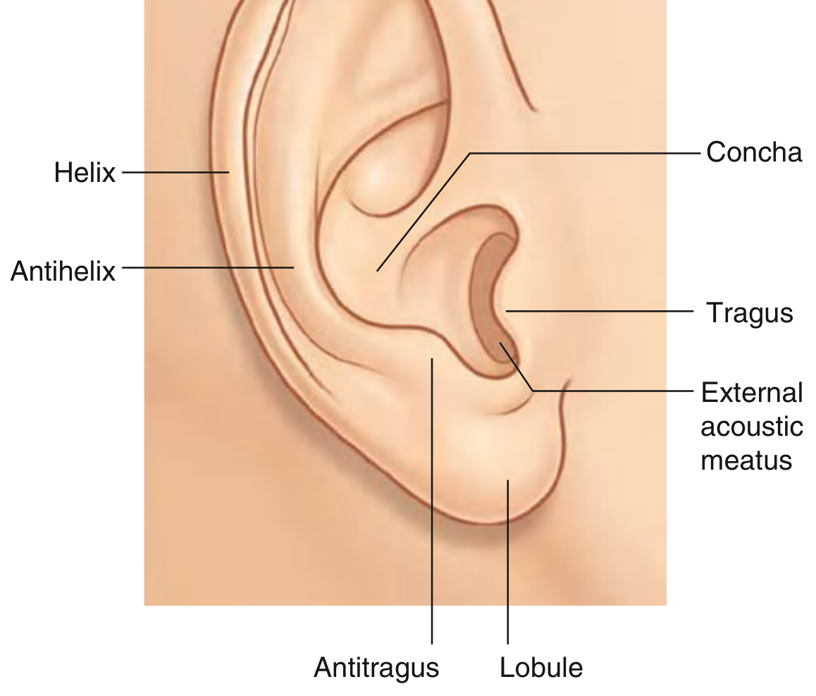
- Auricle (pinna) – the visible projection on the side of the head, made of elastic cartilage covered by skin, shaped to capture sound. Landmarks include the helix (outer rim), antihelix, tragus and antitragus, concha (hollow center), and the lobule (the only part without cartilage support).
- External acoustic meatus – the canal leading from the concha inward to the tympanic membrane, directing sound waves toward the middle ear.
2. Middle Ear (Tympanic Cavity)
- Tympanic membrane – thin, translucent partition that vibrates in response to sound.
- Auditory ossicles – the malleus, incus, and stapes, forming a lever chain that transmits and amplifies vibrations from the tympanic membrane to the oval window of the internal ear. The stapes footplate sits in the oval window.
- Associated muscles – tensor tympani (innervated by CN V3) and stapedius (innervated by CN VII), which dampen excessive ossicular movement (acoustic reflex).
- Pharyngotympanic (Eustachian) tube – connects the middle ear to the nasopharynx, equalizing air pressure across the tympanic membrane.
- Mastoid antrum and air cells – posterior air-filled extension of the middle ear cavity.
- The chorda tympani (a branch of CN VII carrying taste fibers) and the tympanic plexus (CN IX) pass through this cavity.
3. Internal (Inner) Ear
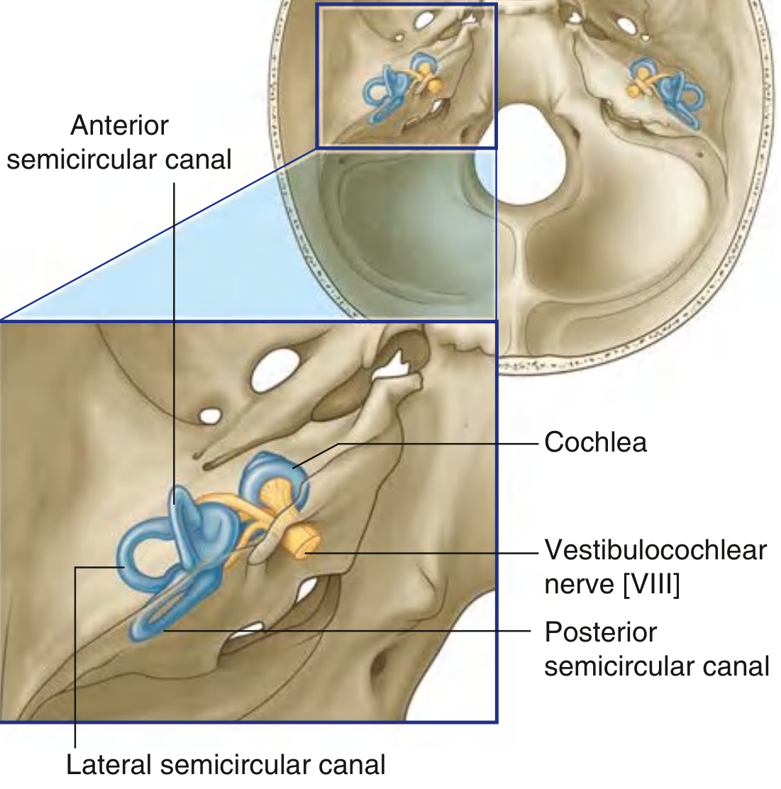
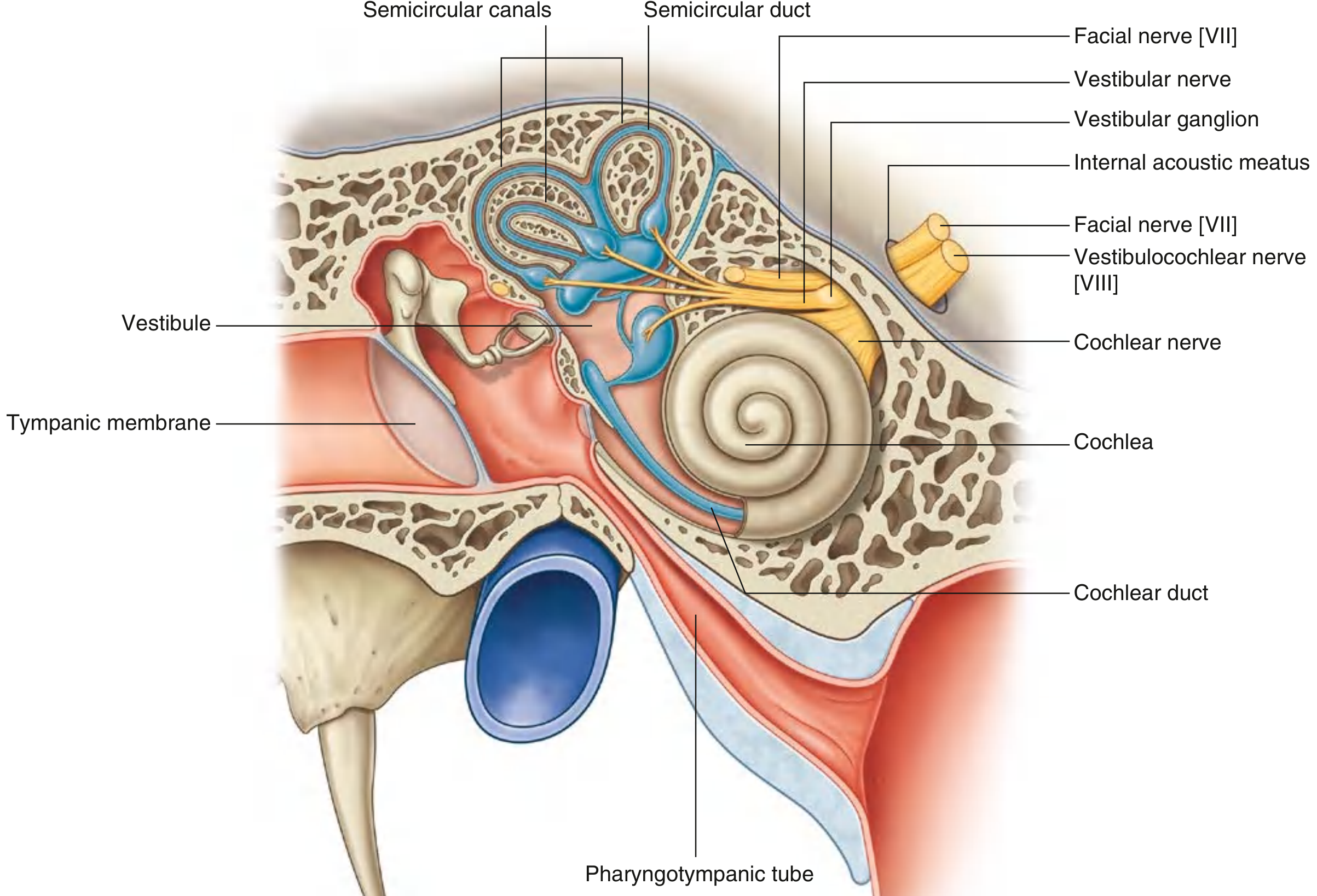
- Bony labyrinth: vestibule, three semicircular canals, and cochlea.
- Membranous labyrinth: semicircular ducts, cochlear duct, and two sacs — the utricle and saccule.
- Vestibular part – balance information from semicircular ducts, utricle, saccule
- Cochlear part – hearing information from the cochlear duct
Composite Overview
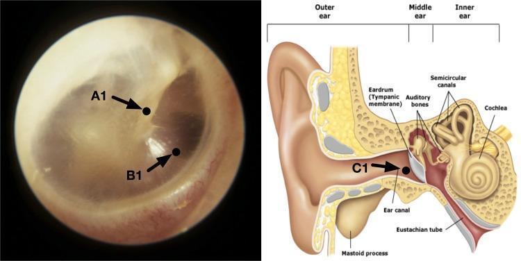
Functional Summary
| Region | Main Structures | Function |
|---|---|---|
| External ear | Auricle, external acoustic meatus | Sound collection and conduction |
| Middle ear | Tympanic membrane, ossicles, Eustachian tube | Sound transmission/amplification, pressure equalization |
| Internal ear | Cochlea, vestibule, semicircular canals | Hearing (cochlea) and balance (vestibular apparatus) |
Convert in pdf
mkdir -p /tmp/workspace/ear-anatomy/assets /tmp/workspace/ear-anatomy/output && ls -la /tmp/workspace/
cd /tmp/workspace/ear-anatomy/assets && \ curl -s -o auricle.png "https://cdn.orris.care/cdss_images/00a54eb5a4673a333c5346b5d2ed1344301c1f3a390bad198477f6ab68cea6ee.png" && \ curl -s -o innervation.png "https://cdn.orris.care/cdss_images/4716d5951d05ab2d79c04a81049cc2d33f308869da768968d9ae3d5d6711f0a7.png" && \ curl -s -o internal_ear_location.png "https://cdn.orris.care/cdss_images/9cb4f2340095f42dbc54a4ec4b9d4c114a910b2e6c596f6ece0f051fee29bc0a.png" && \ curl -s -o internal_ear_relationships.png "https://cdn.orris.care/cdss_images/d02eba7729c051a05f55c7e94923513e843a7f1eafbfba4a0f9dabc611239c08.png" && \ curl -s -o composite_cross_section.jpg "https://cdn.orris.care/cdss_images/pmc_clinical_VQA_3f20ab24d42d04b85ddbd86255917048fc2db8244feb48c751ad719d02bcc441.jpg" && \ ls -la
~/ear-anatomy/build_pdf.py
from reportlab.lib.pagesizes import A4
from reportlab.lib.units import cm
from reportlab.lib.styles import getSampleStyleSheet, ParagraphStyle
from reportlab.lib.enums import TA_CENTER
from reportlab.platypus import (
    SimpleDocTemplate, Paragraph, Spacer, Image, Table, TableStyle,
    PageBreak, ListFlowable, ListItem, KeepTogether
)
from reportlab.lib import colors
from PIL import Image as PILImage
import os

ASSETS = "/tmp/workspace/ear-anatomy/assets"
OUT = "/tmp/workspace/ear-anatomy/output/Anatomy_of_the_Ear.pdf"

styles = getSampleStyleSheet()

title_style = ParagraphStyle(
    "TitleStyle", parent=styles["Title"], fontSize=22, spaceAfter=6, textColor=colors.HexColor("#1a3c6e")
)
subtitle_style = ParagraphStyle(
    "Subtitle", parent=styles["Normal"], fontSize=11, alignment=TA_CENTER, textColor=colors.grey, spaceAfter=18
)
h1 = ParagraphStyle(
    "H1", parent=styles["Heading1"], fontSize=16, textColor=colors.HexColor("#1a3c6e"),
    spaceBefore=14, spaceAfter=8, borderPadding=0
)
h2 = ParagraphStyle(
    "H2", parent=styles["Heading2"], fontSize=13, textColor=colors.HexColor("#2f5d8a"),
    spaceBefore=10, spaceAfter=6
)
body = ParagraphStyle(
    "Body", parent=styles["Normal"], fontSize=10.5, leading=15, spaceAfter=8, alignment=4
)
bullet_style = ParagraphStyle(
    "Bullet", parent=body, leftIndent=14, spaceAfter=4
)
caption_style = ParagraphStyle(
    "Caption", parent=styles["Normal"], fontSize=9, alignment=TA_CENTER, textColor=colors.grey,
    spaceAfter=14, spaceBefore=4, fontName="Helvetica-Oblique"
)
source_style = ParagraphStyle(
    "Source", parent=styles["Normal"], fontSize=8.5, textColor=colors.grey, spaceBefore=20
)

def img_flowable(path, max_width=13*cm, max_height=8*cm):
    with PILImage.open(path) as im:
        w, h = im.size
    ratio = w / h
    width = max_width
    height = width / ratio
    if height > max_height:
        height = max_height
        width = height * ratio
    return Image(path, width=width, height=height)

story = []

# Title page header
story.append(Paragraph("Anatomy of the Ear", title_style))
story.append(Paragraph("External, Middle, and Internal Ear Structures & Function", subtitle_style))

# Intro
story.append(Paragraph(
    "The ear is divided into three anatomical regions: the <b>external (outer) ear</b>, the "
    "<b>middle ear</b>, and the <b>internal (inner) ear</b>. The first two are concerned mainly "
    "with conducting sound, while the internal ear houses the organs of hearing and balance.",
    body
))

# Composite image
story.append(img_flowable(os.path.join(ASSETS, "composite_cross_section.jpg"), max_width=14*cm, max_height=8*cm))
story.append(Paragraph("Labeled cross-section of outer, middle, and inner ear structures", caption_style))

# 1. External Ear
story.append(Paragraph("1. External Ear", h1))
story.append(Paragraph("Consists of two parts:", body))
ext_items = [
    "<b>Auricle (pinna)</b> - the visible projection on the side of the head, made of elastic cartilage covered by skin, shaped to capture sound. Landmarks include the <b>helix</b> (outer rim), <b>antihelix</b>, <b>tragus</b> and <b>antitragus</b>, <b>concha</b> (hollow center), and the <b>lobule</b> (the only part without cartilage support).",
    "<b>External acoustic meatus</b> - the canal leading from the concha inward to the tympanic membrane, directing sound waves toward the middle ear.",
]
story.append(ListFlowable([ListItem(Paragraph(t, bullet_style)) for t in ext_items], bulletType="bullet"))
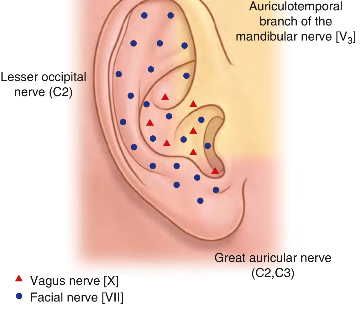
story.append(Paragraph(
    "Both intrinsic and extrinsic auricular muscles are present but are largely vestigial in humans; "
    "they are innervated by the <b>facial nerve (CN VII)</b>. Sensory supply to the auricle comes from "
    "multiple sources: the great auricular and lesser occipital nerves (cervical plexus), the auriculotemporal "
    "nerve (CN V3), and branches of CN VII and CN X.",
    body
))

story.append(img_flowable(os.path.join(ASSETS, "auricle.png"), max_width=9*cm, max_height=7*cm))
story.append(Paragraph("Fig. Auricle - surface landmarks", caption_style))

story.append(img_flowable(os.path.join(ASSETS, "innervation.png"), max_width=9*cm, max_height=7*cm))
story.append(Paragraph("Fig. Sensory innervation of the auricle", caption_style))

# 2. Middle Ear
story.append(Paragraph("2. Middle Ear (Tympanic Cavity)", h1))
story.append(Paragraph(
    "An air-filled space in the petrous temporal bone, situated between the tympanic membrane laterally "
    "and the internal ear medially. Key components:", body
))
mid_items = [
    "<b>Tympanic membrane</b> - thin, translucent partition that vibrates in response to sound.",
    "<b>Auditory ossicles</b> - the <b>malleus</b>, <b>incus</b>, and <b>stapes</b>, forming a lever chain that transmits and amplifies vibrations from the tympanic membrane to the oval window of the internal ear. The stapes footplate sits in the oval window.",
    "<b>Associated muscles</b> - <b>tensor tympani</b> (innervated by CN V3) and <b>stapedius</b> (innervated by CN VII), which dampen excessive ossicular movement (acoustic reflex).",
    "<b>Pharyngotympanic (Eustachian) tube</b> - connects the middle ear to the nasopharynx, equalizing air pressure across the tympanic membrane.",
    "<b>Mastoid antrum and air cells</b> - posterior air-filled extension of the middle ear cavity.",
]
story.append(ListFlowable([ListItem(Paragraph(t, bullet_style)) for t in mid_items], bulletType="bullet"))
story.append(Paragraph(
    "The <b>chorda tympani</b> (a branch of CN VII carrying taste fibers) and the tympanic plexus (CN IX) "
    "pass through this cavity.", body
))

# 3. Internal Ear
story.append(Paragraph("3. Internal (Inner) Ear", h1))
story.append(Paragraph(
    "Located in the petrous part of the temporal bone, medial to the middle ear. It consists of the "
    "<b>bony labyrinth</b> (bony cavities filled with perilymph) containing a <b>membranous labyrinth</b> "
    "(filled with endolymph):", body
))
inner_items = [
    "<b>Bony labyrinth</b>: vestibule, three semicircular canals, and cochlea.",
    "<b>Membranous labyrinth</b>: semicircular ducts, cochlear duct, and two sacs - the <b>utricle</b> and <b>saccule</b>.",
]
story.append(ListFlowable([ListItem(Paragraph(t, bullet_style)) for t in inner_items], bulletType="bullet"))

story.append(Paragraph(
    "<b>Vestibule</b> - the central chamber, contains the oval window and connects anteriorly to the cochlea, "
    "posterosuperiorly to the semicircular canals.", body
))
story.append(Paragraph(
    "<b>Semicircular canals</b> (anterior, posterior, lateral) - each forms two-thirds of a circle, oriented "
    "at right angles to one another, with a dilated <b>ampulla</b> at one end; they detect rotational "
    "(angular) head movements.", body
))
story.append(Paragraph(
    "<b>Cochlea</b> - a coiled bony tube (about 2.5 turns) housing the <b>cochlear duct</b> and the "
    "<b>organ of Corti</b>, the sensory organ of hearing, which converts fluid vibrations into neural signals.",
    body
))
story.append(Paragraph(
    "<b>Utricle and saccule</b> - contain maculae that detect linear acceleration and head position (gravity).",
    body
))
story.append(Paragraph(
    "The <b>vestibulocochlear nerve (CN VIII)</b> enters via the internal acoustic meatus and splits into:",
    body
))
cn8_items = [
    "<b>Vestibular part</b> - balance information from semicircular ducts, utricle, saccule",
    "<b>Cochlear part</b> - hearing information from the cochlear duct",
]
story.append(ListFlowable([ListItem(Paragraph(t, bullet_style)) for t in cn8_items], bulletType="bullet"))

story.append(img_flowable(os.path.join(ASSETS, "internal_ear_location.png"), max_width=9.5*cm, max_height=7.5*cm))
story.append(Paragraph("Fig. Location of the internal ear in the temporal bone", caption_style))

story.append(PageBreak())

story.append(img_flowable(os.path.join(ASSETS, "internal_ear_relationships.png"), max_width=12*cm, max_height=15*cm))
story.append(Paragraph("Fig. Anatomical relationships of internal ear structures", caption_style))

# Functional summary table
story.append(Paragraph("Functional Summary", h1))
table_data = [
    ["Region", "Main Structures", "Function"],
    ["External ear", "Auricle, external acoustic meatus", "Sound collection and conduction"],
    ["Middle ear", "Tympanic membrane, ossicles, Eustachian tube", "Sound transmission/amplification, pressure equalization"],
    ["Internal ear", "Cochlea, vestibule, semicircular canals", "Hearing (cochlea) and balance (vestibular apparatus)"],
]
tbl = Table(table_data, colWidths=[3.2*cm, 6.3*cm, 6.5*cm])
tbl.setStyle(TableStyle([
    ("BACKGROUND", (0, 0), (-1, 0), colors.HexColor("#1a3c6e")),
    ("TEXTCOLOR", (0, 0), (-1, 0), colors.white),
    ("FONTNAME", (0, 0), (-1, 0), "Helvetica-Bold"),
    ("FONTSIZE", (0, 0), (-1, -1), 9),
    ("GRID", (0, 0), (-1, -1), 0.5, colors.grey),
    ("VALIGN", (0, 0), (-1, -1), "TOP"),
    ("ROWBACKGROUNDS", (0, 1), (-1, -1), [colors.whitesmoke, colors.white]),
    ("LEFTPADDING", (0, 0), (-1, -1), 6),
    ("RIGHTPADDING", (0, 0), (-1, -1), 6),
    ("TOPPADDING", (0, 0), (-1, -1), 5),
    ("BOTTOMPADDING", (0, 0), (-1, -1), 5),
]))
story.append(tbl)

story.append(Paragraph(
    "Source: Gray's Anatomy for Students (pp. 1093-1106); Cummings Otolaryngology Head and Neck Surgery.",
    source_style
))

doc = SimpleDocTemplate(
    OUT, pagesize=A4,
    leftMargin=2*cm, rightMargin=2*cm, topMargin=2*cm, bottomMargin=2*cm,
    title="Anatomy of the Ear"
)
doc.build(story)
print("PDF written to", OUT)
cd /tmp/workspace/ear-anatomy && python3 build_pdf.py
cd /tmp/workspace/ear-anatomy/output && python3 -c " from pypdf import PdfReader r = PdfReader('Anatomy_of_the_Ear.pdf') print('Pages:', len(r.pages)) " && ls -la
Anatomy of the Ear
PDF Document · PDF
- Title page with the composite outer/middle/inner ear cross-section diagram
- External ear section with auricle landmarks and innervation diagrams
- Middle ear section (tympanic membrane, ossicles, Eustachian tube)
- Internal ear section with the bony/membranous labyrinth diagrams
- A functional summary table comparing all three regions
Create pdf
Anatomy of the Ear
PDF Document · PDF