
Hey can tell about bacteria classification
Bacteria can be classified in several major ways: by shape (cocci, bacilli, spirilla), by Gram stain (positive vs negative based on cell wall), by oxygen needs (aerobic, anaerobic, facultative), and by metabolism or phylogeny. Gram staining is the most clinically used classification - gram-positive bacteria have thick peptidoglycan walls and stain purple, while gram-negative have thin walls plus an outer membrane and stain pink.
Searching sources...
Reading File
Searching the Web
bacteria classification microbiology overview
Finding Sources
Reading File
Reading File
Here is a thorough overview of bacteria classification, drawn from Jawetz, Melnick & Adelberg's Medical Microbiology (28th Ed.):
Bacteria Classification
Bacteria are classified using several overlapping systems - morphologic, staining, physiologic, genetic, and ecological. The goal of classification is to allow scientists and clinicians to communicate precisely about an organism and predict its behavior and treatment.
1. Taxonomic Hierarchy (Linnaean System)
The formal ranks used for bacteria, from broadest to most specific:
| Rank | Example |
|---|---|
| Kingdom | Bacteria |
| Phylum | Firmicutes |
| Class | Bacilli |
| Order | Lactobacillales |
| Family | Staphylococcaceae |
| Genus | Staphylococcus |
| Species | Staphylococcus aureus |
| Subtype/Serotype | MRSA (O157:H7, etc.) |
The family, genus, and species levels are the most clinically useful. The genus is always capitalized and the species is lowercase - both are written in italics (e.g., Escherichia coli).
2. Classification by Shape (Morphology)
| Shape | Name | Examples |
|---|---|---|
| Spherical | Cocci | Staphylococcus, Streptococcus |
| Rod-shaped | Bacilli | E. coli, Bacillus anthracis |
| Curved rod | Vibrio | Vibrio cholerae |
| Spiral | Spirilla / Spirochetes | Treponema pallidum, Helicobacter pylori |
| Comma-shaped | Vibrio | V. cholerae |
| Pleomorphic | Variable | Mycoplasma |
Arrangement also matters:
- Diplococci - pairs (e.g., Neisseria)
- Streptococci - chains
- Staphylococci - grape-like clusters
- Tetrad - groups of 4
- Sarcinae - cubes of 8
3. Classification by Gram Stain (Cell Wall Structure)
This is the single most important clinical classification tool.
| Feature | Gram-Positive | Gram-Negative |
|---|---|---|
| Cell wall | Thick peptidoglycan | Thin peptidoglycan + outer membrane |
| Stain color | Purple | Pink/Red |
| Outer membrane | Absent | Present (contains LPS/endotoxin) |
| Antibiotic sensitivity | Generally more sensitive to penicillins | More resistant; LPS causes septic shock |
| Examples | Staphylococcus, Streptococcus, Clostridium | E. coli, Pseudomonas, Salmonella |
Atypical bacteria that do not Gram stain well:
- Mycobacteria - use acid-fast (Ziehl-Neelsen) stain due to waxy mycolic acid wall
- Mycoplasma - no cell wall at all
- Chlamydia, Rickettsia - obligate intracellular organisms
4. Classification by Oxygen Requirements
| Type | Description | Examples |
|---|---|---|
| Obligate aerobe | Needs O₂ to grow | Mycobacterium tuberculosis, Pseudomonas |
| Obligate anaerobe | Killed by O₂ | Clostridium, Bacteroides |
| Facultative anaerobe | Grows with or without O₂ | E. coli, Staphylococcus |
| Microaerophilic | Needs low O₂ levels | Helicobacter pylori, Campylobacter |
| Aerotolerant anaerobe | Tolerates O₂ but doesn't use it | Lactobacillus |
5. Classification by Endospore Formation
- Endospore-formers: Can produce dormant, heat/chemical resistant spores
- Gram-positive: Bacillus (aerobic), Clostridium (anaerobic)
- Non-spore-formers: Most other bacteria
Endospores are resistant to boiling, UV, desiccation, and many disinfectants - this is clinically important for sterilization protocols.
6. Classification by Biochemical Tests
Used in clinical labs to distinguish between closely related species:
| Test | What it detects | Clinical use |
|---|---|---|
| Catalase | H₂O₂ breakdown | Staphylococci (+) vs. Streptococci (-) |
| Coagulase | Fibrin clot formation | S. aureus (+) vs. other staph (-) |
| Oxidase | Cytochrome oxidase | Pseudomonas (+) vs. E. coli (-) |
| Urease | Urea hydrolysis | H. pylori (+), Proteus (+) |
| Indole | Tryptophan breakdown | E. coli (+) vs. Klebsiella (-) |
| Hemolysis | RBC lysis on blood agar | Alpha (green), Beta (clear), Gamma (none) |
7. Genetic / Molecular Classification
Modern classification increasingly relies on 16S rRNA gene sequencing - a highly conserved gene that still has enough variation to distinguish species. This is the gold standard for identifying novel or hard-to-culture bacteria.
Other tools:
- DNA-DNA hybridization - measures overall genetic relatedness
- Whole genome sequencing - used in outbreak investigations (replaces older typing methods)
- MALDI-TOF mass spectrometry - rapid protein fingerprinting used in modern clinical labs
8. Classification by Ecological Niche / Pathogenicity
- Pathogens - cause disease (e.g., M. tuberculosis)
- Opportunistic pathogens - cause disease only in immunocompromised hosts (e.g., Pseudomonas aeruginosa)
- Commensals - normal flora, usually harmless (e.g., gut E. coli)
- Obligate intracellular - can only replicate inside host cells (e.g., Chlamydia, Rickettsia)
9. Quick Clinical Summary by Gram Stain
Gram-Positive Cocci: Staphylococcus aureus, Strep. pneumoniae, Enterococcus
Gram-Positive Rods: Bacillus anthracis, Clostridium tetani, Listeria
Gram-Negative Cocci: Neisseria meningitidis, N. gonorrhoeae
Gram-Negative Rods: E. coli, Klebsiella, Salmonella, Pseudomonas, H. pylori
Acid-Fast: Mycobacterium tuberculosis, M. leprae
No Cell Wall: Mycoplasma pneumoniae
Here is a great Ninja Nerd video covering bacteria structure and classification in depth:
Source: Jawetz, Melnick & Adelberg's Medical Microbiology, 28th Ed., Chapter 3 - Classification of Bacteria
A cell cultures using virus
Cell cultures are used to grow and study viruses since viruses are obligate intracellular parasites - they cannot replicate outside a living host cell. The three main types of cell cultures used are primary cell cultures, diploid cell strains, and continuous cell lines (like HeLa or Vero cells).
Searching sources...
Finding Sources
Searching the Web
cell culture techniques for virus cultivation microbiology
Finding Sources
Finding Sources
Reading Web Page
https://pmc.ncbi.nlm.nih.gov/articles/PMC7173560
Finding Sources
Reading File
Reading File
Reading File
Now I have excellent content from the textbook. Let me also fetch the cytopathic effects image for visual clarity.
Loading Image
Here is a thorough explanation of cell cultures used for virus cultivation, sourced directly from Jawetz, Melnick & Adelberg's Medical Microbiology (28th Ed.):
Cell Cultures for Virus Cultivation
Viruses are obligate intracellular parasites - they cannot replicate outside a living cell. Unlike bacteria, they cannot grow on agar plates or in broth. Cell (tissue) culture is the most widely used method for cultivating viruses in the laboratory.
Why Cell Culture?
Viruses need a living host cell to:
- Attach and enter
- Use the cell's ribosomes, energy (ATP), and enzymes
- Replicate their nucleic acid and assemble new virions
- Release progeny viruses
Three Main Types of Cell Cultures
1. Primary Cell Cultures
- Made by dispersing cells (usually with trypsin) from freshly removed host tissues
- Examples: monkey kidney cells, human amnion cells
- Retain normal functions but can only be subcultured a few passages before dying
- Used for: primary isolation of viruses and vaccine production (e.g., polio vaccine)
- Limitation: short lifespan, must be freshly prepared
2. Diploid Cell Lines (Semi-continuous)
- Secondary cultures that underwent a change allowing limited culture (up to ~50 passages)
- Retain the normal diploid chromosome number of the original tissue
- Examples: Human embryonic lung (HEL), Rhesus embryo cell strains, WI-38
- Used for: isolation of fastidious viruses (e.g., CMV, VZV, rhinovirus) and production of viral vaccines
- Stored frozen in liquid nitrogen (-196°C) with DMSO to preserve viability for decades
3. Continuous (Heteroploid) Cell Lines
- Capable of indefinite growth in culture
- Derived from diploid cell lines or malignant tissues (tumors)
- Have altered, irregular chromosome numbers
- Examples:
- HeLa cells - derived from cervical carcinoma (most famous cell line)
- HEp-2 - from larynx carcinoma (used for RSV, HSV)
- Vero cells - African green monkey kidney (reduced interferon production, used for polio, rubella, measles, RSV)
- A-549 - lung carcinoma
- MDCK - Madin-Darby canine kidney (used for influenza)
- Advantage: grow indefinitely, easy to maintain
- Limitation: not always sensitive to all viruses; altered genetics
Types of Cell Culture by Structure
| Type | Description | Use |
|---|---|---|
| Organ culture | Intact organ fragments (e.g., tracheal ring) | Highly specialized viruses like coronaviruses |
| Explant culture | Small tissue fragments, not dispersed | Rarely used |
| Monolayer culture | Cells grow as a single layer on glass/plastic | Most common - used for most viruses |
| Suspension culture | Cells floating in liquid medium | Used for large-scale production |
How to Detect Virus Growth in Cell Culture
Once a virus infects cell culture, growth can be detected by:
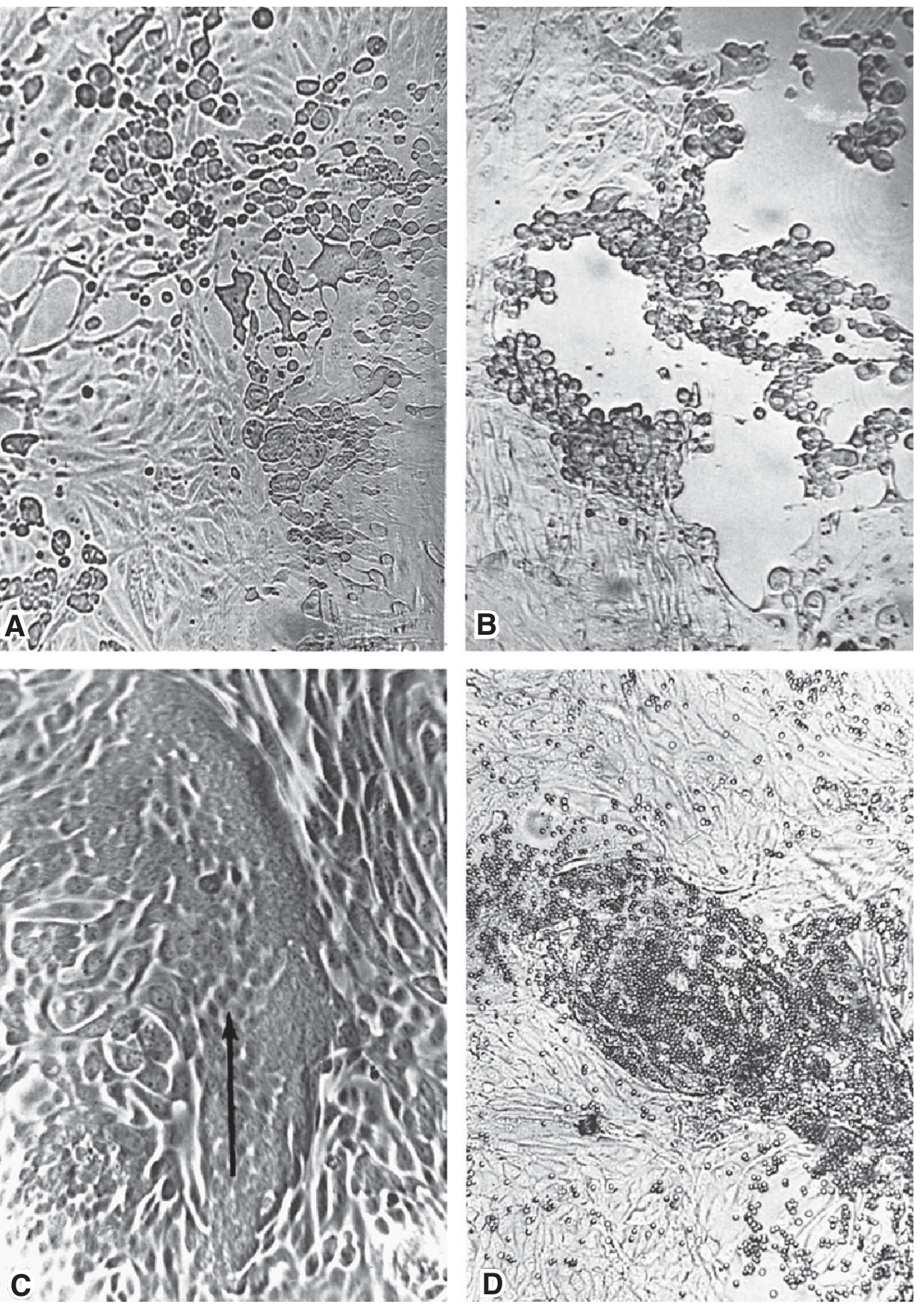
1. Cytopathic Effects (CPE)
Visible morphologic changes in infected cells:
- Cell lysis/necrosis - cells round up and die (e.g., enteroviruses)
- Inclusion body formation - intracellular viral deposits (e.g., herpesvirus, CMV)
- Syncytia/Giant cell formation - cells fuse together (e.g., paramyxovirus, RSV, HIV)
- Cytoplasmic vacuolization
2. Hemadsorption
- Virus-encoded hemagglutinin (e.g., influenza, parainfluenza) inserts into cell membranes
- Red blood cells adhere to infected cell surfaces
- Appears before CPE is visible - useful for early detection
3. Detection of Viral Proteins
- Specific antisera + immunofluorescence to detect viral antigens inside cells
4. Molecular Detection
- PCR - detects virus-specific nucleic acid; rapid, sensitive, and highly specific
Plaque Assay - Quantifying Infectious Virus
- Monolayers of cells are inoculated with diluted virus
- Overlaid with agar or carboxymethylcellulose to prevent virus from spreading freely
- Virus spreads only to neighboring cells, creating a plaque (zone of destroyed cells)
- Each plaque = one plaque-forming unit (PFU) from a single viral particle
- Time to visible plaques: days (poliovirus) to 2 weeks (SV40)
- Used to quantitate viral titer
Susceptible Cell Lines for Common Viruses
| Virus | Preferred Cell Lines |
|---|---|
| Herpes Simplex (HSV) | Vero, HEp-2, human diploid (HEK, HEL) |
| CMV | Human diploid fibroblasts |
| VZV | Human diploid (HEL, HEK) |
| Adenovirus | HEp-2, HEK |
| Poliovirus | MK, Vero, HEp-2, human diploid |
| Influenza A/B | MK, LLC-MK2, MDCK |
| RSV | HEp-2, Vero |
| Mumps | MK, LLC-MK2, HEK, Vero |
| Rhinovirus | Human diploid (HEK, HEL) |
| Measles | MK, HEK |
Other Methods of Virus Cultivation
Cell culture is the preferred method, but two others are also used:
| Method | Description | Used For |
|---|---|---|
| Embryonated egg | Inoculate 8-12 day fertilized egg at specific membranes | Influenza, mumps, smallpox, yellow fever vaccine |
| Animal inoculation | Infect live animals (mice, guinea pigs, primates) | Rabies, some arboviruses; now rarely used |
Here is a video that covers virus cultivation in detail:
Source: Jawetz, Melnick & Adelberg's Medical Microbiology, 28th Ed., Chapter 29 - Cultivation and Detection of Viruses; PMC - Cultivation of viruses
This is a shared conversation. Sign in to Orris to start your own chat.