
Real mitral valve image
Reading File
Now let me search the medical library for mitral valve images.
Finding Sources
Searching the Web
real mitral valve anatomy photograph gross pathology
Let me fetch the best images from the textbook and the web search results in parallel.
Reading File
Loading Image
Loading Image
Loading Image
Let me search in the textbook blocks for actual image URLs related to the mitral valve, and try other image sources.
Finding Sources
Loading Image
Let me look for image URLs directly in the textbook files for the mitral valve anatomy section.
Reading File
Reading File
I found real mitral valve images in the textbooks. Let me fetch them now.
Loading Image
Loading Image
Loading Image
Here are real mitral valve images from authoritative medical textbooks:
Real Mitral Valve - Gross Pathology
Fig. 1 - Gross specimen: Myxomatous degeneration (Mitral Valve Prolapse)
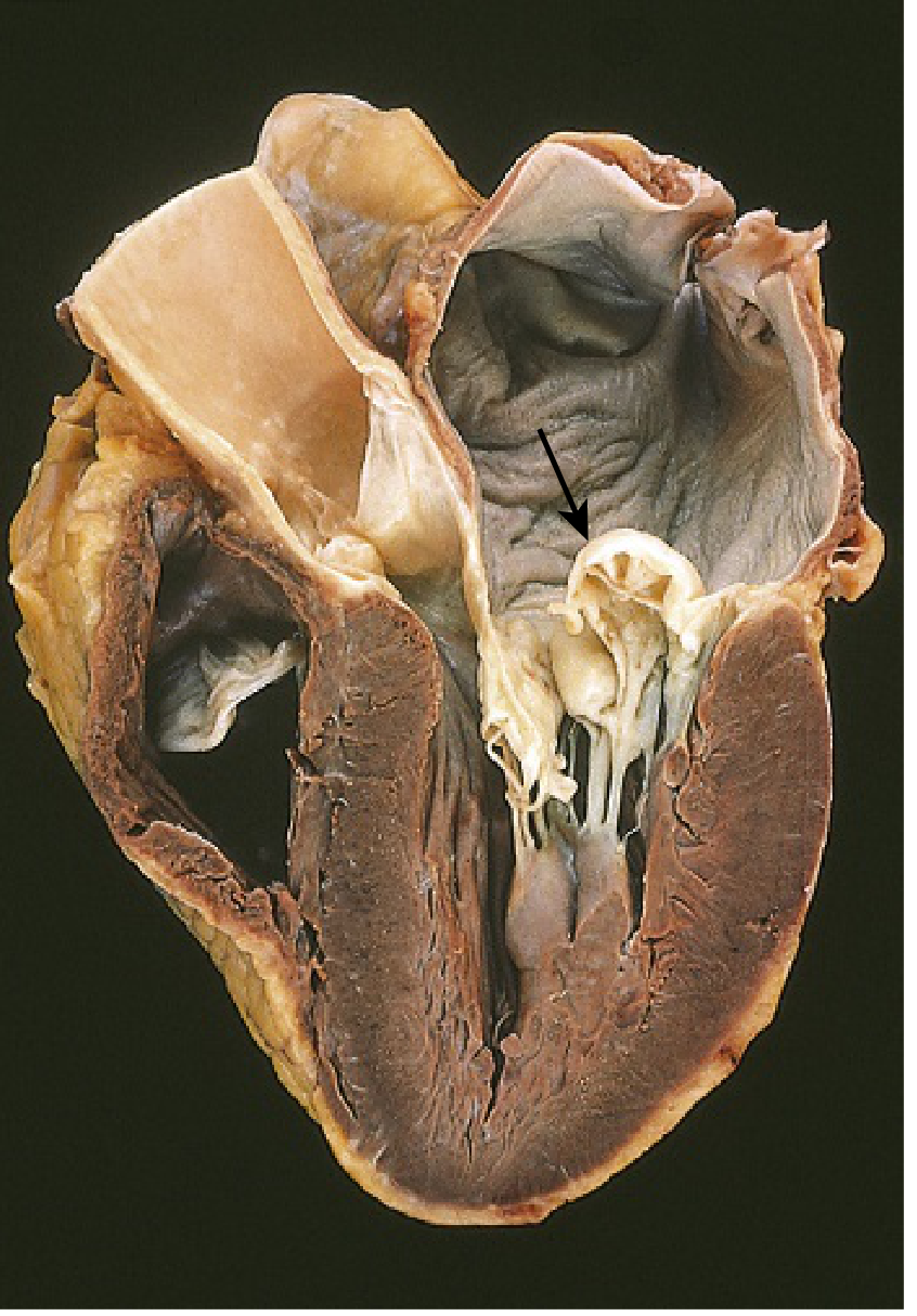
This is an actual dissected heart specimen showing the mitral valve in a four-chamber view. The arrow points to the posterior mitral leaflet, which is ballooning (prolapsing) back into the dilated left atrium - a hallmark of myxomatous mitral valve degeneration (mitral valve prolapse). The left ventricle (brown myocardium) and the chordae tendineae (white cord-like structures) are visible below the leaflets.
Source: Robbins & Kumar Basic Pathology - FIG. 9.18. Courtesy of William D. Edwards, MD, Mayo Clinic.
Mitral Valve Apparatus - Labeled Anatomy Diagram
Fig. 2 - Complete mitral apparatus (lateral view)
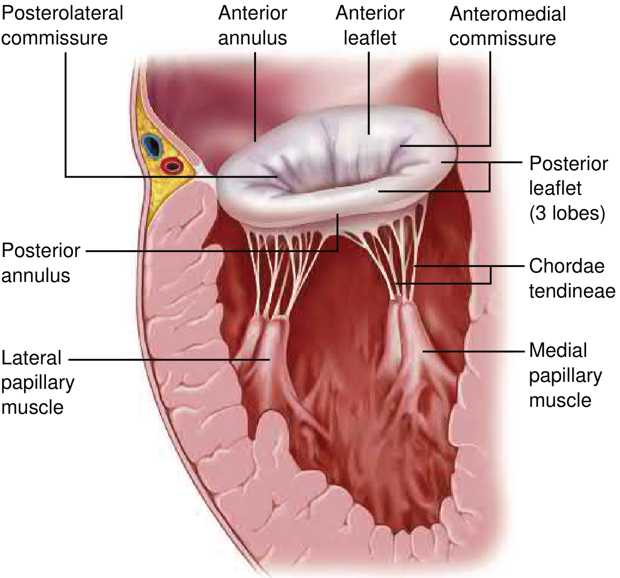
This labeled illustration shows all six components of the mitral apparatus:
- Annulus - anterior (fibrous, in continuity with aortic valve) and posterior (muscular, prone to dilation)
- Anterior leaflet - larger radially, thicker, bears greater tensile load
- Posterior leaflet - 3 lobes (scallops P1, P2, P3), longer circumferentially
- Chordae tendineae - collagen/elastin cords tethering leaflets to papillary muscles
- Lateral and medial papillary muscles - arise from the left ventricular wall
- Commissures (posterolateral and anteromedial) - where the two leaflets meet
Source: Braunwald's Heart Disease - FIGURE 76.1
Leaflet Segmentation (Atrial View)
Fig. 3 - Carpentier segmentation scheme (atrial view looking down)
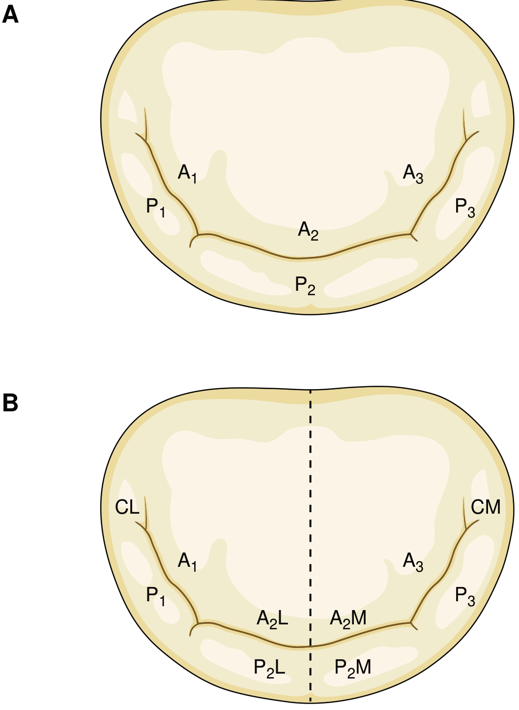
- Panel A (Carpentier's standard scheme): Anterior leaflet divided into A1/A2/A3; posterior leaflet into P1/P2/P3 (three scallops separated by indentations)
- Panel B (Shah's modification): Further divides the middle segment into lateral (A2L, P2L) and medial (A2M, P2M) halves
Source: Braunwald's Heart Disease - FIGURE 76.2
Key anatomy summary
| Structure | Detail |
|---|---|
| Leaflets | 2 - anterior (larger) and posterior (3 scallops) |
| Annulus | Saddle-shaped; anterior part fibrous, posterior part muscular |
| Chordae tendineae | Primary (marginal), secondary (strut), tertiary (basal) |
| Papillary muscles | Anterolateral + posteromedial; arise from LV wall |
| Normal orifice area | ~4-6 cm² |
This is a shared conversation. Sign in to Orris to start your own chat.