
What is auricle
The auricle (also called the pinna) is the visible outer part of the ear - the shell-shaped cartilage structure on the side of the head. It collects and funnels sound waves into the ear canal toward the eardrum. The term "auricle" can also refer to the small ear-shaped pouches on the upper chambers (atria) of the heart.
Searching sources...
Reading File
Finding Sources
Finding Sources
Reading File
Reading File
Loading Image
Loading Image
Loading Image
The Auricle
The auricle (also called the pinna) is the visible, shell-shaped structure on the side of the head that forms the outer ear. It sits on the lateral surface of the head and is the first part of the auditory system to receive sound.
Structure
The auricle consists of elastic fibrocartilage covered by perichondrium and skin. The skin on the lateral (front) surface is firmly attached to the perichondrium, while on the medial (back) surface it is more loosely attached. Its irregular shape creates a series of named landmarks:
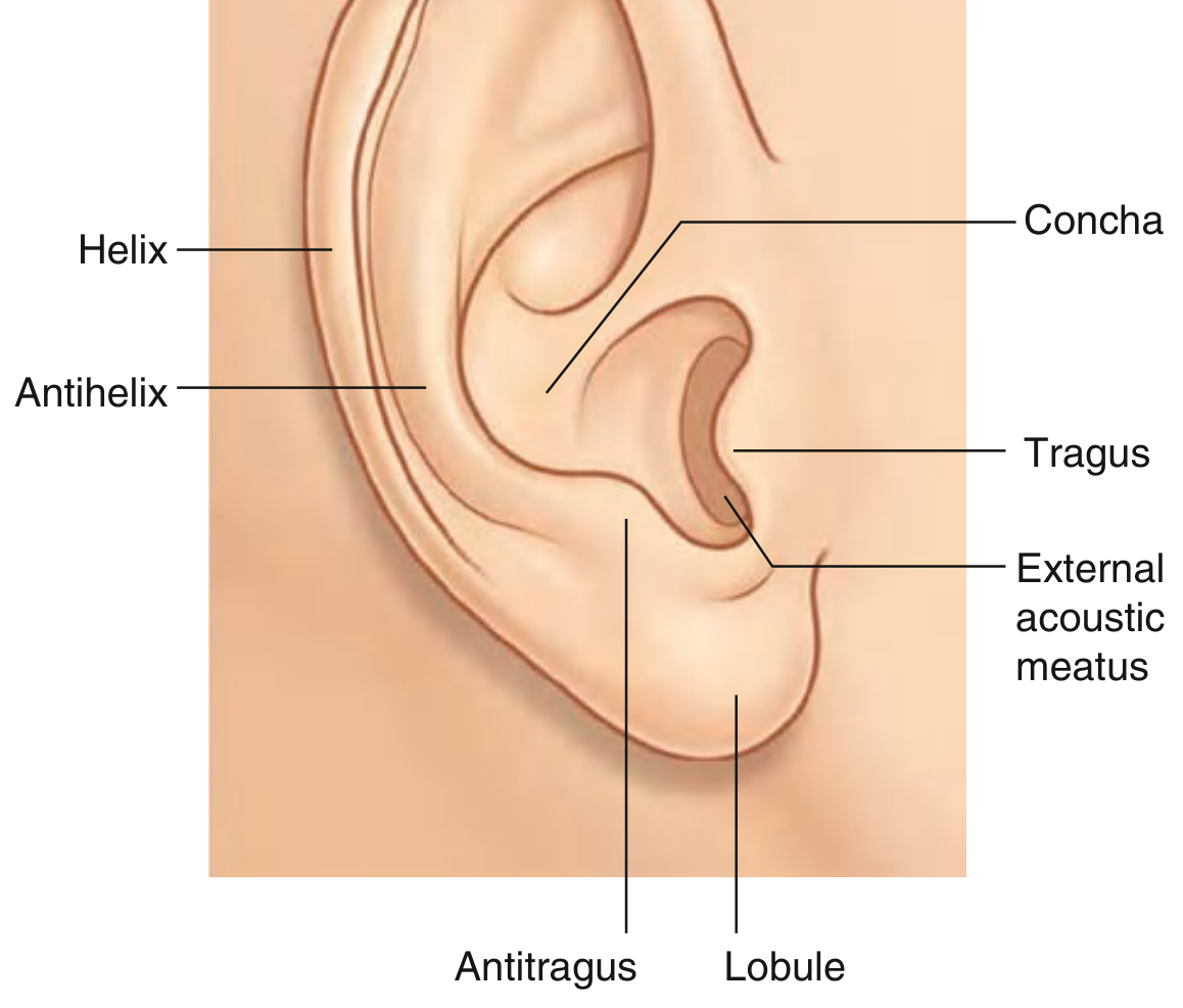
Fig. 8.117 - Gray's Anatomy for Students
| Landmark | Description |
|---|---|
| Helix | The large outer rim of the auricle |
| Antihelix | A smaller curved rim parallel and anterior to the helix |
| Concha | The hollow central bowl; the external acoustic meatus opens from its depths |
| Tragus | An elevation just anterior to the ear canal opening |
| Antitragus | An elevation opposite the tragus, above the lobule |
| Lobule (earlobe) | The inferior fleshy part - the only portion with NO cartilage |
- Gray's Anatomy for Students, p. 1094
Function
The auricle collects and funnels sound waves into the external acoustic meatus (ear canal), directing them toward the tympanic membrane (eardrum). Its irregular contours also help with sound localization.
Muscles
Both intrinsic and extrinsic muscles are associated with the auricle:
- Intrinsic muscles pass between cartilaginous parts and can alter the shape of the auricle.
- Extrinsic muscles (anterior, superior, and posterior auricular muscles) attach from the scalp/skull to the auricle, allowing some positional movement.
- All are innervated by the facial nerve [VII].
Sensory Innervation
The auricle receives innervation from multiple nerves - a clinically important fact:
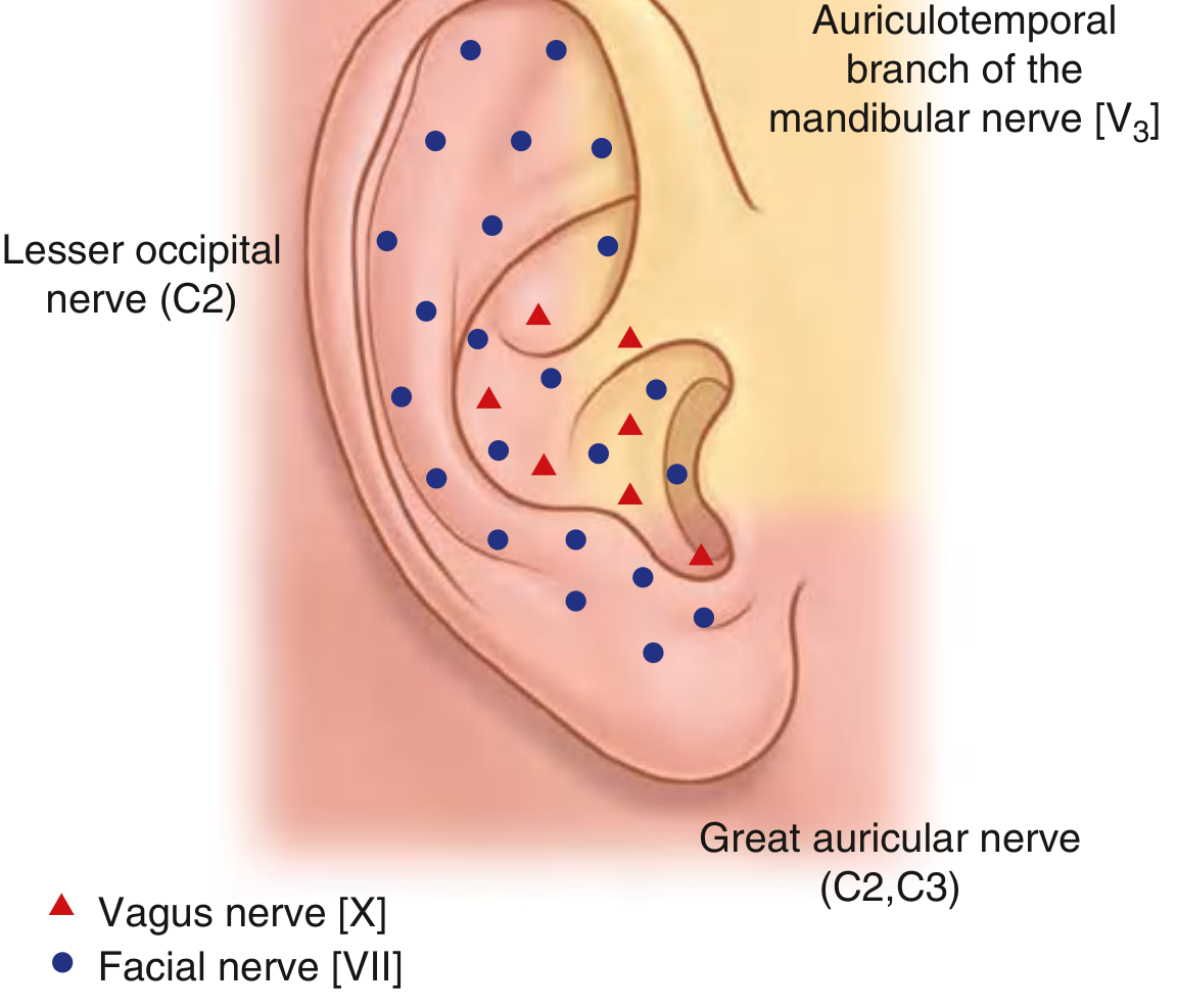
Fig. 8.118 - Gray's Anatomy for Students
| Nerve | Area Supplied |
|---|---|
| Great auricular nerve (C2, C3) | Anterior and posterior inferior surfaces |
| Lesser occipital nerve (C2) | Posterosuperior portion |
| Auriculotemporal nerve (V3) | Anterosuperior portion |
| Vagus nerve [X] | Deeper parts (auricular branch) |
| Facial nerve [VII] | Deeper parts (small branch) |
Embryological Development
The auricle develops from six hillocks of His (mesenchymal condensations) that appear during the 6th week of gestation around the first pharyngeal groove:
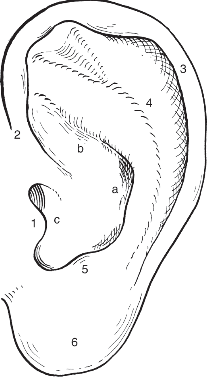
Figure 13-23 - K.J. Lee's Essential Otolaryngology
- Hillocks 1-3: derived from the 1st branchial arch → form the tragus, helical crus, and upper helix
- Hillocks 4-6: derived from the 2nd branchial arch → form the antihelix, antitragus, and lobule/lower helix
- By the 12th week: hillocks fuse to form the auricle
- By the 20th week: adult shape is reached (adult size reached by age 9)
- The concha arises from three areas of the first groove (ectoderm)
Clinical Notes
On examination, the auricle should be inspected for incisions, scarring, congenital abnormalities, trauma, infection, cellulitis, dermatitis, or neoplasia. The postauricular/mastoid region also receives careful attention. - Shambaugh Surgery of the Ear
Note: The term "auricle" in cardiac anatomy refers to the small ear-shaped muscular pouches (appendages) projecting from each atrium of the heart - these are distinct structures from the ear auricle described above.
This is a shared conversation. Sign in to Orris to start your own chat.