
slow or late development of secondary sexual characteristics (e.g. breast) in female
delayed puberty female secondary sexual characteristics Tanner stages
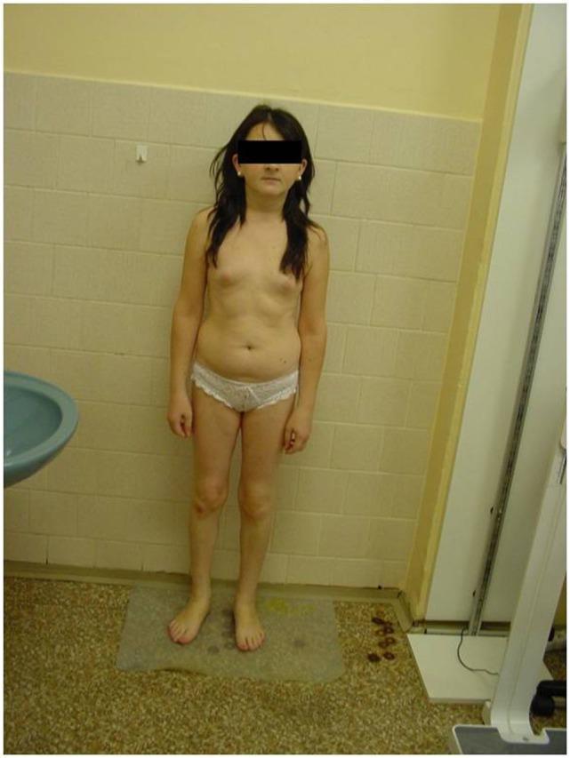
A clinical photograph shows a 27-year-old female patient with a marked childish appearance, characteristic of severe delayed puberty and primary amenorrhea. The patient exhibits short stature and a youthful facial appearance with pale, slightly puffy skin. Morphological assessment reveals significant underdevelopment of secondary sexual characteristics: the breasts are pre-pubertal (Tanner stage B1-B2), and there is an absence of axillary and pubic hair. The physique is slender with an infantile distribution of subcutaneous fat and a slightly rounded abdomen, inconsistent with chronological adulthood. This visual presentation is indicative of hypogonadotropic hypogonadism, which in this case is associated with a PROP1 gene mutation leading to combined pituitary hormone deficiency (CPHD). The photograph serves as a medical educational example of the physical manifestation of untreated pituitary insufficiency and its impact on skeletal growth and sexual maturation.
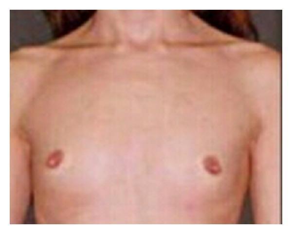
This clinical photograph shows the anterior upper torso of a 21-year-old patient, illustrating a significant lack of secondary sexual characteristics in the breast region. The chest wall appears flat with an absence of glandular breast tissue development (breast hypoplasia or amastia-like appearance). The nipple-areolar complexes are present but small and undeveloped, consistent with a low Tanner stage (B1-B2). The skin over the thorax is unremarkable, with no visible lesions, though the subcutaneous fat layer appears thin. In the context of the provided clinical history, these findings are indicative of delayed puberty or an underlying endocrinological/genetic condition such as primary amenorrhea and Disorders of Sex Development (DSD). The image serves as a clinical reference for evaluating Tanner staging and the physical manifestations of hormonal imbalances or androgen insensitivity syndromes in an adult female-presenting patient.
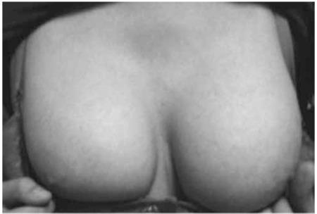
A clinical photograph of the anterior chest of a 15-year-old female patient, demonstrating bilateral Tanner stage V breast development. The image shows mature, round, and symmetrical breasts with significant volume and forward projection. The contour is smooth and continuous, indicating full development of the glandular and fatty tissue, consistent with late puberty. The skin appears uniform in texture without visible lesions, striae, or masses. This visual is significant for pediatric and adolescent medicine when assessing pubertal progression and secondary sexual characteristics. In the clinical context of primary amenorrhea, as noted in the metadata, such normal breast development (Tanner stage V) despite a 46,XY karyotype is a characteristic clinical finding in Swyer syndrome (pure gonadal dysgenesis), where the phenotype is typically female despite the chromosomal genotype.
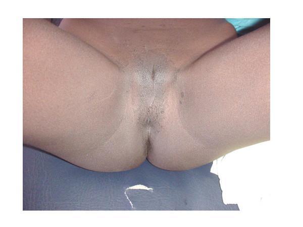
Clinical photograph of the external genitalia of a patient with dark brown skin tone. The image demonstrates a complete absence of pubic hair (sexual infantility), a key finding in cases of delayed puberty and hormonal deficiencies such as low FSH and LH. The genital morphology shows evidence of Type IV Female Genital Mutilation (FGM), characterized by non-excisive procedures such as pricking or incising. Despite the patient's female social assignment, the visual appearance lacks standard secondary sexual characteristics expected for a 21-year-old, correlating with the provided clinical context of a 46, XY karyotype. The skin in the genital area appears smooth without significant hyperpigmentation or lesions. This image is an educational resource for medical specialties including endocrinology, gynecology, and clinical genetics, illustrating the phenotypic presentation of disorders of sex development (DSD) and the anatomical appearance of specific FGM classifications.
Delayed Development of Secondary Sexual Characteristics in Females
Definition & When to Investigate
-
Delayed puberty = absence of secondary sexual characteristics (breast development / thelarche) by age 13 in girls.
-
Normally, breast development is the first sign of estrogen exposure and precedes menarche by ~2 years.
-
Evaluation for primary amenorrhea should be initiated at:
- Age 15-16 if growth and secondary sexual characteristics are otherwise normal
- Age 13 if secondary sexual characteristics are absent or height is below the 3rd percentile
- Within 2 years of breast development if menarche has not occurred
-
Harrison's Principles of Internal Medicine 22E, p. 3174
Clinical Example
Classification & Causes
1. Hypergonadotropic Hypogonadism (High FSH/LH)
- The ovaries fail to respond → no estrogen feedback → pituitary "shouts" with high gonadotropins.
- Accounts for 25-40% of delayed puberty in girls.
| Cause | Details |
|---|---|
| Turner Syndrome (45,X) | Most common cause. Initially normal ovarian development in utero, then accelerated follicle atresia → streak ovaries. Features: short stature, webbed neck, shield chest, cubitus valgus, low hairline, short 4th metacarpal, multiple pigmented nevi |
| Gonadal dysgenesis variants | Partial X deletion, mosaicism (45,X/46,XX), pure gonadal dysgenesis (46,XX or 46,XY with streak gonads) |
| 17α-Hydroxylase / 17,20-Lyase deficiency (CYP17 mutation) | Primary amenorrhea, no secondary sexual characteristics, female phenotype, hypertension and hypokalemia (excess mineralocorticoid) |
| Aromatase deficiency | Cannot convert androgens → estrogen. At puberty: no breast development, primary amenorrhea, virilization, absent growth spurt, multicystic ovaries. Mother may have been virilized during pregnancy |
| Congenital lipoid adrenal hyperplasia (StAR mutations) | Cannot synthesize any steroid from cholesterol; both 46,XX and 46,XY have female external genitalia; 46,XX patients may develop secondary sexual characteristics partially at puberty |
| Galactosemia | Ovarian failure; usually detected on newborn screening |
| Autoimmune oophoritis | |
| Chemotherapy / radiation (prepubertal) | Alkylating agents (e.g., cyclophosphamide), gonadal irradiation |
| Gonadotropin receptor mutations (FSHβ, LHR, FSHR mutations) | Receptor unresponsive to gonadotropins |
- Berek & Novak's Gynecology, pp. 1858-1865
2. Hypogonadotropic Hypogonadism (Low FSH/LH)
- Hypothalamic or pituitary failure → inadequate gonadotropin drive to ovaries.
| Cause | Details |
|---|---|
| Constitutional / Physiologic delay | Most common hypogonadotropic cause. Delayed reactivation of GnRH pulse generator. Normal physiology, just delayed. Positive family history. Self-resolving |
| Kallmann Syndrome | Deficient pulsatile GnRH secretion + anosmia (patient may not notice). Caused by failure of GnRH neuron migration from olfactory placode during fetal development. X-linked (KAL1/ANOS1), autosomal (FGFR1, PROKR2, others) |
| CNS tumors | Craniopharyngioma most common; compresses hypothalamus/pituitary, impairs GnRH/gonadotropin release. Almost always associated with other pituitary hormone deficiencies |
| Functional hypothalamic amenorrhea (HA) | Low energy availability from excessive exercise, dieting, or eating disorders (anorexia nervosa). GnRH pulse suppressed. Accounts for ~25% of delayed puberty. Girls are particularly susceptible |
| Systemic illness | Celiac disease, chronic renal disease, diabetes mellitus, hypothyroidism |
| Prolactinoma | Rare in childhood; hyperprolactinemia suppresses GnRH |
| Congenital hypogonadotropic hypogonadism (CHH) | Multiple gene mutations: GNRHR, GNRH1, TAC3, TACR3, KISS1, KISS1R, etc. |
| 5α-Reductase deficiency | 46,XY with female phenotype and ambiguous genitalia; testes present; virilization at puberty |
- Harrison's Principles of Internal Medicine 22E, p. 3174; Berek & Novak's Gynecology, pp. 1866-1867
Tanner Staging (Breast Development)
| Stage | Description |
|---|---|
| B1 | Prepubertal; elevation of papilla only |
| B2 | Breast bud stage; small mound of breast + areola |
| B3 | Further enlargement; no separation of contours |
| B4 | Areola and papilla project above breast plane |
| B5 | Mature breast; papilla projects, areola recessed |
Initial Evaluation
- LH, FSH (high = hypergonadotropic; low/normal = hypogonadotropic)
- Estradiol
- Bone age (X-ray of left hand/wrist)
- TSH, T4 (rule out hypothyroidism)
- IGF-1, IGFBP-3
- CBC, ESR/CRP, electrolytes, renal and liver function
- DHEAS
-
Karyotype (if FSH elevated - rule out Turner syndrome and variants)
-
GnRH stimulation test / GnRHa stimulation
-
MRI of brain/pituitary (if hypogonadotropic - rule out craniopharyngioma, etc.)
-
Prolactin
-
Harrison's Principles of Internal Medicine 22E (Table 404-2 & 404-3)
Key Differentiating Points
| Feature | Hypergonadotropic | Hypogonadotropic |
|---|---|---|
| FSH/LH | High | Low or inappropriately normal |
| Ovaries | Streak / fibrotic | Small but present |
| Karyotype | May be abnormal (45,X) | Usually 46,XX |
| Anosmia | Absent | May be present (Kallmann) |
| Turner stigmata | May be present | Absent |
| MRI pituitary | Normal | May show mass/abnormality |
| Bone age | Normal or mildly delayed | Often delayed |
Management Overview
- Constitutional delay: Watchful waiting; short-course low-dose estrogen may be used to induce puberty if significantly distressing.
- Hypergonadotropic (gonadal failure): Hormone replacement therapy (HRT) with progressively increasing doses of estrogen, then cyclic estrogen-progesterone to mimic normal puberty and induce/maintain secondary sexual characteristics; protect bone density. If 45,X with Y-cell line mosaicism: gonadectomy (risk of gonadoblastoma).
- Hypogonadotropic (functional): Treat the underlying cause (nutritional rehabilitation, treatment of systemic illness). Pulsatile GnRH or gonadotropin therapy can induce ovulation for fertility.
- Kallmann / CHH: Low-dose estrogen for puberty induction; pulsatile GnRH or exogenous gonadotropins for fertility.
Summary Mnemonic
- Turner syndrome
- Radiation/chemotherapy (gonadal damage)
- Aromatase deficiency / Autoimmune oophoritis
- Constitutional delay / CNS tumors / CHH (Kallmann)
- Karyotypic gonadal dysgenesis variants
Sources: Harrison's Principles of Internal Medicine 22E (2025), Berek & Novak's Gynecology; Medical Physiology (Boron & Boulpaep)