I now have enough foundational information from the medical library. Let me compile a thorough, simple-language answer covering all 9 requested sections.
🩺 Endocrine Disorders - Complete Guide (Simple Language)
1. 📖 Definition
Endocrine disorders are diseases caused by a problem with the endocrine system - the network of glands (like the thyroid, pancreas, adrenal glands, pituitary, ovaries, testes) that produce hormones.
In simple words:
"When any gland makes too much, too little, or the wrong kind of hormone, the body goes out of balance - that is an endocrine disorder."
Common examples include:
- Diabetes mellitus (pancreas - too little insulin)
- Hypothyroidism (thyroid - too little thyroid hormone)
- Hyperthyroidism (thyroid - too much thyroid hormone)
- Cushing's syndrome (adrenal - too much cortisol)
- Addison's disease (adrenal - too little cortisol)
- Hypogonadism (too little sex hormones)
- Hyperparathyroidism (parathyroid - too much PTH)
2. 🔬 Pathophysiology (Step-by-Step)
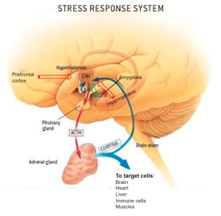
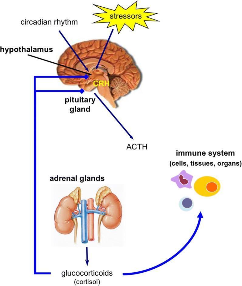
The endocrine system works like a feedback loop - like a thermostat at home. Here is how it normally works and how it breaks:
Normal flow:
Brain (Hypothalamus)
    ↓ sends signal hormone (e.g., CRH, TRH, GHRH)
Pituitary Gland
    ↓ sends stimulating hormone (e.g., ACTH, TSH, FSH)
Target Gland (thyroid / adrenal / gonads / pancreas)
    ↓ releases final hormone (e.g., cortisol, T3/T4, estrogen, insulin)
    ↓ hormone acts on body cells
    ↓ NEGATIVE FEEDBACK → tells brain to stop producing more signals
How disorders develop:
| Step | What Goes Wrong | Example |
|---|
| Step 1 | Gland is destroyed or inflamed | Autoimmune attack on thyroid → Hypothyroidism |
| Step 2 | Gland grows a tumor and overproduces | Adrenal tumor → Cushing's syndrome |
| Step 3 | Pituitary sends wrong signal | Pituitary tumor overproduces TSH → Hyperthyroidism |
| Step 4 | Body cells resist the hormone | Cells ignore insulin → Type 2 Diabetes |
| Step 5 | Feedback loop breaks | No negative feedback → Hormone keeps rising uncontrolled |
| Step 6 | Widespread body effects | High/low hormone levels damage organs, metabolism, and homeostasis |
Key mechanism types:
- Hyposecretion = gland makes too LITTLE hormone (e.g., hypothyroidism)
- Hypersecretion = gland makes too MUCH hormone (e.g., hyperthyroidism)
- Hormone resistance = body cannot use the hormone (e.g., Type 2 DM - insulin resistance)
- Receptor/target organ failure = cells cannot respond to the hormone
3. ⚠️ Risk Factors
| Risk Factor | Explanation |
|---|
| Family history / Genetics | Endocrine disorders often run in families (e.g., Type 1 DM, thyroid disease) |
| Autoimmune disease | Immune system attacks own glands (Hashimoto's, Graves', Addison's) |
| Obesity | Excess fat causes insulin resistance → Type 2 DM |
| Age | Risk increases with aging (menopause, andropause, hypothyroidism) |
| Female sex | Women are 5-8x more likely to develop thyroid disorders |
| Iodine deficiency | Causes goiter and hypothyroidism |
| Chronic stress | Elevates cortisol chronically → adrenal fatigue |
| Medications | Steroids, lithium, amiodarone can disrupt hormone levels |
| Radiation or surgery | Damages glands (e.g., post-thyroidectomy hypothyroidism) |
| Pregnancy | Can trigger gestational diabetes, postpartum thyroiditis |
| Poor diet / Sedentary lifestyle | Promotes insulin resistance and obesity |
| Toxins / Endocrine disruptors | Chemicals (BPA, pesticides) interfere with hormones |
4. 🌡️ Signs & Symptoms
Because different glands are affected, symptoms vary. Here are the most common patterns:
Hypothyroidism (too little thyroid hormone)
- Fatigue, weakness
- Weight gain
- Cold intolerance
- Constipation
- Dry skin, hair loss
- Slow heart rate (bradycardia)
- Depression, poor memory
Hyperthyroidism (too much thyroid hormone)
- Weight loss despite eating more
- Heat intolerance, sweating
- Fast heart rate (tachycardia), palpitations
- Anxiety, irritability, tremor
- Diarrhea
- Bulging eyes (in Graves' disease)
Diabetes Mellitus (insufficient insulin effect)
- Classic 3 Ps: Polyuria (frequent urination), Polydipsia (excessive thirst), Polyphagia (excessive hunger)
- Weight loss (Type 1)
- Fatigue
- Blurry vision
- Slow wound healing
- Recurrent infections
Cushing's Syndrome (too much cortisol)
- Moon face (round, puffy face)
- Buffalo hump (fat pad at back of neck)
- Central obesity
- Purple stretch marks (striae)
- Muscle weakness
- High blood pressure, high blood sugar
Addison's Disease (too little cortisol/aldosterone)
- Severe fatigue, weakness
- Low blood pressure
- Hyperpigmentation (skin darkening, especially in creases)
- Nausea, vomiting, weight loss
- Salt craving
5. 💥 Complications
| Endocrine Disorder | Major Complications |
|---|
| Diabetes | Heart disease, stroke, kidney failure (nephropathy), blindness (retinopathy), nerve damage (neuropathy), foot ulcers/amputation, diabetic ketoacidosis (DKA), hyperosmolar hyperglycemic state (HHS) |
| Hypothyroidism | Myxedema coma (life-threatening), heart disease, infertility, depression, peripheral neuropathy |
| Hyperthyroidism | Thyroid storm (medical emergency), atrial fibrillation, osteoporosis, heart failure |
| Cushing's | Osteoporosis, fractures, infections, Type 2 DM, hypertension, psychiatric disorders |
| Addison's | Adrenal crisis (Addisonian crisis) - life-threatening shock, hypotension, hyperkalemia |
| Hyperparathyroidism | Kidney stones, bone thinning, peptic ulcers, pancreatitis ("bones, stones, groans, psychic moans") |
| General | Metabolic syndrome, cardiovascular disease, infertility, growth abnormalities, osteoporosis |
6. 📊 Disease Pattern
Endocrine disorders generally follow one of these patterns:
| Pattern | Description | Example |
|---|
| Chronic & progressive | Develops slowly over years, worsens without treatment | Type 2 DM, Hypothyroidism, Cushing's |
| Autoimmune (relapsing-remitting) | Periods of flare and remission | Graves' disease, Hashimoto's thyroiditis |
| Acute crisis | Sudden life-threatening decompensation | Thyroid storm, Adrenal crisis, DKA |
| Congenital | Present from birth, often genetic | Congenital hypothyroidism, Congenital Adrenal Hyperplasia |
| Iatrogenic | Caused by treatment (medications, surgery, radiation) | Post-thyroidectomy hypothyroidism, Steroid-induced Cushing's |
| Cyclical/Hormonal | Tied to life stages | Gestational DM in pregnancy, Menopause-related hormonal changes |
Important note: Many endocrine disorders are silent early on - the patient feels fine, but laboratory tests reveal abnormal hormone levels. This is why screening matters.
7. 💊 Medical Management
General Principles
- Identify the cause - Is it hypo or hypersecretion? Primary gland problem or secondary (pituitary) problem?
- Hormone replacement for deficiency states
- Suppression therapy for overproduction
- Treat the underlying cause (remove tumor, treat autoimmune disease)
- Monitor and adjust with regular labs
Specific Medical Treatments
| Disorder | Medical Management |
|---|
| Hypothyroidism | Levothyroxine (T4 replacement) daily; monitor TSH every 6-12 months |
| Hyperthyroidism | Antithyroid drugs (methimazole, propylthiouracil); Radioactive iodine (I-131); Beta-blockers for symptoms (propranolol); Surgery (thyroidectomy) |
| Type 1 Diabetes | Insulin therapy (basal-bolus regimen), blood glucose monitoring, carb counting |
| Type 2 Diabetes | Lifestyle changes first; Metformin (first-line drug); other agents (SGLT2 inhibitors, GLP-1 agonists, sulfonylureas); insulin when needed |
| Cushing's | Remove causative tumor (surgery); Steroidogenesis inhibitors (ketoconazole, metyrapone); Taper exogenous steroids if iatrogenic |
| Addison's disease | Hydrocortisone (glucocorticoid replacement) + Fludrocortisone (mineralocorticoid); Stress dosing during illness |
| Hyperparathyroidism | Surgery (parathyroidectomy) if symptomatic; Cinacalcet (calcimimetic drug); Bisphosphonates for bone protection |
Emergency Management
- DKA: IV fluids, insulin infusion, potassium replacement, treat cause
- Thyroid storm: Beta-blockers, antithyroids, iodine, corticosteroids, cooling, ICU care
- Adrenal crisis: IV hydrocortisone 100 mg STAT, IV saline, treat precipitating cause
8. 🩺 Nursing Diagnosis (Top 3)
#1 - Imbalanced Nutrition: Less/More Than Body Requirements
Related to: Altered metabolism from hormonal imbalance (e.g., increased catabolism in hyperthyroidism; impaired glucose uptake in diabetes)
Evidenced by: Weight changes, fatigue, muscle wasting, abnormal blood glucose or thyroid levels
#2 - Deficient Knowledge
Related to: Lack of information about disease process, medications, self-monitoring, and lifestyle modifications
Evidenced by: Patient verbalization of confusion about condition, non-adherence to treatment, inability to perform self-care (e.g., blood glucose monitoring, insulin injection)
#3 - Risk for Injury / Impaired Tissue Integrity
Related to: Altered metabolism, poor wound healing, peripheral neuropathy, and impaired immune response from hormonal imbalance
Evidenced by (or at risk for): Slow-healing wounds, skin breakdown, falls from neuromuscular dysfunction, hypoglycemic or hypo/hyperadrenal episodes
Bonus: Activity Intolerance (related to fatigue, muscle weakness, slow metabolism) is also very common across most endocrine disorders.
9. 💉 Nursing Interventions
For Nursing Dx #1 - Imbalanced Nutrition
- Monitor daily weight, intake & output, BMI
- Assess eating habits and meal patterns
- Teach carbohydrate counting (for DM patients)
- Collaborate with dietitian for individualized meal plan
- Monitor labs: blood glucose, HbA1c, thyroid function tests, electrolytes
- Administer prescribed medications (insulin, levothyroxine) at correct times
- Teach patient about hypoglycemia signs and how to manage (juice, glucose tablets)
For Nursing Dx #2 - Deficient Knowledge
- Assess current level of understanding about the disease
- Teach in simple language (avoid medical jargon), use teach-back method
- Provide written materials and visual aids about:
- How to take medications correctly (timing, not missing doses)
- How to monitor blood glucose / temperature / weight at home
- Warning signs that need immediate attention (e.g., Addisonian crisis, DKA symptoms)
- Teach proper injection technique for insulin
- Reinforce lifestyle modifications: diet, exercise, stress reduction
- Involve family/caregiver in education
For Nursing Dx #3 - Risk for Injury / Impaired Tissue Integrity
- Inspect skin, especially feet, daily (especially in diabetic patients)
- Keep skin clean, dry, and moisturized
- Use non-restrictive footwear; teach proper foot care
- Assess sensation in extremities (neuropathy screening)
- Pad bony prominences; reposition immobile patients every 2 hours
- Monitor for and report signs of infection early (redness, warmth, swelling)
- Ensure safe environment to prevent falls (especially when patient has weakness or dizziness)
- Teach patient to avoid extreme temperatures (heat/cold injury risk in neuropathy)
- Monitor for hypoglycemia/hyperglycemia, and adrenal crisis signs before/after procedures
📌 Quick Summary Table
| Topic | Key Point |
|---|
| Definition | Disease from too much, too little, or abnormal hormone |
| Pathophysiology | Broken feedback loop → gland dysfunction → systemic imbalance |
| Risk Factors | Genetics, autoimmunity, obesity, age, medications |
| Signs | Depend on gland involved - fatigue, weight change, 3 Ps, skin changes |
| Complications | DKA, thyroid storm, adrenal crisis, neuropathy, CVD |
| Pattern | Mostly chronic & silent early; acute crises possible |
| Medical Mgmt | Hormone replacement OR suppression + treat cause |
| Nursing Dx | Imbalanced nutrition, Deficient knowledge, Risk for injury |
| Nursing Interventions | Monitor, educate, protect skin/safety, medication adherence |
Sources: Goldman-Cecil Medicine; Rosen's Emergency Medicine; Tietz Textbook of Laboratory Medicine; Creasy & Resnik's Maternal-Fetal Medicine