

Give me the full sequence
Thomas test ilio-psoas contracture hip flexion deformity
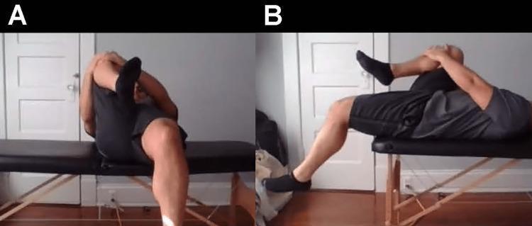
Two clinical photographs demonstrate the performance of the Modified Thomas Test, a physical examination maneuver used to assess hip flexor contractures. Image A shows an anterior view of a patient in a supine position at the edge of an examination table. The patient has one hip and knee fully flexed, held against the chest by the arms to stabilize the pelvis and flatten the lumbar spine. The contralateral leg is allowed to hang freely. This view is used to observe for hip abduction (suggesting tensor fasciae latae involvement) or external rotation (suggesting iliotibial band restriction). Image B provides a lateral view of the same maneuver. This perspective specifically evaluates the degree of hip extension and knee flexion of the hanging leg. A lack of full hip extension indicates psoas or iliopsoas tightness, while an inability to maintain at least 90 degrees of knee flexion suggests rectus femoris contracture. The test is fundamental in musculoskeletal medicine and physical therapy for diagnosing imbalances in the anterior hip and thigh musculature.
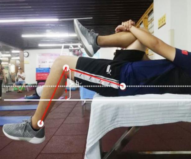
This clinical photograph demonstrates the performance of a Thomas test, a physical examination maneuver used to assess hip flexor contracture and psoas tightness. The subject is positioned supine on an examination table with the sacrum at the table's edge. One hip is maximally flexed toward the chest and held in place by the subject's arms to flatten the lumbar spine. The contralateral leg hangs freely off the table. The image is overlaid with quantitative diagnostic annotations: a horizontal white dotted line represents the pelvic baseline, solid white lines indicate the table level, and red lines delineate the femur and tibia segments. A vertical indicator labeled 'h' measures the elevation of the thigh from the table surface. This visual focuses on the biomechanical assessment of the iliopsoas and rectus femoris muscles, with the failure of the extended thigh to touch the table surface indicating a positive test for hip flexor tightness.
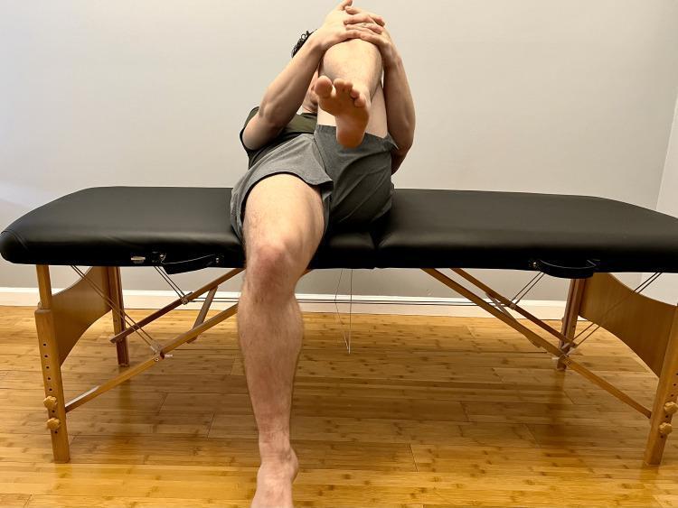
This clinical photograph demonstrates the performance of a Modified Thomas test, a common orthopedic physical examination maneuver used to assess the flexibility of the hip flexors. The patient is positioned supine on a treatment table. One leg (the contralateral limb) is flexed at the hip and knee, with the patient using their hands to pull the knee toward the chest to stabilize the pelvis and flatten the lumbar spine. The test limb is allowed to extend. This visual assessment is used to identify potential muscular tightness or contractures: hip flexion in the test limb suggests psoas involvement, knee extension suggests rectus femoris tightness, and hip abduction or rotation may indicate iliotibial band (ITB) restrictions or tensor fasciae latae (TFL) involvement. This diagnostic maneuver is critical in sports medicine and physical therapy for evaluating hip and knee pathology during a musculoskeletal examination.
Thomas Test - Full Sequence
Position
- Patient lies supine on the examining table
Step 1 - Eliminate compensatory lumbar lordosis (FIRST check)
- Flex both hips maximally toward the abdomen
- This flattens the lumbar lordosis and fixes/stabilizes the pelvis
- Confirm the lumbar spine is flat against the table (obliterate the compensatory lordosis)
- This is the critical first step - without it, a fixed flexion deformity of the hip can be completely masked by an exaggerated lumbar lordosis
Step 2 - Test the affected side
- Hold the contralateral (normal) hip fully flexed against the abdomen
- Lower (extend) the affected limb toward the table and allow it to fall freely
Step 3 - Read the result (LAST - check the angle)
- Negative (Normal): The tested leg lies flat on the table - no contracture
- Positive (Ilio-psoas contracture): The hip of the tested limb remains in flexion and cannot reach the table
- Measure the angle between the posterior aspect of the thigh and the table surface - this is the angle of fixed flexion deformity (F)
Interpretation
| Finding | Meaning |
|---|---|
| Leg lies flat on table | No flexion contracture |
| Leg remains flexed off table | Ilio-psoas contracture present |
| Angle of rise measured | Quantifies degree of fixed flexion deformity |
Why the sequence matters
Give me in a summary manner
Thomas Test – Summary
- Leg lies flat → Negative (normal)
- Leg remains flexed off table → Positive (ilio-psoas contracture)
- Measure the angle of rise = degree of fixed flexion deformity
Key concept: Lumbar lordosis masks a hip flexion contracture. Flattening it first unmasks the true deformity.
Thanks
Give me the procedure for Cozen's test and what is it tested for?
Cozen's test lateral epicondylitis tennis elbow procedure
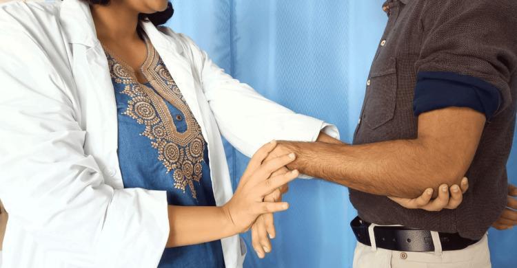
A clinical photograph illustrating the performance of Mill’s test, a provocative physical examination maneuver used to diagnose lateral epicondylitis (tennis elbow). The image shows a healthcare professional in a white coat interacting with a male patient. The clinician is standing, using one hand to palpate the patient's lateral epicondyle at the elbow, while the other hand grasps the patient's hand to maintain the wrist in a flexed position with the forearm pronated. The patient's arm is extended at the elbow. This specific positioning—passive pronation of the forearm, flexion of the wrist, and extension of the elbow—is designed to stretch the extensor carpi radialis brevis muscle, which elicits pain in positive cases of lateral epicondylitis. The educational focus is on orthopedic assessment techniques and musculoskeletal physical examination. The background features a clinical setting with a blue curtain.
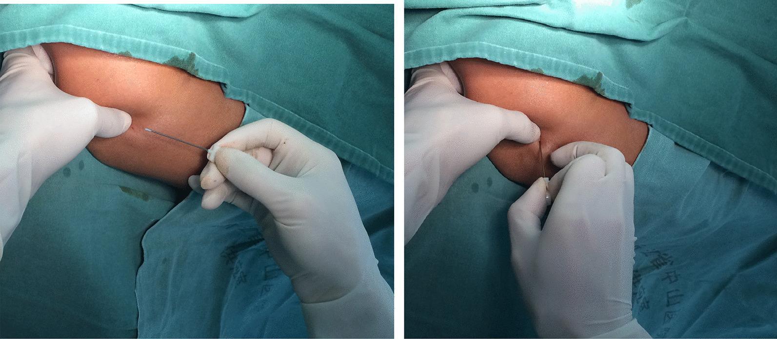
Clinical photograph of an acupotomy procedure for lateral epicondylitis (tennis elbow). The two-panel image demonstrates the surgical technique using a specialized, streamlined 0.6 mm diameter solid needle. The patient's arm is draped with green surgical drapes, exposing the lateral epicondyle region of the humerus. In the left panel, a clinician wearing white sterile gloves is shown preparing for insertion, with one hand stabilizing the soft tissue and the other holding the acupotomy needle. In the right panel, the needle is inserted into the tissue at a 90-degree angle, demonstrating the 'cutting and peeling' or 'transverse oscillation' technique directed toward the bone. This minimally invasive procedure is used to release tendinous tissue and adhesions to relieve pain and improve joint function. The image illustrates a key therapeutic intervention for chronic tendinopathy within an orthopedic or sports medicine context.
Cozen's Test
Procedure
| Step | Action |
|---|---|
| 1 | Patient sits with elbow slightly flexed |
| 2 | Examiner grasps and stabilizes the patient's forearm with one hand |
| 3 | Patient makes a clenched fist and extends the wrist |
| 4 | Examiner resists wrist extension with the other hand |
Result
- Positive: Reproduction of sharp pain at the lateral epicondyle = Cozen's test positive = lateral epicondylitis confirmed
Companion Test - Maudsley's Test
- Patient actively extends the middle finger (3rd digit) against resistance with the elbow extended
- Positive if pain is reproduced over the lateral epicondyle at the ECRB insertion
Key Point
Finkelstein test
Finkelstein test De Quervain tenosynovitis wrist procedure
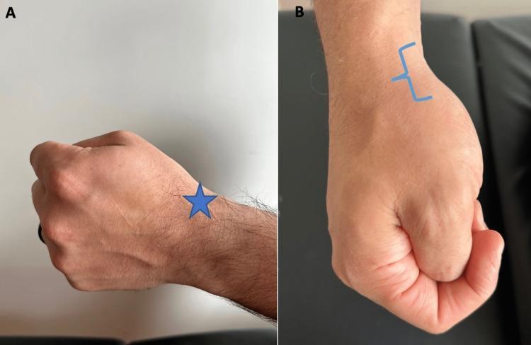
Two clinical photographs demonstrate the positioning for the Finkelstein test, a physical examination maneuver used to diagnose De Quervain tenosynovitis. Image A (lateral view) shows a patient's hand formed into a fist with the thumb tucked inside the fingers. A blue star indicates the first dorsal compartment of the wrist, where patients with the condition typically experience sharp pain during ulnar deviation. Image B (superior view) shows the same fist position from above, with a blue bracket highlighting the anatomic snuffbox region on the radial aspect of the wrist. This visual guide illustrates how clinicians can instruct patients to localize pain or palpate the radial styloid and associated tendons (abductor pollicis longus and extensor pollicis brevis). The maneuver is a key orthopedic assessment tool for evaluating stenosing tenosynovitis of the first extensor compartment.
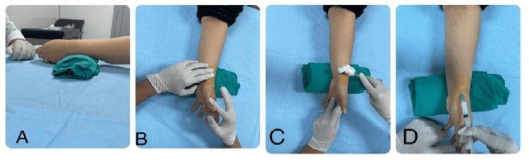
A four-panel clinical photographic sequence (A-D) demonstrating the step-by-step procedure for a corticosteroid injection into the first dorsal extensor compartment of the wrist, typically performed for De Quervain tenosynovitis. (A) Patient positioning: The wrist is placed in a neutral to slight ulnar deviation position, supported by a green folded towel for elevation on a sterile blue drape. (B) Palpation: A gloved clinician identifies the anatomical landmarks, specifically the radial styloid process and the tendons of the abductor pollicis longus (APL) and extensor pollicis brevis (EPB). (C) Site preparation: The radial aspect of the wrist is prepped with a sterile cleaning agent using a swab. (D) Administration: Intralesional injection is shown using a syringe with the needle angled at approximately 45 degrees, directed distal to proximal into the tendon sheath proximal to the radial styloid. The educational focus is on proper anatomical targeting, sterile technique, and needle trajectory for radial wrist interventions.
Finkelstein Test
Procedure
| Step | Action |
|---|---|
| 1 | Patient makes a fist with the thumb tucked inside the fingers |
| 2 | Examiner stabilizes the forearm |
| 3 | Examiner deviates the wrist toward the ulnar side (ulnar deviation) |
Result
- Positive: Sharp pain reproduced over the radial styloid / first dorsal compartment = De Quervain's tenosynovitis
Key Facts
| Feature | Detail |
|---|---|
| Tendons involved | APL + EPB (1st dorsal compartment) |
| Site of pain | Radial styloid / anatomical snuffbox |
| Most common in | Women 30-50 yrs; new mothers (repetitive ulnar deviation in newborn care) |
| Sex ratio | Women affected 6-10x more than men |
| Pathognomonic? | Most pathognomonic sign - but NOT diagnostic alone |
Differentials to exclude
-
Trapeziometacarpal joint arthritis
-
Superficial radial nerve entrapment (Wartenberg syndrome)
-
Intersection syndrome (pain 4-8 cm proximal to radial styloid)
-
Campbell's Operative Orthopaedics 15th Ed 2026; Roberts and Hedges' Clinical Procedures in Emergency Medicine