
Hrct interpretation radiology simplify tome
Reading File
HRCT (High-Resolution CT) interpretation involves recognizing specific patterns in the lung parenchyma. Key patterns include ground-glass opacity, consolidation, honeycombing, reticulation, and nodules - each pointing to different disease categories. Let me pull from the textbooks for a thorough, image-rich guide.
Searching sources...
Finding Sources
Finding Sources
Finding Sources
Finding Sources
Finding Sources
Reading File
Finding Sources
Reading File
Reading File
Reading File
Reading File
Now let me fetch a few key images to include in the answer:
Loading Image
Loading Image
Reading File
Now I have enough material for a comprehensive, well-organized guide. Let me compile it.
HRCT Interpretation - Simplified Guide
Sources: Grainger & Allison's Diagnostic Radiology; Murray & Nadel's Textbook of Respiratory Medicine; Goldman-Cecil Medicine; Washington Manual of Medical Therapeutics
1. What Is HRCT and Why It Matters
HRCT uses thin slices (1-2 mm) at high spatial resolution to image the lung parenchyma with far greater detail than a standard chest CT or plain radiograph. It has become the cornerstone of interstitial lung disease (ILD) evaluation because it:
- Detects early disease that chest X-ray misses
- Can suggest a specific histological diagnosis in certain cases
- Distinguishes reversible (ground-glass, consolidation) from irreversible (fibrosis, honeycombing) changes
- Guides biopsy site selection and treatment response monitoring
(Grainger & Allison's, p. 221)
2. The Four Main HRCT Patterns (Fleischner Glossary Framework)
All diffuse lung abnormalities on HRCT fall into one of four main buckets. Overlap between them is common.
| Pattern | What You See | Think of |
|---|---|---|
| Reticular | Network of lines (fine or coarse) | ILD, fibrosis |
| Nodular | Discrete round opacities | Granulomas, infection, mets |
| Mosaic attenuation | Patchwork of light/dark areas | Air trapping, vascular disease |
| Cystic | Air-filled spaces with definable walls | LAM, LCH, emphysema |
(Grainger & Allison's, p. 224)
3. Pattern Deep-Dives
A. Reticular Pattern
A reticular (net-like) pattern almost always represents significant ILD.
Types of reticulation:
| Type | Appearance | Causes |
|---|---|---|
| Interlobular septal thickening - smooth | Clean straight lines at lung edges | Pulmonary edema, alveolar proteinosis |
| Interlobular septal thickening - irregular/beaded | Lumpy, nodular septa | Lymphangitic carcinomatosis, sarcoidosis |
| Intralobular lines | Fine lace-like net within the lobule | Fibrotic lung disease (IPF, NSIP) |
| Honeycombing | Stacked cystic air spaces with thick, irregular walls | End-stage fibrosis (UIP/IPF) |
Key concept - honeycombing vs. reticulation:
Honeycombing = cystic air spaces (3-10 mm) with thick fibrotic walls, typically subpleural and basal. It is the hallmark of UIP/IPF and signals irreversible disease. Distinguishing it from mere reticulation changes the diagnosis and prognosis.
Traction bronchiectasis often accompanies honeycombing - bronchi dragged outward by surrounding fibrosis, appearing as dilated, irregular airways in the periphery.
(Grainger & Allison's, p. 224; Murray & Nadel's, p. 470)
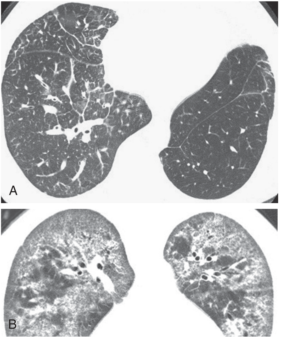
B. Nodular Pattern
The single most useful step: classify nodules by their location within the secondary pulmonary lobule (SPL).
Secondary Pulmonary Lobule = the smallest unit of lung anatomy
- Center: centrilobular artery + terminal bronchiole
- Periphery: interlobular septa + subpleural lymphatics
Three nodule distributions:
1. Perilymphatic Nodules
- Located along interlobular septa, subpleural surfaces, and peribronchovascular bundles
- Fissure involvement is a KEY clue
- Causes: Sarcoidosis (classic), lymphangitic carcinomatosis, silicosis
2. Random Nodules
- Evenly scattered throughout the lung - no lobular pattern preference
- Causes: Miliary TB, hematogenous metastases, fungal infection
3. Centrilobular Nodules
- Located a few mm away from the pleura (important - they spare the pleural surface and fissures)
- Can be well-defined or poorly defined
- Tree-in-bud = centrilobular nodules with branching pattern = impacted small bronchioles
- Causes: Endobronchial infection (TB, NTM, bronchopneumonia), aspiration
| Distribution | Spares Pleura? | Key Disease |
|---|---|---|
| Perilymphatic | No (affects it) | Sarcoidosis, lymphangitic ca |
| Random | No | Miliary TB, hematogenous mets |
| Centrilobular | YES | Hypersensitivity pneumonitis, bronchopneumonia |
(Murray & Nadel's, p. 468-469)
C. Mosaic Attenuation Pattern
The lung looks like a patchwork quilt - alternating areas of different density.
The key question: which component is abnormal - the dark areas or the bright areas?
- Pathological dark areas (increased black):
- Air trapping from small airway obstruction (confirm with expiratory CT - dark areas persist)
- Reduced blood flow from chronic vascular disease (e.g. CTEPH)
- Pathological gray/bright areas:
- Increased interstitial density from inflammation or edema (the dark areas are actually normal)
Expiratory CT is the most useful discriminator - air trapping causes the dark areas to remain dark on expiration, while perfusion-related differences equalize.
(Grainger & Allison's, p. 224)
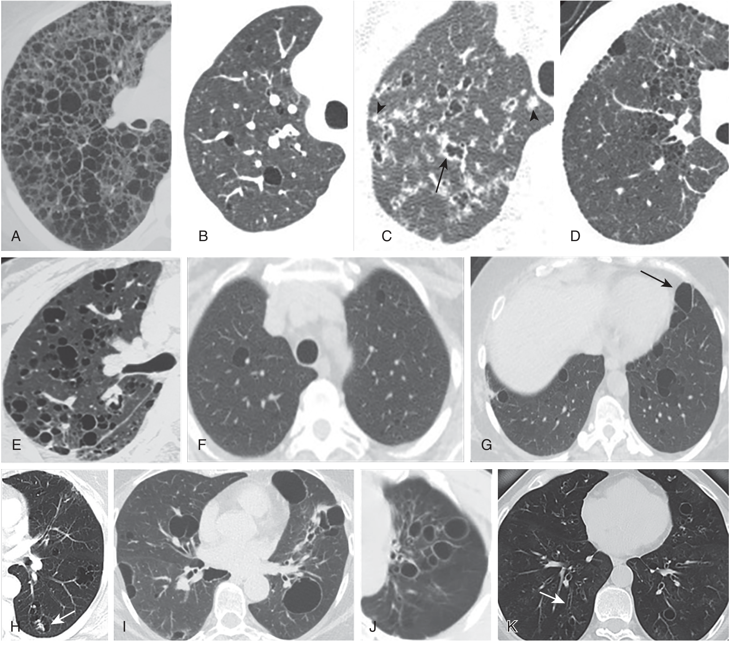
D. Cystic Pattern
Cysts = air-filled spaces with a definable wall (distinguishing them from emphysema which has no wall, and cavities which have thicker walls).
| Disease | Cyst Features | Distribution | Clues |
|---|---|---|---|
| LAM (Lymphangioleiomyomatosis) | Thin-walled, round, uniform | Diffuse, bilateral | Young women, chylothorax |
| LCH (Langerhans Cell Histiocytosis) | Irregular/bizarre shapes, thick-walled early | Upper lobe predominant | Smoker + nodules also present |
| LIP (Lymphoid Interstitial Pneumonia) | Thin-walled, scattered | Perivascular | Sjögren's syndrome, HIV |
| Birt-Hogg-Dubé | Oblong, septated | Lower lobe, subpleural, paradiastinal | Renal tumors, skin fibrofolliculomas |
| Emphysema | No perceptible wall | Upper (centrilobular), lower (paraseptal) | Smoking history |
(Murray & Nadel's, p. 474)
4. Ground-Glass Opacity (GGO) - A Special Finding
GGO = hazy increased lung attenuation that does not obscure the underlying vessels (unlike consolidation, which does).
Key concept: GGO = potentially reversible. Honeycombing + reticulation = likely irreversible.
| GGO alone | GGO + reticulation ("crazy paving") | GGO + traction bronchiectasis |
|---|---|---|
| Active alveolitis, DIP, HP, edema, drug reaction | Alveolar proteinosis, NSIP, Pneumocystis | Organizing phase - fibrosis setting in |
- Consolidation = complete air space replacement - vessels obscured. Causes: pneumonia, COP, AIP, malignancy, edema.
- Crazy paving = GGO + smooth interlobular septal thickening = classic for pulmonary alveolar proteinosis (PAP), also Pneumocystis jirovecii, lipoid pneumonia.
(Murray & Nadel's, p. 474; Grainger & Allison's, p. 224)
5. Reading an HRCT Systematically - A 5-Step Approach
Step 1 - Distribution
- Upper vs. lower lobe predominance?
- Central (peribronchovascular) vs. peripheral (subpleural)?
- Bilateral or unilateral?
Step 2 - Pattern
- Assign to one of the 4 main patterns (reticular / nodular / mosaic / cystic)
- Note GGO, consolidation, honeycombing if present
Step 3 - Nodule location (if nodules present)
- Centrilobular / perilymphatic / random?
Step 4 - Associated findings
- Traction bronchiectasis (fibrosis)
- Lymphadenopathy (sarcoid, lymphoma, infection)
- Pleural effusion / thickening
- Air trapping on expiratory CT
Step 5 - Reversibility assessment
| Reversible | Irreversible |
|---|---|
| Ground-glass opacity | Honeycombing |
| Consolidation | Coarse reticulation |
| Centrilobular nodules | Traction bronchiectasis |
| Architectural distortion |
(Grainger & Allison's, p. 225)
6. Quick-Reference: Common ILDs and Their HRCT Fingerprints
| Disease | Pattern | Distribution | Key Feature |
|---|---|---|---|
| IPF (UIP) | Reticulation + honeycombing | Basal, subpleural, bilateral | Temporal heterogeneity; honeycomb |
| NSIP | GGO + fine reticulation | Basal, subpleural, bilateral | Subpleural sparing of ~1 cm |
| Sarcoidosis | Perilymphatic nodules | Upper/mid lobe, peribronchovascular | Fissure involvement; lymphadenopathy |
| Hypersensitivity pneumonitis (subacute) | Centrilobular nodules + GGO + air trapping | Upper/mid lobe | Mosaic attenuation; spares bases |
| COP | Consolidation ± GGO | Peripheral, peribronchial | Migratory; "reversed halo sign" |
| PAP | Crazy paving | Geographic/diffuse | GGO + smooth septa |
| LAM | Thin-walled cysts | Diffuse | Young woman; chylothorax |
| DIP | Diffuse GGO | Lower zone, peripheral | Smoker |
| RB-ILD | Centrilobular GGO nodules | Upper lobe | Smoker; air trapping |
| LCH | Nodules + irregular cysts | Upper lobe | Heavy smoker; cysts evolve from nodules |
| Miliary TB | Random nodules | Diffuse, uniform | Tiny (1-3 mm), no lobular pattern |
7. Reversibility - The Clinically Crucial Axis
The whole point of HRCT over CXR is its ability to differentiate:
- "Active" / treatable disease = GGO, consolidation, centrilobular nodules - these may improve with steroids/immunosuppression
- "Fibrotic" / irreversible disease = honeycombing, traction bronchiectasis, architectural distortion - no recovery expected
This directly guides whether to offer treatment vs. transplant evaluation.
(Goldman-Cecil Medicine, p. 63)
Summary Mental Map
HRCT FINDING
│
├── RETICULAR ──► Fine = NSIP/early IPF
│ Coarse + honeycomb = UIP/IPF (basal, subpleural)
│ Smooth septal thickening = Edema / Lymphangitic ca
│
├── NODULAR ──► Perilymphatic = Sarcoidosis / Lymphangitic ca
│ Random = Miliary TB / Hematogenous mets
│ Centrilobular = HP / Bronchopneumonia / Tree-in-bud
│
├── MOSAIC ──► Dark component abnormal = Air trapping / CTEPH
│ Gray component abnormal = Infiltrative disease
│ Confirm with EXPIRATORY CT
│
└── CYSTIC ──► Round, uniform, diffuse = LAM
Irregular, upper lobe + nodules = LCH
Lower lobe, paradiastinal = Birt-Hogg-Dubé
No wall = Emphysema
No shortcuts please , at least mention I. The bracket
You want the full names spelled out in brackets wherever abbreviations are used. Let me redo the entire guide with every abbreviation expanded.
Refining...
Reading File
Reading File
Reading File
Reading File
Loading Image
Loading Image
Here is the fully expanded guide with every abbreviation written out in full on first use, and the short form kept in brackets afterwards for reference.
High-Resolution Computed Tomography [HRCT] Interpretation - Complete Simplified Guide
Sources: Grainger & Allison's Diagnostic Radiology; Murray & Nadel's Textbook of Respiratory Medicine; Goldman-Cecil Medicine; Washington Manual of Medical Therapeutics
1. What Is High-Resolution Computed Tomography [HRCT] and Why It Matters
High-Resolution Computed Tomography [HRCT] uses very thin slices (1-2 mm) and a high-spatial-frequency reconstruction algorithm to image the lung parenchyma in far greater detail than standard Computed Tomography [CT] or a plain chest radiograph.
It has become the cornerstone of Interstitial Lung Disease [ILD] evaluation because it:
- Detects early disease that a chest X-ray misses
- Can suggest a specific histological diagnosis in certain conditions
- Distinguishes reversible changes (ground-glass opacity, consolidation) from irreversible changes (fibrosis, honeycombing)
- Guides biopsy site selection and monitors response to treatment
The chest radiograph remains part of the initial assessment of Interstitial Lung Disease [ILD], but its pattern is often non-specific, observer variation is considerable, and it has low sensitivity for early disease. High-Resolution Computed Tomography [HRCT] has transformed this, enabling early detection and providing insights into disease reversibility and prognosis.
(Grainger & Allison's Diagnostic Radiology, p. 221)
2. The Lung Anatomy You Must Know First: The Secondary Pulmonary Lobule [SPL]
The Secondary Pulmonary Lobule [SPL] is the smallest discrete unit of lung anatomy visible on High-Resolution Computed Tomography [HRCT], roughly 1-2 cm in size and polyhedral in shape. Everything in HRCT interpretation anchors to it.
Secondary Pulmonary Lobule [SPL] anatomy:
┌─────────────────────────────────────────┐
│ Center: │
│ - Centrilobular artery │
│ - Terminal bronchiole │
│ │
│ Periphery: │
│ - Interlobular septa │
│ - Subpleural lymphatics │
│ - Visceral pleural surface │
└─────────────────────────────────────────┘
The interstitium of the lung supports all these structures and includes:
- The intralobular interstitium beneath the alveolar epithelium (inside the lobule)
- The interlobular septa (walls between lobules)
- The peribronchovascular interstitium (around airways and vessels)
Thickening of any of these compartments produces the patterns we read on High-Resolution Computed Tomography [HRCT].
3. The Four Main High-Resolution Computed Tomography [HRCT] Patterns
According to the Fleischner Society Glossary (the international standard terminology for chest imaging), all diffuse lung abnormalities on High-Resolution Computed Tomography [HRCT] can be broadly classified into four main patterns. Overlap between patterns is common.
| Pattern | What You See | Broad Category of Disease |
|---|---|---|
| Reticular | Network of lines, fine or coarse | Interstitial Lung Disease [ILD], fibrosis |
| Nodular | Discrete round opacities | Granulomas, infection, metastases |
| Mosaic attenuation | Patchwork of light and dark areas | Air trapping, vascular disease, infiltration |
| Cystic | Air-filled spaces with definable walls | Lymphangioleiomyomatosis [LAM], Langerhans Cell Histiocytosis [LCH], emphysema |
(Grainger & Allison's, p. 224)
4. Pattern 1 - The Reticular Pattern
A reticular (net-like) pattern on Computed Tomography [CT] almost always represents significant Interstitial Lung Disease [ILD].
What Causes It?
Morphologically, a reticular pattern is caused by:
- Thickened intralobular or interlobular septa
- Honeycombing (end-stage fibrotic destruction)
Types of Reticular Findings
A. Interlobular Septal Thickening
These are visible lines at the lung periphery outlining the lobule walls.
| Appearance | Cause |
|---|---|
| Smooth septal thickening | Pulmonary edema, Pulmonary Alveolar Proteinosis [PAP] |
| Irregular / beaded septal thickening | Lymphangitis Carcinomatosa [LC], sarcoidosis (rare) |
Numerous thickened interlobular septa indicate an extensive interstitial abnormality - caused by infiltration with fluid (e.g. pulmonary edema) or abnormal cells (e.g. Lymphangitis Carcinomatosa [LC]).
B. Intralobular Lines
Fine lines within the Secondary Pulmonary Lobule [SPL] separated by a few millimeters, creating a fine lace-like or net-like pattern. This is the hallmark of fibrotic lung disease - seen in Idiopathic Pulmonary Fibrosis [IPF] and Non-Specific Interstitial Pneumonia [NSIP].
C. Honeycombing
Severe pulmonary fibrosis produces a coarse reticular pattern made up of interlacing irregular linear opacities, eventually creating cystic air spaces (3-10 mm) surrounded by irregular, thick fibrotic walls - this is honeycombing.
Key features:
- Subpleural and basal predominance
- Stacked in layers ("stack of coins" appearance)
- The defining feature for Usual Interstitial Pneumonia [UIP] / Idiopathic Pulmonary Fibrosis [IPF]
- Represents irreversible destruction - no treatment can reverse it
D. Traction Bronchiectasis / Bronchiolectasis
The extensive fibrosis accompanying honeycombing distorts normal lung architecture, dragging the walls of segmental and subsegmental airways outward. This appears as dilated, irregular, non-tapering airways in the lung periphery. It is a marker of established fibrosis.
(Grainger & Allison's, p. 224; Murray & Nadel's, p. 470)
5. Pattern 2 - The Nodular Pattern
A nodular pattern appears in both interstitial and airspace diseases. The most important step is to classify nodules by their location within the Secondary Pulmonary Lobule [SPL]. This single step generates the differential diagnosis.
The Three Nodule Distributions
Distribution 1 - Perilymphatic Nodules
- Located along the interlobular septa, subpleural surfaces, and peribronchovascular bundles
- Follow the path of lymphatic vessels
- Involvement of the fissures is a key diagnostic clue - fissures are covered by visceral pleura which contains lymphatics, so nodules here are strongly perilymphatic
- These nodules touch the pleura
| Disease | Additional Clues |
|---|---|
| Sarcoidosis | Upper/mid lobe, bilateral hilar lymphadenopathy, peribronchovascular clustering |
| Lymphangitis Carcinomatosa [LC] | Unilateral possible, beaded septal thickening, history of known primary cancer |
| Silicosis / Coal Workers' Pneumoconiosis [CWP] | Upper lobe, subpleural and centrilobular regions, occupational history |
Distribution 2 - Random Nodules
- Scattered evenly and diffusely throughout both lungs with no preference for pleura or center
- Defined by their broad, fairly uniform pattern - present throughout lung parenchyma in a roughly equal manner
- They approach but also clearly contact the pleural surface (unlike centrilobular) and also appear centrally (unlike pure perilymphatic)
| Disease | Additional Clues |
|---|---|
| Miliary Tuberculosis [TB] | 1-3 mm tiny nodules, acute presentation, fever |
| Hematogenous metastases | Larger and variable-sized nodules, history of primary malignancy |
| Miliary fungal infection (histoplasmosis, coccidioidomycosis) | Travel/exposure history |
Distribution 3 - Centrilobular Nodules
- Located in the center of the Secondary Pulmonary Lobule [SPL], a few millimeters from the centrilobular artery and terminal bronchiole
- Critically: they spare the pleural surface and fissures - this is the key distinguishing feature from perilymphatic nodules
- Represent small airway or peribronchiolar disease
| Sub-type | Disease | Additional Clues |
|---|---|---|
| Well-defined centrilobular nodules | Silicosis, Pulmonary Langerhans Cell Histiocytosis [PLCH], Coal Workers' Pneumoconiosis [CWP] | Occupational history, smoking |
| Poorly defined / ground-glass centrilobular nodules | Hypersensitivity Pneumonitis [HP], Respiratory Bronchiolitis-Interstitial Lung Disease [RB-ILD], pulmonary edema | Antigen exposure, smoking |
| Tree-in-bud pattern | Endobronchial infection (Tuberculosis [TB], Non-Tuberculous Mycobacteria [NTM], bacterial bronchopneumonia), aspiration | Centrilobular nodules + branching linear densities = impacted/inflamed small bronchioles |
Side-by-Side Visual Comparison of Nodule Distributions
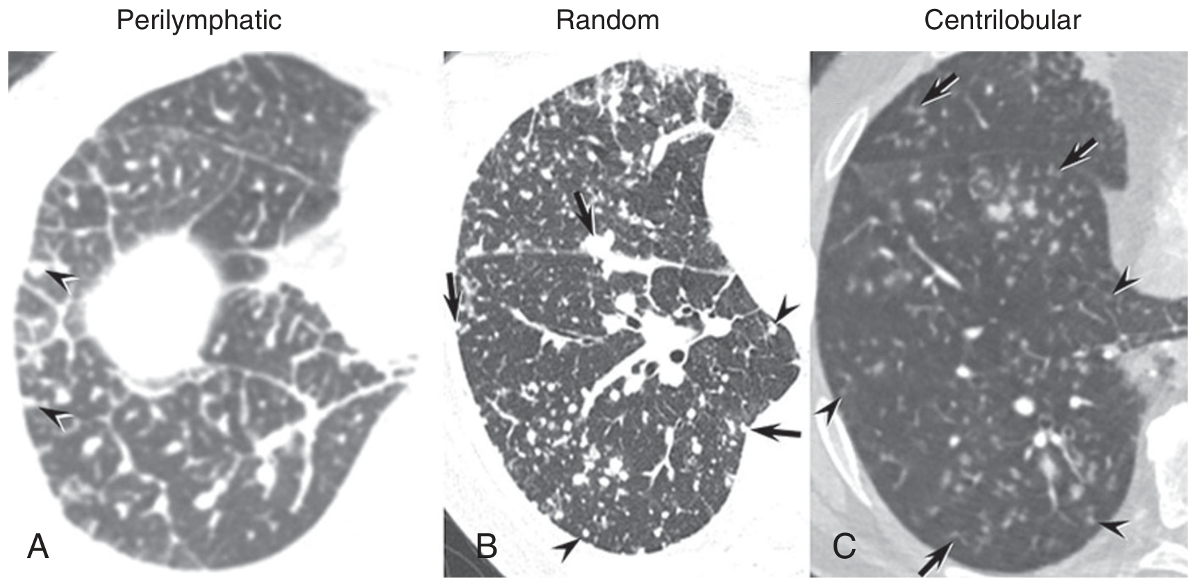
(Murray & Nadel's, p. 468-469; Grainger & Allison's, p. 224)
6. Pattern 3 - Mosaic Attenuation Pattern
The lung looks like a patchwork quilt - alternating areas of different radiodensity with well-defined borders corresponding to interlobular septa.
The key question is: which component is abnormal?
The attenuation of a lung area depends on the amount of blood, parenchymal tissue, and air it contains. A mosaic pattern is the dominant abnormality in three completely different diseases:
Three Causes of Mosaic Attenuation
A. Small Airways Disease (obstructive)
- The dark (low-attenuation) areas are abnormal - they contain too much air because diseased small airways trap it
- Look for: bronchial wall thickening, associated bronchiectasis
- Causes: Obliterative bronchiolitis [OB], Chronic Obstructive Pulmonary Disease [COPD], asthma, Cystic Fibrosis [CF]
- Confirm with expiratory High-Resolution Computed Tomography [HRCT]: the dark areas remain dark (persist) on expiration because air cannot escape - called air trapping
B. Chronic Occlusive Vascular Disease
- The dark areas are abnormal - they are underperfused because pulmonary vessels are blocked
- The lung hypoperfusion causes reflex hypoxic vasoconstriction and bronchodilatation in that area
- Look for: enlarged pulmonary trunk, abnormal artery-to-bronchus ratio (>1), no air trapping on expiratory High-Resolution Computed Tomography [HRCT]
- Cause: Chronic Thromboembolic Pulmonary Hypertension [CTEPH]
C. Infiltrative Lung Disease
- The gray/bright areas are abnormal - they contain increased interstitial or alveolar material
- The dark areas are actually normal lung
- Borders between areas may be less well-defined than in vascular causes
- No vascular caliber differences are present
- Causes: subacute Hypersensitivity Pneumonitis [HP], Acute Respiratory Distress Syndrome [ARDS], Non-Specific Interstitial Pneumonia [NSIP]
Summary Table
| Cause | Abnormal Component | Expiratory High-Resolution Computed Tomography [HRCT] | Clue |
|---|---|---|---|
| Small airways disease | Dark areas | Air trapping persists | Bronchial wall thickening |
| Chronic Thromboembolic Pulmonary Hypertension [CTEPH] | Dark areas | No air trapping | Enlarged pulmonary trunk |
| Infiltrative lung disease | Gray/bright areas | Equalizes | Ground-glass opacity pattern |
(Grainger & Allison's, p. 224)
7. Pattern 4 - The Cystic Pattern
Cysts on High-Resolution Computed Tomography [HRCT] are defined as air-filled spaces with a clearly definable, perceptible wall (even if thin). This distinguishes them from:
- Emphysema: areas of low attenuation with NO perceptible wall
- Cavities: thick-walled (>4 mm) air spaces inside a solid lesion
- Bullae: >1 cm air spaces, usually in emphysema
| Disease | Full Name | Cyst Morphology | Distribution | Key Clinical Clues |
|---|---|---|---|---|
| Lymphangioleiomyomatosis [LAM] | Lymphangioleiomyomatosis | Thin-walled, round, uniform in size | Diffuse, bilateral, no zonal predominance | Young women; chylothorax; may have Tuberous Sclerosis Complex [TSC] |
| Pulmonary Langerhans Cell Histiocytosis [PLCH] | Pulmonary Langerhans Cell Histiocytosis | Bizarre/irregular shapes, variable wall thickness; early lesions are nodules that cavitate | Upper and mid lobe predominant, spares Costophrenic [CP] angles | Heavy smoker; nodules coexist with cysts |
| Lymphoid Interstitial Pneumonia [LIP] | Lymphoid Interstitial Pneumonia | Thin-walled, uniform, peribronchovascular | Mid-lung, not predominantly subpleural | Sjögren Syndrome [SjS], Human Immunodeficiency Virus [HIV] infection |
| Birt-Hogg-Dubé Syndrome [BHD] | Birt-Hogg-Dubé Syndrome | Oblong, septated, unusual shapes | Lower lobe, subpleural, paradiastinal | Renal tumors, skin fibrofolliculomas |
| Emphysema | Emphysema | NO perceptible wall - just areas of low attenuation | Centrilobular: upper lobe; Panlobular: lower lobe; Paraseptal: subpleural | Smoking history; Chronic Obstructive Pulmonary Disease [COPD] |
(Murray & Nadel's, p. 474)
8. Ground-Glass Opacity [GGO] - The Most Common Finding Explained
Ground-Glass Opacity [GGO] = a hazy, increased lung opacity that does not obscure the underlying pulmonary vessels or bronchial walls (if it does obscure them, it becomes consolidation).
At a microscopic level, Ground-Glass Opacity [GGO] results from:
- Partial filling of the air spaces
- Thickening of the interstitium
- Or both simultaneously
- Essentially: subtotal displacement of air from the lung
Ground-Glass Opacity [GGO] can reflect active (potentially reversible) disease or early fibrosis below the spatial resolution of High-Resolution Computed Tomography [HRCT].
Reading Ground-Glass Opacity [GGO] in Context
| Ground-Glass Opacity [GGO] Pattern | Interpretation | Key Diseases |
|---|---|---|
| Pure Ground-Glass Opacity [GGO] alone | Active alveolitis, likely reversible | Hypersensitivity Pneumonitis [HP], Desquamative Interstitial Pneumonia [DIP], pulmonary edema, drug reaction |
| Ground-Glass Opacity [GGO] + smooth interlobular septal thickening = "Crazy Paving" | Combined airspace and septal involvement | Pulmonary Alveolar Proteinosis [PAP] (classic), Pneumocystis jirovecii Pneumonia [PJP], lipoid pneumonia |
| Ground-Glass Opacity [GGO] + traction bronchiectasis | Organizing/fibrotic phase - less reversible | Non-Specific Interstitial Pneumonia [NSIP], Acute Interstitial Pneumonia [AIP] (organizing phase) |
| Ground-Glass Opacity [GGO] + coarse reticulation + architectural distortion | Likely early fibrosis masquerading as Ground-Glass Opacity [GGO] | Idiopathic Pulmonary Fibrosis [IPF] (fibrotic Non-Specific Interstitial Pneumonia [NSIP] overlap) |
| Bilateral diffuse Ground-Glass Opacity [GGO] + consolidation (dependent) | Diffuse Alveolar Damage [DAD] | Acute Respiratory Distress Syndrome [ARDS], Acute Interstitial Pneumonia [AIP] |
Consolidation vs. Ground-Glass Opacity [GGO]
| Feature | Ground-Glass Opacity [GGO] | Consolidation |
|---|---|---|
| Vessels visible? | Yes | No (obscured) |
| Air bronchograms? | No | Yes (often) |
| Reversibility | Often reversible | Variable |
| Typical cause | Alveolitis, early edema | Pneumonia, Cryptogenic Organizing Pneumonia [COP], edema, malignancy |
(Murray & Nadel's, p. 474; Grainger & Allison's, p. 225)
9. The Idiopathic Interstitial Pneumonias [IIPs] and Their High-Resolution Computed Tomography [HRCT] Fingerprints
The Idiopathic Interstitial Pneumonias [IIPs] are a group of disorders with no known cause, each with a distinct histological and radiological appearance. The 2013 American Thoracic Society [ATS] / European Respiratory Society [ERS] consensus classification requires a multidisciplinary team approach - clinicians, radiologists, and pathologists together.
Usual Interstitial Pneumonia [UIP] / Idiopathic Pulmonary Fibrosis [IPF]
Idiopathic Pulmonary Fibrosis [IPF] is applied when the clinical, imaging, and (when available) histological features all show a pattern of Usual Interstitial Pneumonia [UIP]. However, the Usual Interstitial Pneumonia [UIP] pattern can also be secondary to Connective Tissue Disease [CTD] (especially Rheumatoid Arthritis [RA]), Chronic Hypersensitivity Pneumonitis [HP], asbestosis, and drugs.
High-Resolution Computed Tomography [HRCT] features of Usual Interstitial Pneumonia [UIP]:
- Reticulation (fine intralobular lines) - bilateral
- Honeycombing - subpleural, basal predominant
- Traction bronchiectasis - within fibrotic areas
- Temporal heterogeneity - areas of different stages of fibrosis (fresh fibroblastic foci next to dense scar) in the same scan - this is unique to Usual Interstitial Pneumonia [UIP] and is macroscopically depicted by High-Resolution Computed Tomography [HRCT]
- Subpleural and basilar predominance is required for a "definite Usual Interstitial Pneumonia [UIP]" diagnosis on High-Resolution Computed Tomography [HRCT]
- Ground-Glass Opacity [GGO] is minimal and always less than the reticulation
Non-Specific Interstitial Pneumonia [NSIP]
High-Resolution Computed Tomography [HRCT] features:
- Bilateral Ground-Glass Opacity [GGO] + fine reticulation
- Subpleural sparing (~1 cm rim of normal lung at the pleural edge) - this is the single most useful distinguishing feature from Usual Interstitial Pneumonia [UIP] / Idiopathic Pulmonary Fibrosis [IPF]
- Basal predominance
- Traction bronchiectasis in fibrotic Non-Specific Interstitial Pneumonia [NSIP] (fibrosing pattern)
- Commonly associated with Connective Tissue Disease [CTD] - especially Systemic Sclerosis [SSc] / scleroderma
Cryptogenic Organizing Pneumonia [COP]
High-Resolution Computed Tomography [HRCT] features:
- Bilateral peripheral, peribronchial consolidation
- Ground-Glass Opacity [GGO] surrounding consolidation
- Reversed Halo Sign (also called "atoll sign") - area of Ground-Glass Opacity [GGO] surrounded by a rim of denser consolidation - classic for Cryptogenic Organizing Pneumonia [COP]
- Migratory pattern over time (lesions appear, disappear, and reappear elsewhere)
Acute Interstitial Pneumonia [AIP]
Acute Interstitial Pneumonia [AIP] is the idiopathic form of Acute Respiratory Distress Syndrome [ARDS]. The histological pattern is Diffuse Alveolar Damage [DAD].
High-Resolution Computed Tomography [HRCT] features:
- Bilateral Ground-Glass Opacity [GGO] + consolidation (consolidation mainly in dependent lung)
- Traction bronchiectasis and reticulation appear after several days (organizing phase)
- Relative sparing of non-dependent areas (anterior)
- Anterior non-dependent fibrotic damage from barotrauma in survivors
Respiratory Bronchiolitis-Interstitial Lung Disease [RB-ILD]
Caused by cigarette smoking - macrophage accumulation within respiratory bronchioles and adjacent alveoli.
High-Resolution Computed Tomography [HRCT] features:
- Patchy Ground-Glass Opacity [GGO] (from macrophage accumulation in alveolar spaces and ducts)
- Poorly defined, low-attenuation centrilobular nodules
- Upper lobe centrilobular emphysema (limited extent)
- Areas of air trapping (from bronchiolitic component)
- Scattered interlobular septal thickening
Desquamative Interstitial Pneumonia [DIP]
Also smoking-related, closely related to Respiratory Bronchiolitis-Interstitial Lung Disease [RB-ILD] but more diffuse and severe.
High-Resolution Computed Tomography [HRCT] features:
- Diffuse Ground-Glass Opacity [GGO] is the dominant and characteristic feature
- Lower zone, peripheral distribution
- May be patchy or geographic
- Limited architectural distortion in some patients
- The term "Smoking-Related Interstitial Lung Disease [SR-ILD]" now encompasses Desquamative Interstitial Pneumonia [DIP], Respiratory Bronchiolitis-Interstitial Lung Disease [RB-ILD], Pulmonary Langerhans Cell Histiocytosis [PLCH], and smoking-related interstitial fibrosis
Lymphoid Interstitial Pneumonia [LIP]
Classically occurs with autoimmune disease, most often Sjögren Syndrome [SjS], Human Immunodeficiency Virus [HIV], or dysproteinaemias.
High-Resolution Computed Tomography [HRCT] features:
- Ground-Glass Opacity [GGO] (patchy)
- Indistinct nodules of varying sizes
- Thickened bronchovascular bundles
- Interlobular septal thickening
- Thin-walled cysts (1-30 mm) - discrete, sometimes clustered, tend not to be subpleural
(Grainger & Allison's, p. 228-232)
10. Sarcoidosis on High-Resolution Computed Tomography [HRCT]
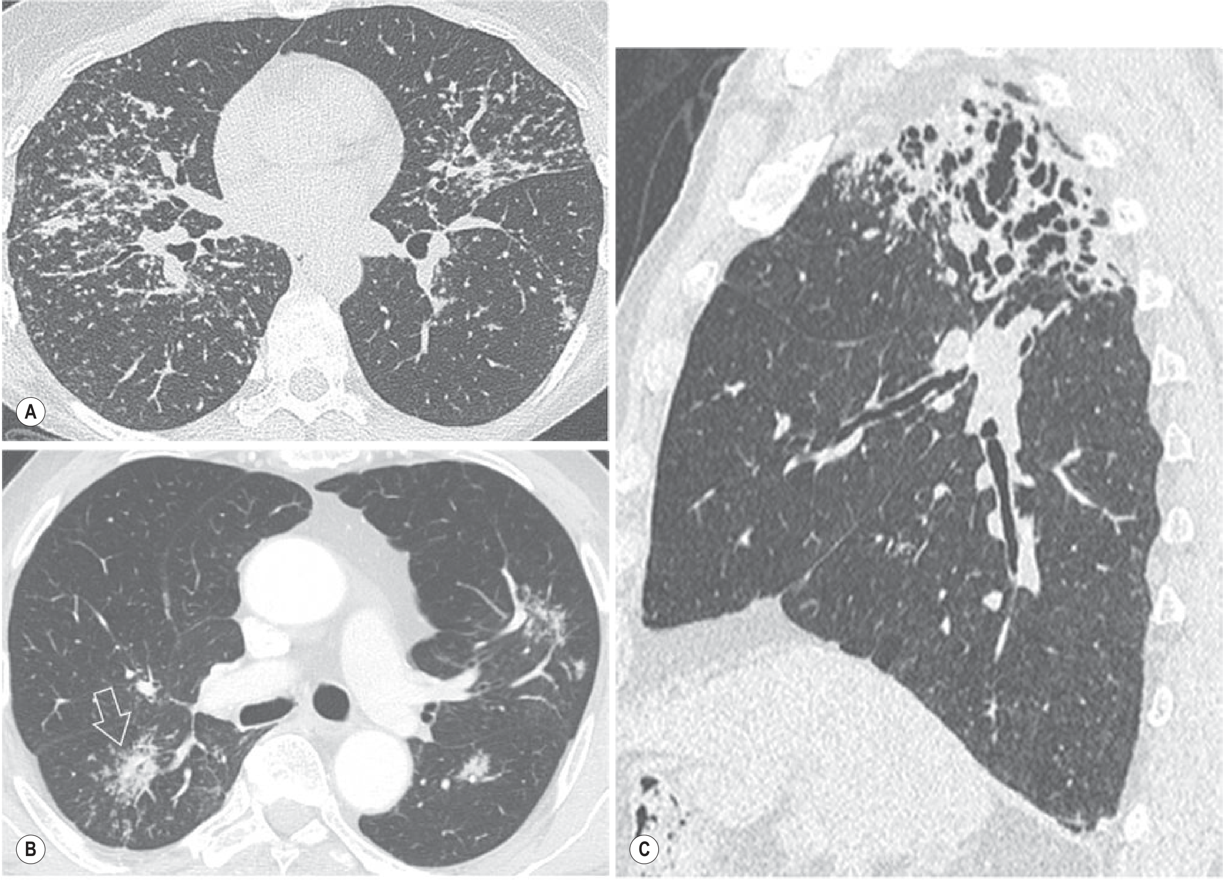
Hypersensitivity Pneumonitis [HP] (also called Extrinsic Allergic Alveolitis [EAA]) is an immunologically mediated lung disease triggered by inhaled organic antigens (bird proteins, moulds, farmer's lung antigens).
High-Resolution Computed Tomography [HRCT] features of sarcoidosis:
- Well-defined, smooth or irregular nodules 2-4 mm in perilymphatic distribution - mainly along interlobar fissures, peribronchovascular interstitium, and interlobular septa
- Most extensive in the upper and mid lobes
- Nodules may cluster into the Galaxy Sign (large opacity made of many small nodules)
- Bilateral hilar and mediastinal lymphadenopathy - highly characteristic
- Fibrotic stage: linear opacities radiating from the hilum, upper lobe volume loss, traction bronchiectasis, honeycombing concentrated in upper zones
- Ground-Glass Opacity [GGO] and consolidation in active disease are reversible; reticulation and architectural distortion are not
(Grainger & Allison's, p. 215)
11. Hypersensitivity Pneumonitis [HP] on High-Resolution Computed Tomography [HRCT]
Subacute Hypersensitivity Pneumonitis [HP] High-Resolution Computed Tomography [HRCT] features:
- Profuse bilateral, poorly defined centrilobular nodules
- Ground-Glass Opacity [GGO]
- Mosaic attenuation with air trapping
- Upper and mid lobe predominance (spares bases - distinguishing from Idiopathic Pulmonary Fibrosis [IPF])
- Air trapping on expiratory High-Resolution Computed Tomography [HRCT] - reflects bronchiolitic component
Chronic / Fibrotic Hypersensitivity Pneumonitis [HP] High-Resolution Computed Tomography [HRCT] features:
- Reticulation + honeycombing (can mimic Usual Interstitial Pneumonia [UIP]/Idiopathic Pulmonary Fibrosis [IPF])
- Bronchocentric distribution of fibrotic changes (unlike the purely peripheral Usual Interstitial Pneumonia [UIP])
- Relative mid-upper lobe involvement (unlike the basal Usual Interstitial Pneumonia [UIP])
- Mosaic attenuation and air trapping remain as clues
(Grainger & Allison's, p. 216)
12. Reversibility - The Clinically Critical Axis
The entire purpose of High-Resolution Computed Tomography [HRCT] over a chest radiograph is distinguishing what might respond to treatment from what will not.
| Reversible (Potentially Treatable) | Irreversible (No Recovery) |
|---|---|
| Ground-Glass Opacity [GGO] | Honeycombing |
| Consolidation | Coarse reticulation with architectural distortion |
| Centrilobular nodules | Traction bronchiectasis / bronchiolectasis |
| Mosaic Ground-Glass Opacity [GGO] | Volume loss |
This directly guides the clinical decision: treat with steroids/immunosuppression vs. refer for lung transplantation evaluation.
(Goldman-Cecil Medicine, p. 63)
13. A 5-Step Systematic Approach to Reading Every High-Resolution Computed Tomography [HRCT]
Step 1 - Distribution
- Which lobes? Upper vs. lower predominance?
- Which zone? Central (peribronchovascular) vs. peripheral (subpleural)?
- Symmetry? Bilateral symmetric vs. unilateral vs. bilateral asymmetric?
- Ask on expiratory High-Resolution Computed Tomography [HRCT]: does any area retain low attenuation?
Step 2 - Dominant Pattern
- Assign to one of the four main patterns: Reticular / Nodular / Mosaic attenuation / Cystic
- Note Ground-Glass Opacity [GGO], consolidation, or honeycombing if present alongside
Step 3 - Nodule Classification (if nodules are present)
- Perilymphatic (touches fissures/pleura)?
- Centrilobular (spares pleura)?
- Random (uniform, diffuse, both perilymphatic and centrilobular locations)?
- Tree-in-bud? (branching centrilobular)
Step 4 - Associated Findings
- Traction bronchiectasis / bronchiolectasis (fibrosis marker)
- Mediastinal / hilar lymphadenopathy
- Pleural effusion or pleural thickening
- Pneumothorax
- Pulmonary artery enlargement (pulmonary hypertension)
- Chest wall / skeletal findings
Step 5 - Reversibility Assessment
- Classify each finding as reversible or irreversible (see table above)
- This guides prognosis and management
14. Quick-Reference Master Table - Common Diseases and Their High-Resolution Computed Tomography [HRCT] Fingerprints
| Disease (Full Name) | Short Form | Dominant Pattern | Distribution | Single Most Specific Feature |
|---|---|---|---|---|
| Idiopathic Pulmonary Fibrosis | IPF | Reticulation + honeycombing | Bilateral, basal, subpleural | Temporal heterogeneity; definite honeycombing |
| Non-Specific Interstitial Pneumonia | NSIP | Ground-Glass Opacity + fine reticulation | Bilateral, basal, subpleural | Subpleural sparing (~1 cm) |
| Sarcoidosis | - | Perilymphatic nodules | Upper/mid, peribronchovascular | Fissure nodules; bilateral hilar adenopathy |
| Hypersensitivity Pneumonitis | HP | Centrilobular nodules + Ground-Glass Opacity | Upper/mid, bronchocentric | Mosaic attenuation + air trapping |
| Cryptogenic Organizing Pneumonia | COP | Consolidation | Peripheral, peribronchial | Reversed Halo / Atoll Sign; migratory |
| Pulmonary Alveolar Proteinosis | PAP | Crazy paving | Geographic/diffuse | Ground-Glass Opacity + smooth septa |
| Lymphangioleiomyomatosis | LAM | Thin-walled round cysts | Diffuse, bilateral | Young woman; chylothorax; uniform cysts |
| Pulmonary Langerhans Cell Histiocytosis | PLCH | Nodules + irregular cysts | Upper lobe | Smoker; bizarre-shaped cysts; nodules → cysts |
| Desquamative Interstitial Pneumonia | DIP | Diffuse Ground-Glass Opacity | Lower zone, peripheral | Smoker; extensive diffuse Ground-Glass Opacity |
| Respiratory Bronchiolitis-Interstitial Lung Disease | RB-ILD | Centrilobular Ground-Glass Opacity nodules | Upper lobe | Smoker; air trapping |
| Acute Interstitial Pneumonia | AIP | Ground-Glass Opacity + consolidation | Bilateral diffuse, dependent consolidation | Acute onset; Diffuse Alveolar Damage pattern |
| Lymphoid Interstitial Pneumonia | LIP | Ground-Glass Opacity + cysts | Mid-lung | Sjögren Syndrome; thin-walled peribronchovascular cysts |
| Miliary Tuberculosis | Miliary TB | Random nodules | Diffuse, uniform | 1-3 mm uniform tiny nodules; no zonal bias |
| Lymphangitis Carcinomatosa | LC | Irregular septal thickening + perilymphatic nodules | Peribronchovascular, septal | Beaded septa; known primary malignancy |
| Chronic Thromboembolic Pulmonary Hypertension | CTEPH | Mosaic attenuation | Bilateral, no zonal pattern | No air trapping on expiratory scan; enlarged pulmonary trunk |
15. Summary Mental Map
HIGH-RESOLUTION COMPUTED TOMOGRAPHY [HRCT] FINDING
│
├── RETICULAR ──► Fine intralobular lines
│ = Non-Specific Interstitial Pneumonia [NSIP] (+ subpleural sparing)
│ or early Idiopathic Pulmonary Fibrosis [IPF]
│
│ Coarse + honeycombing (basal, subpleural)
│ = Usual Interstitial Pneumonia [UIP] / Idiopathic Pulmonary Fibrosis [IPF]
│
│ Smooth septal thickening
│ = Pulmonary edema / Pulmonary Alveolar Proteinosis [PAP]
│
│ Beaded/irregular septal thickening
│ = Lymphangitis Carcinomatosa [LC]
│
├── NODULAR ──► Perilymphatic (touches pleura/fissures)
│ = Sarcoidosis / Lymphangitis Carcinomatosa [LC]
│
│ Random (uniform, diffuse)
│ = Miliary Tuberculosis [TB] / Hematogenous metastases
│
│ Centrilobular (spares pleura)
│ = Hypersensitivity Pneumonitis [HP] / Bronchopneumonia
│
│ Centrilobular + branching = Tree-in-bud
│ = Endobronchial Tuberculosis [TB] / Non-Tuberculous Mycobacteria [NTM]
│
├── MOSAIC ──► Dark areas abnormal + air trapping on expiry
│ = Small airways disease (Obliterative Bronchiolitis [OB], asthma)
│
│ Dark areas abnormal + no air trapping + enlarged pulmonary trunk
│ = Chronic Thromboembolic Pulmonary Hypertension [CTEPH]
│
│ Gray/bright areas abnormal
│ = Infiltrative lung disease / Ground-Glass Opacity [GGO]
│
└── CYSTIC ──► Round, thin-walled, uniform, diffuse
= Lymphangioleiomyomatosis [LAM]
Irregular, bizarre-shaped, upper lobe + nodules
= Pulmonary Langerhans Cell Histiocytosis [PLCH]
Lower lobe, paradiastinal, oblong/septated
= Birt-Hogg-Dubé Syndrome [BHD]
Peribronchovascular, Sjögren Syndrome [SjS] / Human Immunodeficiency Virus [HIV]
= Lymphoid Interstitial Pneumonia [LIP]
No wall, upper lobe (smoker)
= Centrilobular emphysema (Chronic Obstructive Pulmonary Disease [COPD])
This is a shared conversation. Sign in to Orris to start your own chat.