
Make presintation about Stitching methods, types and instruments
What context is this presentation for? · Who is the target audience?
surgical suture stitching methods types instruments needle
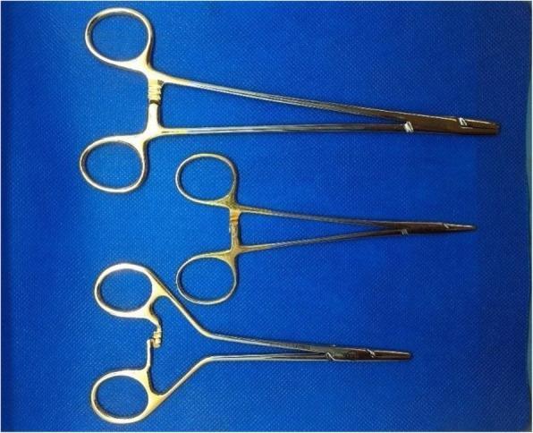
A clinical photograph of three different surgical needle holders used in specialized suturing procedures, displayed against a blue surgical drape. All three instruments are made of stainless steel with a metallic finish and feature ratcheted locking mechanisms and slender jaws designed for gripping suture needles. The top instrument is a 20.5 cm Stille Mayo-Hegar Needle Holder (LMH), a standard larger-format instrument for heavy tissue suturing. The middle instrument is a 16 cm Stille Mayo-Hegar Needle Holder (SMH), providing a more compact profile for refined manipulation. The bottom instrument is a 15 cm Stille Frimand Duo-Grip Needle Holder (FR), which distinguishes itself through a unique ergonomic handle design; it features offset finger rings and a curved lever arm leading to the ratchet, intended to enhance grip control and tactile feedback. These instruments represent various mechanical designs optimized for surgical anastomoses, where precise control over needle placement is critical.
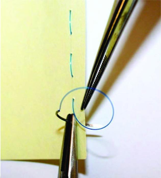
This clinical educational photograph demonstrates basic surgical suturing techniques using a simulated model. The image depicts a yellow synthetic material serving as a tissue analog, showing two parallel lines of previously placed green interrupted sutures. Two primary surgical instruments are visible: a needle holder (hemostat) and a pair of serrated tissue forceps (Adson or similar). The needle holder is shown grasping a curved surgical needle which has passed through the material, trailing a blue monofilament suture. The tissue forceps are positioned to stabilize the edge of the material and manipulate the suture loop to facilitate knot tying. This visual is designed for medical training to illustrate hand-instrument coordination, suture placement, and the mechanics of instrument-assisted knot tying in a controlled, non-biological environment. It serves as a foundational resource for teaching surgical skills and wound closure principles to medical students and surgical residents.
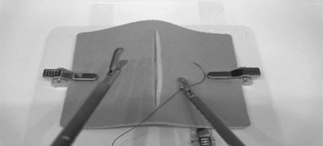
This monochrome image displays a still frame from a laparoscopic surgical simulator, specifically designed for intracorporeal suturing training. The focus is on a central suturing pad with a linear vertical defect that mimics a surgical incision or wound. Two laparoscopic instruments are active within the simulated field: a grasper on the left side and a needle holder on the right. The needle holder is currently manipulating a curved surgical needle attached to a suture thread, demonstrating the initial phase of wound closure. The suturing pad is secured to a white platform by lateral metal clamps. This setup is used in medical education to develop psychomotor skills, depth perception, and manual dexterity required for minimally invasive surgery. The image serves as a clinical simulation tool for teaching advanced surgical techniques, such as continuous or interrupted suturing, within a controlled augmented reality environment.
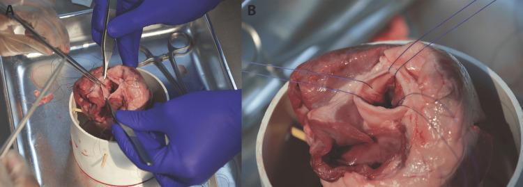
This clinical photograph displays a side-by-side comparison (labeled A and B) of a cardiac surgical simulation involving a porcine heart specimen. Panel A illustrates the active phase of suture placement at the aortic annulus. Visible instruments include needle drivers, surgical forceps, and scissors being used by a surgeon in blue gloves to place sutures at the nadir of the aortic valve cusps. The suture threads appear loose and unorganized at this stage. Panel B shows the same specimen after initial suture placement. The surgical instruments are removed, and the blue suture threads are organized and pulled taut, radiating superiorly from the annulus. This configuration creates guide paths intended to facilitate the descent of a prosthetic valve into the aortic lumen. The images demonstrate key technical steps in valve replacement surgery, focusing on annular suturing and thread management to ensure proper prosthetic seating.
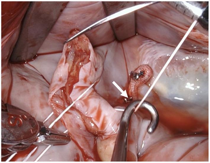
Intraoperative clinical photograph showing a close-up view of a cardiac surgical field, likely during a valve repair or replacement procedure. The image depicts reddish-pink cardiac tissue, possibly a valve leaflet, being manipulated. Key surgical instruments include a stainless steel suture hook in the foreground, applying light traction to a white, braided suture thread to maintain tension. In the lower-left corner, a surgical needle holder or clamp is visible, grasping the other end of the suture. In the center-right background, a small, textured, pinkish-orange prosthetic or pledget-like material is anchored to the tissue with a metallic fastener (indicated by a white arrow). Retractors are positioned at the top of the frame to maintain surgical exposure. The image demonstrates precise tissue handling and suture management techniques essential in cardiovascular surgery.
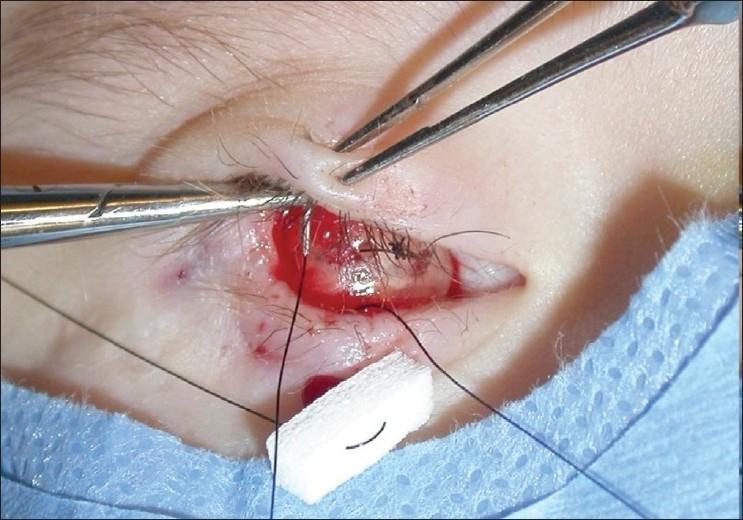
Clinical photograph of a periocular surgical procedure, specifically a suture tarsorrhaphy being performed over a hemisphere expander. The image shows the right ocular region of a patient, likely pediatric, with surgical instruments in use: a needle driver is seen placing a dark, non-absorbable suture through the upper eyelid margin, while fine-toothed forceps (such as Bishop-Harmon or Castroviejo) are used to stabilize the tissue. The palpebral fissure is partially closed by pre-existing sutures. Visible anatomical structures include the superior and inferior eyelids, eyelashes, and the palpebral conjunctiva. Surgical materials present include dark suture threads and a white absorbent gauze bolster or pad near the inferior margin to manage localized bleeding. The procedure is indicated for the management of congenital anophthalmia or microphthalmia, where the tarsorrhaphy provides external support to maintain the position of an orbital volume expander (hydrogel hemisphere) within the conjunctival socket to prevent extrusion and encourage orbital growth.
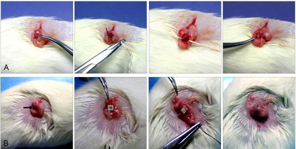
Clinical photograph series documenting a comparative surgical procedure in a rodent model, specifically used in oncological research for chemical induction of ovarian cancer. Row A illustrates the 'Sutures' (S) group: an exposed reddish-pink rat ovary is manipulated with surgical forceps and a needle driver to insert a 3-0 silk suture coated with 7,12-dimethylbenz(a)anthracene (DMBA). The process involves deep tissue penetration to deliver the carcinogen. Row B illustrates the 'Cloth Strip' (CS) group: a small square of DMBA-coated cloth (indicated by a white asterisk) is carefully packed onto the surface of the ovary after dissecting the membranous pouch. Key instruments shown include curved forceps, needle drivers, and sutures. The images demonstrate the variation in mechanical delivery methods—deep insertion via suture versus surface packing via cloth strip—designed to evaluate the developmental origins of ovarian epithelial adenocarcinomas versus other non-epithelial histological types.
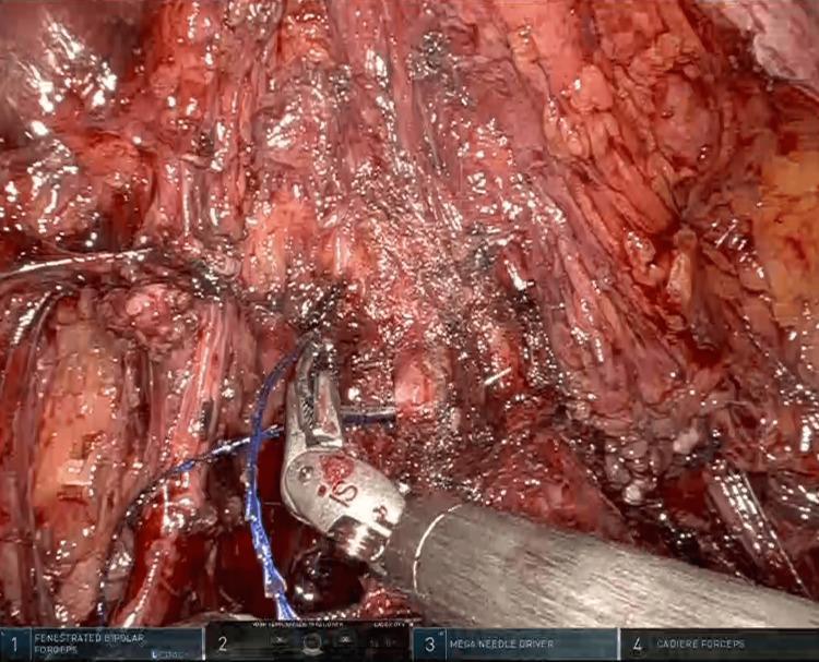
An intraoperative clinical photograph captured during a robotic-assisted gynecological procedure, specifically a radical hysterectomy. The image illustrates the preparation of the vaginal cuff for closure. Centrally, robotic surgical instruments, including a Mega Needle Driver and Fenestrated Bipolar Forceps marked with 'IS' branding, are manipulating a blue, monofilament, barbed suture (Stratafix). The suture is being passed through the anterior and lateral vaginal walls to create a circular purse-string suture. The surgical field displays moist, hyperemic pelvic tissues with visible dissection planes within the vesicovaginal and rectovaginal septa. The educational focus of the image is on the technique of intracorporeal vaginal cuff closure using knotless, absorbable barbed suturing in a minimally invasive robotic surgery context. This visual demonstrates key steps in pelvic surgery, emphasizing instrument handling, tissue traction, and the application of advanced suture materials for secure closure.
suture types absorbable non-absorbable monofilament braided
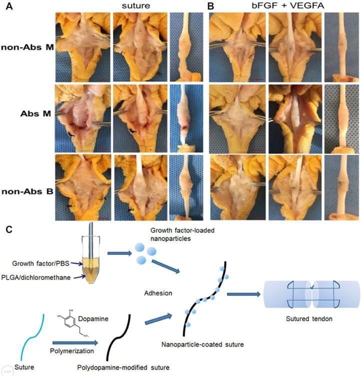
This composite educational image illustrates experimental methods for tendon repair and adhesion prevention. Panels A and B contain clinical photographs from an animal model (chicken flexor tendon) comparing surgical outcomes across three suture types: non-absorbable monofilament (non-Abs M), absorbable monofilament (Abs M), and non-absorbable braided (non-Abs B). Group A (Suture) shows control results with standard sutures, demonstrating significant peritendinous adhesions, disorganized tissue structure, and in the Abs M group, visible gaps and focal redness at the repair site. Group B (bFGF + VEGFA) shows tendons treated with nanoparticle-coated sutures loaded with basic fibroblast growth factor and vascular endothelial growth factor A. These specimens exhibit smoother surfaces, reduced scarring, and improved longitudinal tissue integration. Panel C is a pathophysiology and bioengineering diagram detailing the suture modification process: growth factors are encapsulated in PLGA nanoparticles and adhered to dopamine-polymerized sutures. The final schematic shows the modified suture applied to a transected tendon, aiming to facilitate organized healing and reduce post-operative adhesions.
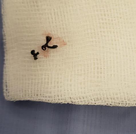
A clinical photograph displaying a retained foreign body removed from a patient. The image shows a dark, likely black, non-absorbable monofilament or braided suture material that has been surgically excised. The suture exhibits two visible surgical knots and is resting on a sterile, white, open-weave gauze dressing. A localized faint reddish-pink serosanguinous stain is visible on the gauze fibers surrounding one of the knots, indicating previous contact with wound tissue or fluid. This visual represents a common complication of post-operative wound healing, specifically a 'suture granuloma' or chronic non-healing wound caused by a retained suture. The image is pedagogically significant for illustrating iatrogenic causes of wound dehiscence or chronic inflammation, highlighting the importance of thorough wound exploration when a patient presents with a persistent, non-healing surgical site.
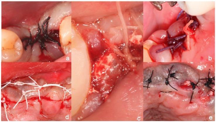
A composite of five clinical intraoral photographs (a–e) demonstrating different surgical suture materials used for wound closure in dental or maxillofacial surgery. Each panel illustrates specific material properties and knotting techniques: (a) Black braided silk suture showing a multifilament texture with multiple throws; (b) Violet-colored Vicryl (polyglactin 910), a coated synthetic absorbable suture; (c) Translucent gut suture, a natural collagenous material; (d) White PTFE (polytetrafluoroethylene) monofilament suture, characterized by its non-wicking, hydrophobic appearance and simple interrupted knots; (e) Dark-colored monofilament polyamide (nylon) suture. The images focus on tissue approximation, suture bite size, and the physical interaction between the thread and the oral mucosa. This visual guide serves as a pedagogical resource for identifying suture types by color, texture, and handling characteristics while highlighting clinical application in periodontal and surgical sites.
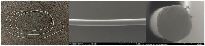
This composite figure provides a multi-scale visual characterization of a P(3HB-co-16 mol% 4HB) absorbable monofilament suture, relevant to surgical biomaterials and wound closure. The left panel is a clinical photograph showing the suture's macroscopic appearance as a thin, translucent, and flexible strand arranged in loose loops. The center panel is a side-view Scanning Electron Microscopy (SEM) image at 40x magnification, revealing a highly uniform, smooth longitudinal surface characteristic of synthetic monofilaments. The right panel displays a cross-sectional SEM image at 250x magnification, demonstrating a predominantly circular morphology with a solid, non-porous internal structure. Minor surface irregularities and small peripheral fragments are visible at this higher resolution. These images serve to document the structural integrity and surface quality of the polyhydroxyalkanoate-based suture, which is designed for slow-healing wounds due to its durability and biocompatibility.
interrupted suture continuous running mattress suture technique diagram
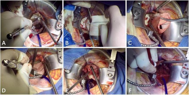
A composite of six intraoperative clinical photographs (A-F) documenting the key stages of a Right Minithoracotomy Bentall Procedure. (A, B) Focus on the aortic annulus, demonstrating the placement of interrupted mattress sutures using 2-0 poly(ethylene terephthalate) with gaskets to secure the proximal graft. (C) Shows the coronary anastomosis of the left main artery to the vascular graft performed in a continuous running fashion with 5-0 polypropylene. (D) Illustrates the subsequent right main coronary artery anastomosis, also utilizing a running 5-0 polypropylene suture technique. (E) Depicts the distal anastomosis between the vascular graft and the native ascending aorta using 4-0 polypropylene in a running fashion, with surgical tools like a nerve hook used for suture tension management. (F) Captures the final de-airing process of the left ventricle and aortic root graft using a venting needle prior to the removal of the aortic cross-clamp. This series illustrates a minimally invasive approach to aortic root replacement including valve and ascending aorta reconstruction.
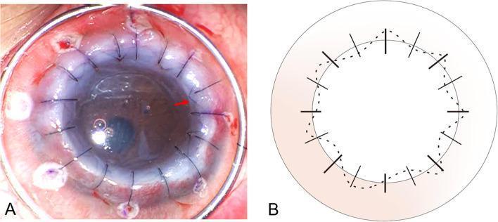
This dual-panel image features an intraoperative clinical photograph (A) and a matching schematic diagram (B) detailing a combined suturing technique for penetrating keratoplasty (PK). Panel A shows a large-diameter corneal graft in situ. The recipient cornea appears thickened and opaque with significant stromal edema. A combination of radial interrupted sutures and a continuous intrastromal running suture (highlighted by a red arrow) is visible across the graft-host junction. Small fluid-filled bullae are present on the corneal surface. Panel B provides a schematic representation of the surgical technique: coarse solid lines indicate the initial eight radial interrupted sutures, a dotted line represents the continuous intrastromal suture passing through deep stromal layers, and fine solid lines depict eight additional interrupted sutures used for reinforcement. The diagram illustrates how the needle entry and exit points for the continuous suture coincide to ensure deep tissue placement, bypassing the epithelial layer to maximize graft stability in complex cases like advanced keratoconus with hydrops.
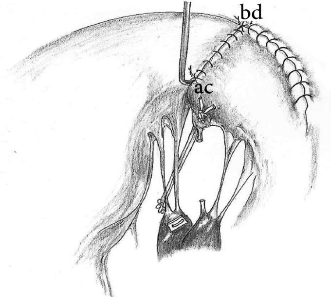
This medical illustration depicts a surgical technique for mitral valve repair, specifically focusing on the reconstruction of the posterior leaflet and subvalvular apparatus. The diagram illustrates a quadrangular resection followed by the reattachment of the posterior leaflet remnants (P2 and P1 segments) to the mitral annulus using a continuous running suture pattern. Interrupted sutures are visible where the P2 remnant (marked 'c-d') is affixed to the commissure remnant (marked 'a-b'). A critical feature shown is the neochordae formation, where an artificial cord (likely PTFE) is anchored from a pledget-reinforced site on the anterior papillary head of the posterior papillary muscle to the free edge of the high P2 remnant. The subvalvular anatomy is clearly represented, including the papillary muscles and native chordae tendineae. This illustration is intended for cardiothoracic surgery education, demonstrating techniques to manage posterior leaflet prolapse while maintaining optimal coaptation and ventricular-mitral continuity.
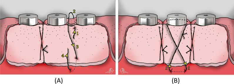
This medical illustration depicts two schematic views (A and B) of suturing techniques for securing a free gingival graft (FGG) around three dental implants in the mandible. Figure A illustrates the 'Double Vertical Interrupted Suture' technique used in the inter-implant areas. The diagram uses numbered yellow arrows (1–4) to show the needle's path: entering at the coronal aspect of the graft, passing through the lingual/palatal tissue, and re-entering apically to engage the periosteum before being tied at the graft base. This technique focuses on graft stabilization and adaptation to the underlying bed. Figure B demonstrates the 'Suspensory Cross-Mattress Suture' applied over each implant. This suture crosses diagonally over the implant abutment, forming an 'X' shape to ensure firm adaptation of the graft against the recipient bed, which is critical for establishing vascular supply. The illustration serves as an educational guide for periodontal or oral surgery, specifically for treating peri-implantitis and augmenting keratinized mucosa and vestibular depth.
mkdir -p /tmp/workspace/stitching-pptx && ls /tmp/workspace/
~/stitching-pptx/build.py
import subprocess, json, base64
from io import BytesIO
from pptx import Presentation
from pptx.util import Inches, Pt, Emu
from pptx.dml.color import RGBColor
from pptx.enum.text import PP_ALIGN, MSO_ANCHOR
from pptx.oxml.ns import qn
from lxml import etree

# ── Colour palette ─────────────────────────────────────────────────────────────
NAVY     = RGBColor(0x0D, 0x2B, 0x55)   # deep navy - title bg
TEAL     = RGBColor(0x00, 0x7A, 0x87)   # teal accent
GOLD     = RGBColor(0xF0, 0xA5, 0x00)   # warm gold highlight
WHITE    = RGBColor(0xFF, 0xFF, 0xFF)
LIGHT_BG = RGBColor(0xF4, 0xF8, 0xFB)   # slide background
DARK_TXT = RGBColor(0x1A, 0x1A, 0x2E)
MID_GREY = RGBColor(0x55, 0x6B, 0x82)
TEAL_LIGHT = RGBColor(0xE0, 0xF5, 0xF7)

# ── Presentation setup ─────────────────────────────────────────────────────────
prs = Presentation()
prs.slide_width  = Inches(13.333)
prs.slide_height = Inches(7.5)
blank = prs.slide_layouts[6]

# ── Helper: solid fill ─────────────────────────────────────────────────────────
def solid_fill(shape, color):
    fill = shape.fill
    fill.solid()
    fill.fore_color.rgb = color

# ── Helper: add textbox ────────────────────────────────────────────────────────
def tb(slide, x, y, w, h, text, size, bold=False, color=WHITE,
       align=PP_ALIGN.LEFT, wrap=True, italic=False):
    txb = slide.shapes.add_textbox(Inches(x), Inches(y), Inches(w), Inches(h))
    tf  = txb.text_frame
    tf.word_wrap = wrap
    tf.margin_left = tf.margin_right = tf.margin_top = tf.margin_bottom = 0
    p = tf.paragraphs[0]
    p.alignment = align
    r = p.add_run()
    r.text = text
    r.font.size  = Pt(size)
    r.font.bold  = bold
    r.font.italic = italic
    r.font.color.rgb = color
    r.font.name = "Calibri"
    return txb

# ── Helper: add rect ───────────────────────────────────────────────────────────
def rect(slide, x, y, w, h, color):
    s = slide.shapes.add_shape(1, Inches(x), Inches(y), Inches(w), Inches(h))
    solid_fill(s, color)
    s.line.fill.background()
    return s

# ── Helper: multi-paragraph textbox ───────────────────────────────────────────
def tb_bullets(slide, x, y, w, h, title, bullets, title_size=14,
               bullet_size=11, title_color=NAVY, bullet_color=DARK_TXT,
               bg_color=None, padding=0.15):
    if bg_color:
        rect(slide, x, y, w, h, bg_color)
    txb = slide.shapes.add_textbox(Inches(x+padding), Inches(y+padding),
                                   Inches(w-padding*2), Inches(h-padding*2))
    tf  = txb.text_frame
    tf.word_wrap = True
    tf.margin_left = tf.margin_right = tf.margin_top = tf.margin_bottom = 0
    # Title
    p = tf.paragraphs[0]
    r = p.add_run()
    r.text = title
    r.font.size  = Pt(title_size)
    r.font.bold  = True
    r.font.color.rgb = title_color
    r.font.name = "Calibri"
    # Bullets
    for b in bullets:
        p = tf.add_paragraph()
        r = p.add_run()
        r.text = f"• {b}"
        r.font.size  = Pt(bullet_size)
        r.font.color.rgb = bullet_color
        r.font.name = "Calibri"
    return txb

# ── Helper: add image from URL ────────────────────────────────────────────────
def add_img_url(slide, url, x, y, w, h):
    try:
        result = json.loads(subprocess.check_output(
            ["python", "/tmp/skills/shared/scripts/fetch_images.py", url],
            timeout=30
        ))
        if result and result[0].get("base64"):
            fmt, b64 = result[0]["base64"].split(";base64,")
            raw = base64.b64decode(b64)
            slide.shapes.add_picture(BytesIO(raw), Inches(x), Inches(y),
                                     Inches(w), Inches(h))
            return True
    except Exception as e:
        print(f"Image load failed: {e}")
    return False

# ── Helper: slide header strip ────────────────────────────────────────────────
def header(slide, title_text, subtitle_text=None):
    rect(slide, 0, 0, 13.333, 7.5, LIGHT_BG)       # background
    rect(slide, 0, 0, 13.333, 1.05, NAVY)            # header bar
    rect(slide, 0, 0.95, 13.333, 0.06, GOLD)         # gold accent line
    tb(slide, 0.35, 0.12, 12.5, 0.75, title_text,
       28, bold=True, color=WHITE, align=PP_ALIGN.LEFT)
    if subtitle_text:
        tb(slide, 0.35, 0.75, 12.5, 0.3, subtitle_text,
           12, color=GOLD, align=PP_ALIGN.LEFT, italic=True)

# ── SLIDE 1 – Title slide ─────────────────────────────────────────────────────
s1 = prs.slides.add_slide(blank)
rect(s1, 0, 0, 13.333, 7.5, NAVY)
# Decorative gold band
rect(s1, 0, 3.5, 13.333, 0.12, GOLD)
rect(s1, 0, 6.9, 13.333, 0.6, TEAL)
# Side teal strip
rect(s1, 0, 0, 0.18, 7.5, TEAL)
tb(s1, 0.5, 1.2, 12.0, 1.5,
   "STITCHING METHODS,\nTYPES & INSTRUMENTS",
   40, bold=True, color=WHITE, align=PP_ALIGN.LEFT)
tb(s1, 0.5, 3.7, 12.0, 0.6,
   "A Comprehensive Overview for Medical Students & Professionals",
   16, color=GOLD, align=PP_ALIGN.LEFT, italic=True)
tb(s1, 0.5, 4.45, 12.0, 0.55,
   "Covering: Suture Materials  |  Stitch Techniques  |  Wound Closure Methods  |  Surgical Instruments",
   13, color=RGBColor(0xB0, 0xC8, 0xE0), align=PP_ALIGN.LEFT)
# Bottom bar text
tb(s1, 0.5, 7.05, 8.0, 0.4, "Based on Tintinalli's Emergency Medicine & Dermatology 5e",
   10, color=WHITE, italic=True)

# ── SLIDE 2 – Overview / Table of Contents ────────────────────────────────────
s2 = prs.slides.add_slide(blank)
header(s2, "Table of Contents", "What we will cover today")

topics = [
    ("01", "Introduction to Wound Closure",      "Principles, goals, closure timing"),
    ("02", "Suture Materials",                    "Absorbable vs. non-absorbable, monofilament vs. braided"),
    ("03", "Basic Stitch Techniques",             "Simple interrupted, continuous, subcuticular"),
    ("04", "Advanced Stitch Techniques",          "Mattress, figure-of-eight, purse-string"),
    ("05", "Other Wound Closure Methods",         "Staples, tissue adhesives, steri-strips, staples"),
    ("06", "Surgical Instruments",                "Needle holders, forceps, scissors, scalpels"),
    ("07", "Choosing the Right Technique",        "Wound type decision table"),
    ("08", "Wound Healing & Complications",       "Healing phases, dehiscence, infection"),
]
cols = [(0.35, 3.6), (6.85, 6.1)]
for i, (num, title, desc) in enumerate(topics):
    col_x, col_w = cols[i // 4]
    row = i % 4
    y = 1.35 + row * 1.5
    bg = TEAL_LIGHT if i % 2 == 0 else RGBColor(0xE8, 0xF0, 0xF8)
    r_shape = rect(s2, col_x, y, col_w - 0.2, 1.3, bg)
    # Accent left strip
    acc = rect(s2, col_x, y, 0.18, 1.3, TEAL)
    tb(s2, col_x + 0.28, y + 0.08, 0.7, 0.4, num, 22, bold=True, color=TEAL)
    tb(s2, col_x + 1.0, y + 0.08, col_w - 1.4, 0.45, title, 13, bold=True, color=NAVY)
    tb(s2, col_x + 1.0, y + 0.52, col_w - 1.4, 0.6, desc, 10, color=MID_GREY)

# ── SLIDE 3 – Introduction to Wound Closure ───────────────────────────────────
s3 = prs.slides.add_slide(blank)
header(s3, "01 | Introduction to Wound Closure",
       "Principles of wound healing and closure timing")

rect(s3, 0.35, 1.25, 6.1, 5.85, TEAL_LIGHT)
left_bullets = [
    "Restore anatomical continuity and function",
    "Minimise infection and scarring",
    "Provide structural support during healing",
    "Achieve haemostasis at wound edges",
]
tb_bullets(s3, 0.5, 1.3, 5.9, 1.95,
           "Goals of Wound Closure",
           left_bullets, bg_color=None, title_color=TEAL, bullet_color=DARK_TXT)

timing_bullets = [
    "Primary closure: within 6 h (clean wounds)",
    "Delayed primary closure: 2–5 days post-op",
    "Secondary intention: heavily contaminated",
    "Tertiary (delayed primary): granulation then close",
]
tb_bullets(s3, 0.5, 3.4, 5.9, 2.2,
           "Closure Timing",
           timing_bullets, bg_color=None, title_color=TEAL, bullet_color=DARK_TXT)

tension_bullets = [
    "Static tension (at rest) vs. dynamic (with motion)",
    "High-tension wounds → deep dermal sutures first",
    "Undermine tissue to relieve skin tension",
    "Keloid-prone patients → tapes or adhesives preferred",
    "Small clean hand lacerations <2 cm may heal without sutures",
]
rect(s3, 6.75, 1.25, 6.2, 3.0, RGBColor(0xE8, 0xF0, 0xF8))
tb_bullets(s3, 6.9, 1.3, 6.0, 2.95,
           "Tension Considerations",
           tension_bullets, bg_color=None, title_color=NAVY, bullet_color=DARK_TXT)

method_bullets = [
    "Sutures (strongest, most precise)",
    "Staples (fastest, good for scalp/trunk)",
    "Tissue adhesives (low-tension facial wounds)",
    "Adhesive strips (Steri-Strips) – low tension only",
    "Wound clips (skin staples)",
]
rect(s3, 6.75, 4.45, 6.2, 2.65, TEAL_LIGHT)
tb_bullets(s3, 6.9, 4.5, 6.0, 2.55,
           "Wound Closure Methods",
           method_bullets, bg_color=None, title_color=TEAL, bullet_color=DARK_TXT)

# ── SLIDE 4 – Suture Materials Overview ──────────────────────────────────────
s4 = prs.slides.add_slide(blank)
header(s4, "02 | Suture Materials", "Classification and properties")

# 2-column layout
rect(s4, 0.35, 1.25, 6.1, 5.85, TEAL_LIGHT)
abs_bullets = [
    "Lose tensile strength within 60 days",
    "Natural: plain gut, chromic gut",
    "Synthetic: Vicryl (polyglactin 910), Monocryl\n  (poliglecaprone 25), PDS (polydioxanone)",
    "Uses: deep dermal, fascial, mucosal layers",
    "Monocryl – similar handling to nylon;\n  ideal for subcuticular closure",
    "PDS – long-lasting (180 days), used for fascia",
]
tb_bullets(s4, 0.5, 1.3, 5.9, 5.7,
           "ABSORBABLE SUTURES",
           abs_bullets, bg_color=None, title_color=TEAL,
           bullet_color=DARK_TXT, title_size=15)

rect(s4, 6.75, 1.25, 6.2, 5.85, RGBColor(0xE8, 0xF0, 0xF8))
nonabs_bullets = [
    "Retain tensile strength ≥60 days",
    "Natural: silk, linen (rarely used now)",
    "Synthetic: Nylon (ethilon), Prolene\n  (polypropylene), Polybutester",
    "Uses: skin closure, tendon repair, vascular",
    "Nylon / Prolene preferred – low tissue reactivity",
    "Polybutester: elastic – good when swelling\n  is anticipated",
    "Silk: excellent handling; causes tissue reaction",
]
tb_bullets(s4, 6.9, 1.3, 6.0, 5.7,
           "NON-ABSORBABLE SUTURES",
           nonabs_bullets, bg_color=None, title_color=NAVY,
           bullet_color=DARK_TXT, title_size=15)

# Gold divider label
rect(s4, 6.55, 1.25, 0.2, 5.85, GOLD)

# ── SLIDE 5 – Suture Structure (Mono vs Braided) ──────────────────────────────
s5 = prs.slides.add_slide(blank)
header(s5, "02 | Suture Structure", "Monofilament vs. Multifilament (Braided)")

cards = [
    ("MONOFILAMENT", TEAL, [
        "Single strand – smooth surface",
        "Low tissue drag and less bacterial\n  adherence (lower infection risk)",
        "Stiffer, harder to handle/knot",
        "Examples: Nylon, Prolene, Monocryl, PDS",
        "Preferred for vascular and skin closure",
        "Poor knot security → needs more throws",
    ]),
    ("BRAIDED / MULTIFILAMENT", NAVY, [
        "Multiple strands twisted or braided together",
        "Excellent handling and knot security",
        "Higher capillarity → wicks fluid/bacteria",
        "Higher tissue reactivity / inflammation",
        "Examples: Vicryl, Silk, Polyester (Ethibond)",
        "Useful for deep tissues, fascia, GI anastomosis",
    ]),
    ("PSEUDOMONOFILAMENT", TEAL, [
        "Coated braided sutures",
        "Handling of braided + surface of mono",
        "Examples: Coated Vicryl, Coated Prolene",
        "Reduce tissue drag of braided sutures",
        "Increasingly used in practice",
    ]),
    ("SIZING SYSTEM", NAVY, [
        "USP sizing: 11-0 (finest) → 5 (heaviest)",
        "More zeros = finer suture (e.g. 6-0 < 4-0)",
        "Face/eye: 5-0 to 7-0",
        "Skin general: 3-0 to 4-0",
        "Fascia / deep tissue: 0 to 2",
        "Choice based on tissue tensile strength",
    ]),
]
positions = [(0.35, 1.25), (6.85, 1.25), (0.35, 4.3), (6.85, 4.3)]
for idx, ((x, y), (title, color, bullets)) in enumerate(zip(positions, cards)):
    rect(s5, x, y, 6.1, 2.8, TEAL_LIGHT if idx % 2 == 0 else RGBColor(0xE8, 0xF0, 0xF8))
    rect(s5, x, y, 6.1, 0.38, color)
    tb(s5, x + 0.15, y + 0.04, 5.8, 0.32, title, 13, bold=True, color=WHITE)
    txb = slide_shapes_text = s5.shapes.add_textbox(
        Inches(x+0.2), Inches(y+0.45), Inches(5.7), Inches(2.25))
    tf = slide_shapes_text.text_frame
    tf.word_wrap = True
    tf.margin_left = tf.margin_right = tf.margin_top = tf.margin_bottom = 0
    for b in bullets:
        p = tf.add_paragraph() if tf.paragraphs[0].text else tf.paragraphs[0]
        if tf.paragraphs[0].text:
            p = tf.add_paragraph()
        r = p.add_run()
        r.text = f"• {b}"
        r.font.size = Pt(10.5)
        r.font.color.rgb = DARK_TXT
        r.font.name = "Calibri"

# ── SLIDE 6 – Basic Stitch Techniques ────────────────────────────────────────
s6 = prs.slides.add_slide(blank)
header(s6, "03 | Basic Stitch Techniques",
       "Fundamental suture patterns used in wound closure")

techniques = [
    ("Simple Interrupted", TEAL, [
        "Most versatile stitch – each bite tied separately",
        "Failure of one stitch does not compromise whole closure",
        "Allows selective tension adjustment",
        "Optimal for irregular lacerations",
        "Disadvantage: time-consuming",
    ]),
    ("Simple Continuous (Running)", NAVY, [
        "Single suture runs the length of the wound",
        "Faster than interrupted; shares tension evenly",
        "Risk: if suture breaks → entire closure fails",
        "Best for long linear lacerations",
        "Variants: running locked (haemostatic effect)",
    ]),
    ("Subcuticular (Intradermal)", TEAL, [
        "Runs horizontally within dermis",
        "No surface punctures → excellent cosmesis",
        "Absorbable: buried, not removed",
        "Non-absorbable: pulled out at follow-up",
        "Used for elective facial/cosmetic closures",
    ]),
    ("Deep Dermal (Buried Interrupted)", NAVY, [
        "Suture is buried with knot facing deep",
        "Relieves tension from skin surface layer",
        "Prevents surface suture from cutting through",
        "Used with absorbable sutures (Vicryl, Monocryl)",
        "Placed before skin closure in layered approach",
    ]),
]
xs = [0.35, 6.85, 0.35, 6.85]
ys = [1.25, 1.25, 4.3, 4.3]
for idx, (x, y, (title, color, bullets)) in enumerate(zip(xs, ys, techniques)):
    rect(s6, x, y, 6.1, 2.9, TEAL_LIGHT if idx % 2 == 0 else RGBColor(0xE8, 0xF0, 0xF8))
    rect(s6, x, y, 6.1, 0.4, color)
    tb(s6, x + 0.15, y + 0.05, 5.8, 0.34, title, 13, bold=True, color=WHITE)
    txb2 = s6.shapes.add_textbox(
        Inches(x+0.2), Inches(y+0.48), Inches(5.7), Inches(2.3))
    tf2 = txb2.text_frame
    tf2.word_wrap = True
    tf2.margin_left = tf2.margin_right = tf2.margin_top = tf2.margin_bottom = 0
    for b in bullets:
        p = tf2.paragraphs[0] if not tf2.paragraphs[0].text else tf2.add_paragraph()
        r = p.add_run()
        r.text = f"• {b}"
        r.font.size = Pt(10.5)
        r.font.color.rgb = DARK_TXT
        r.font.name = "Calibri"

# ── SLIDE 7 – Advanced Stitch Techniques ─────────────────────────────────────
s7 = prs.slides.add_slide(blank)
header(s7, "04 | Advanced Stitch Techniques",
       "Specialised patterns for challenging wound types")

adv_techniques = [
    ("Vertical Mattress", TEAL, [
        "Two throws: deep (tension) + superficial (eversion)",
        "Distributes tension at two levels of depth",
        "Excellent wound edge eversion → good cosmesis",
        "Shorthand variant: faster to place",
        "Used for high-tension wounds (back, scalp)",
    ]),
    ("Horizontal Mattress", NAVY, [
        "Needle enters/exits 0.5–1 cm from wound edge",
        "Parallel bites run horizontally across wound",
        "Strong – used for fascia, tendon repair",
        "Risk of edge ischaemia if too tight",
        "Good haemostasis at wound edges",
    ]),
    ("Figure-of-Eight", TEAL, [
        "Crosses diagonally – creates an 'X' pattern",
        "Combines tension distribution of both mattress types",
        "Used for fascial closure, muscle, tendon",
        "Strong mechanical hold; fewer knots needed",
        "Common in abdominal wall closure",
    ]),
    ("Purse-String Suture", NAVY, [
        "Continuous circular suture drawing edges together",
        "Used to invert appendix stump, ostomy creation",
        "Robotic surgery: vaginal cuff closure",
        "Creates a drawstring-like seal around opening",
        "Barbed sutures used for knotless variants",
    ]),
    ("Half-Buried Horizontal Mattress", TEAL, [
        "Preserves blood supply to flap tips",
        "Needle passes through dermis of tip (not epidermis)",
        "Avoids strangulation of flap tip vasculature",
        "Ideal for stellate lacerations and skin flaps",
        "Also used at corners of wound edges",
    ]),
    ("Continuous Locking (Running Locked)", NAVY, [
        "Running suture locked around itself at each pass",
        "Enhanced haemostatic effect vs. simple running",
        "Good for scalp lacerations (high vascularity)",
        "Maintains even tension throughout wound",
        "Should not be used in high-swelling areas",
    ]),
]
xs2 = [0.35, 4.75, 9.15, 0.35, 4.75, 9.15]
ys2 = [1.25, 1.25, 1.25, 4.3, 4.3, 4.3]
for idx, (x, y, (title, color, bullets)) in enumerate(zip(xs2, ys2, adv_techniques)):
    rect(s7, x, y, 3.9, 2.9, TEAL_LIGHT if idx % 2 == 0 else RGBColor(0xE8, 0xF0, 0xF8))
    rect(s7, x, y, 3.9, 0.4, color)
    tb(s7, x + 0.12, y + 0.05, 3.7, 0.34, title, 11.5, bold=True, color=WHITE)
    txb3 = s7.shapes.add_textbox(
        Inches(x+0.15), Inches(y+0.48), Inches(3.6), Inches(2.3))
    tf3 = txb3.text_frame
    tf3.word_wrap = True
    tf3.margin_left = tf3.margin_right = tf3.margin_top = tf3.margin_bottom = 0
    for b in bullets:
        p = tf3.paragraphs[0] if not tf3.paragraphs[0].text else tf3.add_paragraph()
        r = p.add_run()
        r.text = f"• {b}"
        r.font.size = Pt(9.5)
        r.font.color.rgb = DARK_TXT
        r.font.name = "Calibri"

# ── SLIDE 8 – Other Wound Closure Methods ────────────────────────────────────
s8 = prs.slides.add_slide(blank)
header(s8, "05 | Other Wound Closure Methods",
       "Staples, tissue adhesives, strips, and negative pressure")

other_methods = [
    ("Skin Staples", TEAL, [
        "Fastest closure method available",
        "Cost-effective for long linear lacerations",
        "Least precise wound approximation",
        "Best for: scalp, trunk, extremities",
        "Avoid on face – poor cosmetic result",
        "Removed at 5–10 days depending on site",
    ]),
    ("Tissue Adhesives (Glue)", NAVY, [
        "Cyanoacrylate polymers (Dermabond, Histocryl)",
        "Water-impermeable; low infection rate",
        "No removal required",
        "Best for small low-tension facial lacerations",
        "Not suitable for deep or high-tension wounds",
        "Meta-analysis: equal cosmesis to sutures",
    ]),
    ("Adhesive Skin Tapes (Steri-Strips)", TEAL, [
        "Minimal tissue trauma; no needles",
        "Reinforces wound after suture/adhesive closure",
        "Not suitable for high-tension, wet, or hairy areas",
        "Good for pretibial thin-skin lacerations",
        "Useful adjunct post-suture removal",
        "Patient preference: low anxiety",
    ]),
    ("Negative Pressure Wound Therapy", NAVY, [
        "Applied to open/contaminated wounds",
        "Promotes granulation tissue formation",
        "Reduces wound oedema and bacterial load",
        "Facilitates delayed primary closure",
        "Used when immediate closure is not possible",
        "Vacuum-assisted closure (VAC) device",
    ]),
]
xs3 = [0.35, 6.85, 0.35, 6.85]
ys3 = [1.25, 1.25, 4.35, 4.35]
for idx, (x, y, (title, color, bullets)) in enumerate(zip(xs3, ys3, other_methods)):
    rect(s8, x, y, 6.1, 2.85, TEAL_LIGHT if idx % 2 == 0 else RGBColor(0xE8, 0xF0, 0xF8))
    rect(s8, x, y, 6.1, 0.4, color)
    tb(s8, x + 0.15, y + 0.05, 5.8, 0.34, title, 13, bold=True, color=WHITE)
    txb4 = s8.shapes.add_textbox(
        Inches(x+0.2), Inches(y+0.48), Inches(5.7), Inches(2.27))
    tf4 = txb4.text_frame
    tf4.word_wrap = True
    tf4.margin_left = tf4.margin_right = tf4.margin_top = tf4.margin_bottom = 0
    for b in bullets:
        p = tf4.paragraphs[0] if not tf4.paragraphs[0].text else tf4.add_paragraph()
        r = p.add_run()
        r.text = f"• {b}"
        r.font.size = Pt(10.5)
        r.font.color.rgb = DARK_TXT
        r.font.name = "Calibri"

# ── SLIDE 9 – Surgical Instruments (with image) ───────────────────────────────
s9 = prs.slides.add_slide(blank)
header(s9, "06 | Surgical Instruments",
       "Essential tools for suturing and wound closure")

instruments = [
    ("Needle Holders", [
        "Mayo-Hegar: standard workhorse holder",
        "Webster: delicate; facial/dermatology work",
        "Stille Frimand Duo-Grip: ergonomic handle",
        "Castroviejo: ophthalmic/microsurgery",
        "Grip needle at 2/3 from tip; lock mechanism",
    ]),
    ("Tissue Forceps", [
        "Adson toothed: general skin handling",
        "Bishop-Harmon: ophthalmic/fine skin",
        "Castroviejo (0.9 mm): delicate facial work",
        "Plain (non-toothed): fragile/mucosal tissue",
        "Never crush tissue – grasp gently",
    ]),
    ("Scissors", [
        "Iris scissors (curved): general suture cutting",
        "Metzenbaum: blunt undermining scissors",
        "Supercut Shea: fine undermining",
        "Mayo: heavy fascial/suture scissors",
        "Suture scissors: dedicated for cutting thread",
    ]),
    ("Scalpels & Other", [
        "#15 blade: most common – gentle curve",
        "#10 blade: heavy tissue (back, thick dermis)",
        "#11 blade: stab incisions, drainage",
        "Skin hooks (single/double-pronged): gentle retraction",
        "Mosquito haemostats: control bleeding points",
    ]),
]
rect(s9, 0.35, 1.25, 8.15, 5.85, LIGHT_BG)
xs4 = [0.5, 4.3, 0.5, 4.3]
ys4 = [1.3, 1.3, 4.1, 4.1]
for idx, (x, y, (title, bullets)) in enumerate(zip(xs4, ys4, instruments)):
    c = TEAL if idx % 2 == 0 else NAVY
    rect(s9, x, y, 3.6, 2.65, TEAL_LIGHT if idx % 2 == 0 else RGBColor(0xE8, 0xF0, 0xF8))
    rect(s9, x, y, 3.6, 0.38, c)
    tb(s9, x + 0.12, y + 0.04, 3.4, 0.32, title, 12, bold=True, color=WHITE)
    txb5 = s9.shapes.add_textbox(
        Inches(x+0.15), Inches(y+0.45), Inches(3.4), Inches(2.1))
    tf5 = txb5.text_frame
    tf5.word_wrap = True
    tf5.margin_left = tf5.margin_right = tf5.margin_top = tf5.margin_bottom = 0
    for b in bullets:
        p = tf5.paragraphs[0] if not tf5.paragraphs[0].text else tf5.add_paragraph()
        r = p.add_run()
        r.text = f"• {b}"
        r.font.size = Pt(9.5)
        r.font.color.rgb = DARK_TXT
        r.font.name = "Calibri"

# Image panel on right
rect(s9, 8.65, 1.25, 4.3, 5.85, RGBColor(0xE8, 0xF0, 0xF8))
tb(s9, 8.75, 1.35, 4.1, 0.4, "NEEDLE HOLDERS", 11, bold=True, color=NAVY)
add_img_url(s9,
    "https://cdn.orris.care/cdss_images/pmc_clinical_VQA_fb3f93d5556dbbbdcdb998188926b46df33f7a3396ec37d41524fa225593d75c.jpg",
    8.75, 1.8, 4.0, 2.6)
tb(s9, 8.75, 4.5, 4.1, 0.4, "SUTURING TECHNIQUE", 11, bold=True, color=NAVY)
add_img_url(s9,
    "https://cdn.orris.care/cdss_images/pmc_clinical_VQA_78072f3a45877df87dd8d78aa699a3a3c7094a704f788253807b845e4faf9aaa.jpg",
    8.75, 4.95, 4.0, 2.0)

# ── SLIDE 10 – Decision Table ─────────────────────────────────────────────────
s10 = prs.slides.add_slide(blank)
header(s10, "07 | Choosing the Right Technique",
       "Wound type decision guide – based on Tintinalli's Emergency Medicine")

rect(s10, 0.35, 1.25, 12.65, 5.85, LIGHT_BG)

# Table header
rect(s10, 0.35, 1.25, 12.65, 0.5, NAVY)
cols_x = [0.45, 3.2, 5.85, 8.5, 10.95]
col_w_list = [2.65, 2.55, 2.55, 2.35, 2.05]
headers_ = ["Wound Type", "Recommended Stitch", "Suture Material", "Tension", "Notes"]
for x, w, h in zip(cols_x, col_w_list, headers_):
    tb(s10, x, 1.3, w, 0.4, h, 11, bold=True, color=WHITE)

rows = [
    ("Short linear (face)", "Simple interrupted", "5-0 / 6-0 Nylon", "Low", "Best cosmesis"),
    ("Long linear (trunk)", "Running / staples", "3-0 / 4-0 Nylon", "Low–moderate", "Fastest closure"),
    ("High-tension wound", "Vertical mattress\n+ deep dermal", "2-0 Vicryl deep;\n3-0 Nylon skin", "High", "Layer by layer"),
    ("Scalp laceration", "Staples / horizontal\nmattress", "3-0 Nylon", "Moderate", "Haemostatic"),
    ("Flap tip / corner", "Half-buried horiz.\nmattress", "4-0 / 5-0 Nylon", "Low–moderate", "Preserves vascularity"),
    ("Deep cavity / fascia", "Figure-of-eight\nor running PDS", "0 / 1 PDS", "High", "Long-lasting strength"),
    ("Skin graft / thin skin", "Adhesive tape /\ngentle interrupted", "4-0 / 5-0", "Very low", "Avoid strangulation"),
    ("Contaminated wound", "Leave open (secondary\nintention or delayed)", "VAC / packing", "N/A", "Primary closure avoided"),
]
row_colors = [TEAL_LIGHT, RGBColor(0xE8, 0xF0, 0xF8)]
for i, row in enumerate(rows):
    y = 1.85 + i * 0.62
    rect(s10, 0.35, y, 12.65, 0.60, row_colors[i % 2])
    if i % 2 == 0:
        rect(s10, 0.35, y, 0.08, 0.60, TEAL)
    for x, w, cell in zip(cols_x, col_w_list, row):
        tb(s10, x, y + 0.05, w, 0.52, cell, 9.5, color=DARK_TXT)

# ── SLIDE 11 – Needles ────────────────────────────────────────────────────────
s11 = prs.slides.add_slide(blank)
header(s11, "06b | Surgical Needles",
       "Types, geometry, and clinical selection")

needle_cards = [
    ("By Shape", TEAL, [
        "Straight (Keith): hand suturing, GI, skin",
        "1/4 circle: ophthalmic surgery",
        "3/8 circle: most common – superficial tissue",
        "1/2 circle: deep cavities, limited access",
        "5/8 circle: urological, orthopaedic",
        "Compound curve: pelvis, cardiovascular",
    ]),
    ("By Tip (Point) Type", NAVY, [
        "Round/Taper point: soft tissue, GI, vascular",
        "Cutting: skin and tough tissue – triangular tip",
        "Reverse cutting: preferred for skin (edge faces out)",
        "Blunt taper: friable tissue (liver, kidney)",
        "Trocar point: calcified / tough fascia",
        "Side-cutting (spatula): ophthalmic procedures",
    ]),
    ("By Attachment", TEAL, [
        "Swaged (atraumatic): suture factory-attached\n  – no eye, minimal trauma, single use",
        "Eyed needle: suture threaded manually,\n  double-strand passes through tissue",
        "Control-release: breaks free with gentle tug\n  – speeds interrupted suturing",
        "Swaged preferred in all modern surgery",
    ]),
    ("Needle Material & Coding", NAVY, [
        "Most needles: stainless steel alloy",
        "Size coded by wire gauge and chord length",
        "Ethibond: larger braided polyester needles",
        "Needle holder positioned at 2/3 from tip",
        "Never re-use needles – sharps safety protocol",
        "Blunt needles: reduce needle-stick injury",
    ]),
]
xs5 = [0.35, 6.85, 0.35, 6.85]
ys5 = [1.25, 1.25, 4.3, 4.3]
for idx, (x, y, (title, color, bullets)) in enumerate(zip(xs5, ys5, needle_cards)):
    rect(s11, x, y, 6.1, 2.85, TEAL_LIGHT if idx % 2 == 0 else RGBColor(0xE8, 0xF0, 0xF8))
    rect(s11, x, y, 6.1, 0.4, color)
    tb(s11, x + 0.15, y + 0.05, 5.8, 0.34, title, 13, bold=True, color=WHITE)
    txb6 = s11.shapes.add_textbox(
        Inches(x+0.2), Inches(y+0.48), Inches(5.7), Inches(2.27))
    tf6 = txb6.text_frame
    tf6.word_wrap = True
    tf6.margin_left = tf6.margin_right = tf6.margin_top = tf6.margin_bottom = 0
    for b in bullets:
        p = tf6.paragraphs[0] if not tf6.paragraphs[0].text else tf6.add_paragraph()
        r = p.add_run()
        r.text = f"• {b}"
        r.font.size = Pt(10.5)
        r.font.color.rgb = DARK_TXT
        r.font.name = "Calibri"

# ── SLIDE 12 – Wound Healing & Complications ──────────────────────────────────
s12 = prs.slides.add_slide(blank)
header(s12, "08 | Wound Healing & Complications",
       "Phases of healing and common post-closure complications")

# Three healing phases
phases = [
    ("Phase 1\nHAEMOSTASIS\n0–24 hrs", TEAL,
     "Vasoconstriction → platelet plug → fibrin clot formation. Wound edges held by suture tension."),
    ("Phase 2\nINFLAMMATION\n1–4 days", NAVY,
     "Neutrophils then macrophages clean debris. Redness, swelling, warmth normal. Sutures should not be too tight."),
    ("Phase 3\nPROLIFERATION\n4–21 days", TEAL,
     "Fibroblasts deposit collagen. Angiogenesis occurs. Wound gains tensile strength – sutures can be removed."),
    ("Phase 4\nREMODELLING\n21 days – 2 yrs", NAVY,
     "Collagen reorganises. Scar matures and contracts. Final tensile strength reaches 80% of original tissue."),
]
for idx, (title, color, desc) in enumerate(phases):
    x = 0.35 + idx * 3.22
    rect(s12, x, 1.3, 3.1, 1.6, color)
    tb(s12, x + 0.1, 1.35, 2.9, 1.5, title, 12, bold=True, color=WHITE, align=PP_ALIGN.CENTER)
    # Arrow
    if idx < 3:
        tb(s12, x + 3.0, 1.9, 0.3, 0.6, "→", 20, bold=True, color=GOLD)
    rect(s12, x, 3.0, 3.1, 1.5, TEAL_LIGHT if idx % 2 == 0 else RGBColor(0xE8, 0xF0, 0xF8))
    txb7 = s12.shapes.add_textbox(
        Inches(x+0.15), Inches(3.05), Inches(2.85), Inches(1.4))
    tf7 = txb7.text_frame
    tf7.word_wrap = True
    tf7.margin_left = tf7.margin_right = tf7.margin_top = tf7.margin_bottom = 0
    p = tf7.paragraphs[0]
    r = p.add_run()
    r.text = desc
    r.font.size = Pt(9.5)
    r.font.color.rgb = DARK_TXT
    r.font.name = "Calibri"

# Complications row
rect(s12, 0.35, 4.7, 12.65, 2.4, RGBColor(0xFF, 0xF0, 0xE8))
rect(s12, 0.35, 4.7, 12.65, 0.4, GOLD)
tb(s12, 0.5, 4.74, 12.0, 0.34,
   "COMMON COMPLICATIONS OF SUTURED WOUNDS", 13, bold=True, color=DARK_TXT)

comps = [
    ("Wound Dehiscence", "Sutures too tight/loose; infection; poor technique; malnutrition"),
    ("Surgical Site Infection", "Contaminated wound; inadequate debridement; immunosuppression"),
    ("Suture Granuloma", "Foreign body reaction; non-absorbable suture in deep tissue"),
    ("Stitch Abscess", "Suture acts as a nidus; treat with removal of suture"),
    ("Scarring / Keloid", "Excess tension; genetic predisposition; dark skin phototypes"),
    ("Track Marks", "Sutures left in too long; tight stitches causing ischaemia"),
]
for ci, (ctitle, cdesc) in enumerate(comps):
    cx = 0.45 + ci * 2.15
    tb(s12, cx, 5.18, 2.0, 0.3, ctitle, 9, bold=True, color=NAVY)
    tb(s12, cx, 5.52, 2.0, 0.8, cdesc, 8.5, color=DARK_TXT)

# ── SLIDE 13 – Summary & Key Points ──────────────────────────────────────────
s13 = prs.slides.add_slide(blank)
rect(s13, 0, 0, 13.333, 7.5, NAVY)
rect(s13, 0, 0, 13.333, 1.05, TEAL)
rect(s13, 0, 0.95, 13.333, 0.06, GOLD)
tb(s13, 0.35, 0.12, 12.5, 0.75,
   "Summary & Key Take-Away Points",
   26, bold=True, color=WHITE, align=PP_ALIGN.LEFT)

key_points = [
    ("Suture Selection", "Match suture absorbability and size to tissue type and closure layer."),
    ("Technique Choice", "Consider wound tension, site, and cosmetic requirements."),
    ("Absorbable vs Non-Absorbable", "Absorbable for deep layers; non-absorbable (or slowly absorbable) for skin."),
    ("Monofilament Preferred", "Lower infection risk; better for contaminated or vascular wounds."),
    ("Instruments Matter", "Use appropriate needle holder size; delicate instruments for facial work."),
    ("Needle Geometry", "Reverse cutting for skin; round-body for soft internal tissues."),
    ("Timing Is Critical", "Primary closure safe within 6 hrs; contaminated wounds left open."),
    ("Complications Are Preventable", "Correct tension, layer closure, and sterile technique minimise risk."),
]
cols_kp = [(0.35, 5.85), (6.85, 6.1)]
for i, (kp_title, kp_desc) in enumerate(key_points):
    col_x = cols_kp[i // 4][0]
    col_w = cols_kp[i // 4][1]
    row = i % 4
    y = 1.2 + row * 1.5
    bg = RGBColor(0x0F, 0x3A, 0x6E) if i % 2 == 0 else RGBColor(0x0A, 0x4A, 0x5A)
    rect(s13, col_x, y, col_w - 0.2, 1.35, bg)
    rect(s13, col_x, y, 0.14, 1.35, GOLD)
    tb(s13, col_x + 0.25, y + 0.08, col_w - 0.55, 0.38, kp_title, 12, bold=True, color=GOLD)
    tb(s13, col_x + 0.25, y + 0.5, col_w - 0.55, 0.75, kp_desc, 10, color=RGBColor(0xCC, 0xDD, 0xEE))

tb(s13, 0.35, 7.12, 10.0, 0.3,
   "Sources: Tintinalli's Emergency Medicine | Dermatology 5e | Sabiston Textbook of Surgery | Campbell's Operative Orthopaedics",
   8.5, color=RGBColor(0x88, 0xAA, 0xCC), italic=True)

# ── Save ──────────────────────────────────────────────────────────────────────
out = "/tmp/workspace/stitching-pptx/Stitching_Methods_Types_Instruments.pptx"
buf = BytesIO()
prs.save(buf)
with open(out, "wb") as f:
    f.write(buf.getvalue())
print(f"Saved: {out}")
print(f"Slides: {len(prs.slides)}")
cd /tmp/workspace/stitching-pptx && python build.py 2>&1
~/stitching-pptx/build.py
Successfully replaced 1 instance(s) of the string in '/tmp/workspace/stitching-pptx/build.py'
cd /tmp/workspace/stitching-pptx && python build.py 2>&1
Stitching Methods Types Instruments
Presentation · PPTX
What's inside (13 slides)
| Slide | Title |
|---|---|
| 1 | Title Slide - dark navy/teal design |
| 2 | Table of Contents - 8 topic overview cards |
| 3 | Introduction to Wound Closure - goals, timing, tension considerations |
| 4 | Suture Materials - Absorbable vs Non-absorbable (Vicryl, PDS, Nylon, Prolene, Silk…) |
| 5 | Suture Structure - Monofilament vs Braided vs Pseudomonofilament + USP sizing system |
| 6 | Basic Stitch Techniques - Simple interrupted, continuous running, subcuticular, deep dermal |
| 7 | Advanced Stitch Techniques - Vertical mattress, horizontal mattress, figure-of-eight, purse-string, half-buried, running locked |
| 8 | Other Closure Methods - Staples, tissue adhesives (Dermabond), Steri-Strips, NPWT/VAC |
| 9 | Surgical Instruments - Needle holders (Mayo-Hegar, Webster, Castroviejo), forceps, scissors, scalpels + clinical photos |
| 10 | Decision Table - 8-row wound-type guide mapping wound → technique → material |
| 11 | Surgical Needles - Shape (3/8, 1/2 circle…), tip type (cutting, reverse cutting, round body…), attachment (swaged), sizing |
| 12 | Wound Healing & Complications - 4 phases (haemostasis → remodelling) + 6 complications (dehiscence, granuloma, keloid…) |
| 13 | Summary - 8 key take-away points |
Plantar fasciitis