
Salivary gland fistula
Reading File
Now let me search the medical library for salivary gland fistula content in parallel.
Finding Sources
Finding Sources
Searching PubMed
"salivary gland fistula" management treatment
Let me search more broadly across the textbooks:
Finding Sources
Finding Sources
Excellent finds. Now let me read the most relevant sections in parallel:
Reading File
Reading File
Reading File
Now let me read the dedicated "Sialocele and Salivary Fistula" section from Cummings and the S Das manual section:
Reading File
Reading File
Reading File
I now have comprehensive material. Let me compile the full answer.
Salivary Gland Fistula
Definition
A salivary gland fistula is an abnormal communication between the salivary gland parenchyma or duct and the skin surface (external fistula) or between the gland/duct and another mucosal surface (internal fistula), allowing saliva to escape through an unintended pathway.
Classification
Based on etiology, fistulas are classified as:
| Type | Cause |
|---|---|
| Traumatic | Post-operative (parotidectomy, submandibular gland excision, Sistrunk procedure) or accidental injury (penetrating trauma, vehicular accidents, blasts) |
| Inflammatory | Salivary gland abscess rupturing to the exterior |
| Malignant | Advanced carcinoma infiltrating surrounding structures |
| Congenital | Rare; associated with branchial arch anomalies |
The parotid gland and its duct are most commonly involved due to their superficial location. - S Das: A Manual on Clinical Surgery, 13th Ed
Etiology
1. Traumatic / Post-surgical - the most common cause:
- Parotidectomy (most common source of case-series data)
- Submandibular gland surgery
- Post-Sistrunk procedure (thyroglossal duct cyst excision) - salivary fistula can develop from minor salivary glands near the tongue base musculature
- Total laryngectomy - pharyngocutaneous fistula (salivary leak from pharyngeal closure)
- Penetrating facial trauma: parotid duct injuries are often missed and manifest later as a fistula
- Accidental lacerations, blast injuries, vehicular accidents
2. Inflammatory / Infective:
- Salivary gland abscess pointing and draining to skin
3. Ductal injury: An unrecognized parotid duct injury initially mimics soft tissue edema, glandular swelling, or hematoma, then evolves into a fistula. - Cummings Otolaryngology Head and Neck Surgery
Pathophysiology
When the glandular parenchyma or duct is disrupted, saliva accumulates in the wound bed (sialocele). If this collection communicates with the skin, a fistula tract forms - typically lined by granulation tissue. The constant flow of amylase-rich saliva impairs wound healing and promotes infection and irregular scarring.
Clinical Features
- Skin opening with clear or slightly turbid fluid discharge (saliva)
- Discharge increases after meals (gustatory stimulation)
- Associated local inflammation, swelling, or scarring
- May be preceded by a sialocele (fluctuant subcutaneous swelling)
- In pharyngocutaneous fistula after laryngectomy: increased turbid drain output, erythema, edema around wound closure, draining purulent material and saliva (occurring 1-6 weeks post-operatively)
- Possible associated facial nerve palsy or sialocele depending on the extent of injury
Diagnosis
Clinical:
- History of trauma or surgery to the parotid/submandibular region
- Characteristic meal-related increase in discharge
- Amylase estimation of the discharge fluid (elevated amylase confirms salivary source)
- Aspiration of fluid - sent for amylase levels
Special tests:
- Methylene blue injection - dilute methylene blue injected transorally into the parotid duct; blue exudate from the fistula tract confirms the diagnosis (though tissue staining may complicate subsequent exploration)
- Methylene blue swallow test / Gastrografin swallow - for pharyngocutaneous fistula
- Sialography - to assess parotid duct integrity; useful in deep penetrating injuries or when the wound cannot be easily explored; technically demanding (cannulation of the duct)
- Sialendoscopy - emerging technique; may facilitate identification of the site of duct injury and aid repair
- CT / MRI - rarely helpful in acute setting; CT angiography may be needed if vascular injury suspected
- Ultrasound - guides botulinum toxin injections
Management
Conservative (first-line)
Most posttraumatic salivary fistulas resolve with conservative therapy. - Cummings Otolaryngology
- Pressure dressings - reduces salivary collection and encourages closure from inside out
- Repeated needle aspiration (especially for sialoceles) - can be repeated as needed
- Nothing by mouth / dietary modification - reduces salivary stimulation
- Antiseptic gauze packing of the fistula tract
- Antibiotics - broad-spectrum coverage given the risk of infection in contaminated wound beds; culture-directed
- Antiseptic oral rinse - 0.25% acetic acid by mouth (10 mL, 3-4 times daily) to "sterilize" the fistula from within
- Anticholinergic agents to reduce saliva production:
- Propantheline
- Scopolamine
- Glycopyrrolate
- Tricyclic antidepressants (cholinergic blockade as a side effect)
Botulinum Toxin A (emerging adjunct)
- Direct injection into the parenchyma of the involved gland
- Ultrasound guidance recommended to limit side effects (masticator or facial weakness if injected too anteriorly)
- Increasing case reports with favorable outcomes, though no large controlled trials yet
- Used as adjunct to conservative therapy - Cummings Otolaryngology
Surgical Management (when conservative therapy fails)
Timing:
- If missed duct injury is identified early (before significant fibrosis) - wound can be reopened and duct anastomosis attempted
Duct repair options:
- Within 72 hours: end-to-end anastomosis over a cannula/Silastic stent using fine monofilament sutures
- If duct tissue loss >1 cm: proximal end cannulated for marsupialisation into the oral cavity OR duct rerouting
- Extensive parenchyma + duct injury: ductal ligation + gland excision - Bailey and Love's Short Practice of Surgery, 28th Ed
For pharyngocutaneous fistula (post-laryngectomy):
- Salivary bypass tube - diverts saliva away from the pharyngeal defect; reduces incidence and severity of fistula; can be used at initial operation or postoperatively for high-output fistulas
- Pedicled muscle flap (pectoralis major, trapezius, or latissimus dorsi) - interposed between pharyngeal and skin defects; provides excellent blood supply and antibacterial benefit in an avascular, infected bed
- Combined anterolateral thigh free flap + salivary bypass tube for complex reconstruction
- Control of esophageal reflux is important in prevention and management
Complications of Untreated / Poorly Managed Fistula
- Impaired wound healing
- Wound infection and abscess
- Irregular scarring
- Salivary gland atrophy
- Fistula tract fibrosis making late repair more difficult
- Salivary bypass complications if untreated after laryngectomy (carotid blowout in irradiated patients)
Key Points for Exams
- Most common cause: post-parotidectomy (iatrogenic)
- Diagnosis confirmation: amylase in discharge fluid; methylene blue dye test
- Most fistulas close conservatively with pressure dressing ± anticholinergics
- Botulinum toxin A is an effective adjunct with growing evidence
- Sialendoscopy is the emerging tool for duct assessment
- Pharyngocutaneous fistula post-laryngectomy: pectoralis major flap is the workhorse for surgical closure
- In trauma: duct repair within 72 hours gives the best chance of primary anastomosis
Sources: Cummings Otolaryngology Head and Neck Surgery | Bailey and Love's Short Practice of Surgery, 28th Ed | Fischer's Mastery of Surgery, 8th Ed | S Das: A Manual on Clinical Surgery, 13th Ed
Nasolabial flaps
Reading File
This is a broad surgical/anatomical topic. Let me run parallel searches across the textbook library.
Finding Sources
Finding Sources
Excellent results. Let me now read the dedicated nasolabial flap sections in parallel:
Reading File
Reading File
Reading File
Good material. Now let me get the remaining sections - the Scott-Brown continuation (including the Note flap / oral cavity uses), plus oral cancer and lip reconstruction uses:
Reading File
Reading File
Finding Sources
Now let me get the Dermatology section on nasolabial transposition flap, and also fetch the key clinical images:
Reading File
Finding Sources
Loading Image
Now let me fetch the interpolation flap image and the transposition flap diagram, and also check for Cummings content on nasolabial flap in oral/cheek reconstruction:
Loading Image
Loading Image
Finding Sources
I now have comprehensive content from multiple authoritative textbooks with excellent images. Let me compile the full answer.
Nasolabial Flaps
Definition
The nasolabial flap (also called the melolabial flap) is a versatile local fasciocutaneous/myocutaneous flap harvested from the nasolabial fold and adjacent medial cheek skin. It uses the naturally redundant skin of this region and the resulting donor scar is concealed within the nasolabial fold, making it one of the most cosmetically favourable flaps for facial reconstruction.
Anatomy and Blood Supply
The nasolabial fold is supplied by perforating branches of the facial and angular arteries (terminal branch of the facial artery). This robust axial/random pattern supply allows the flap to be raised either superiorly or inferiorly based, as an island flap, or as a myocutaneous unit.
- The superiorly based flap is supplied predominantly by the angular artery (superior labial and angular branches of the facial artery)
- The inferiorly based flap is supplied by inferior labial and submental branches
The skin here has excellent color and texture match with adjacent nasal and lip skin.
Types of Nasolabial Flaps
1. Nasolabial Transposition Flap
- A single-stage random-pattern transposition flap
- Used for alar crease, distal nasal sidewall, and lateral alar defects
- The flap is incised from the nasofacial sulcus, dissected, transposed, and the distal flap is aggressively defatted to recreate the alar rim contour
- When used for through-and-through alar defects, the flap can be folded on itself to reconstruct the inner lining and outer skin simultaneously
- A Burow's triangle (standing cone deformity correction) is excised on the nasal sidewall
- Cartilage strut graft (auricular cartilage) is placed along the alar rim and anchored with periosteal sutures to the undersurface of the advancing flap
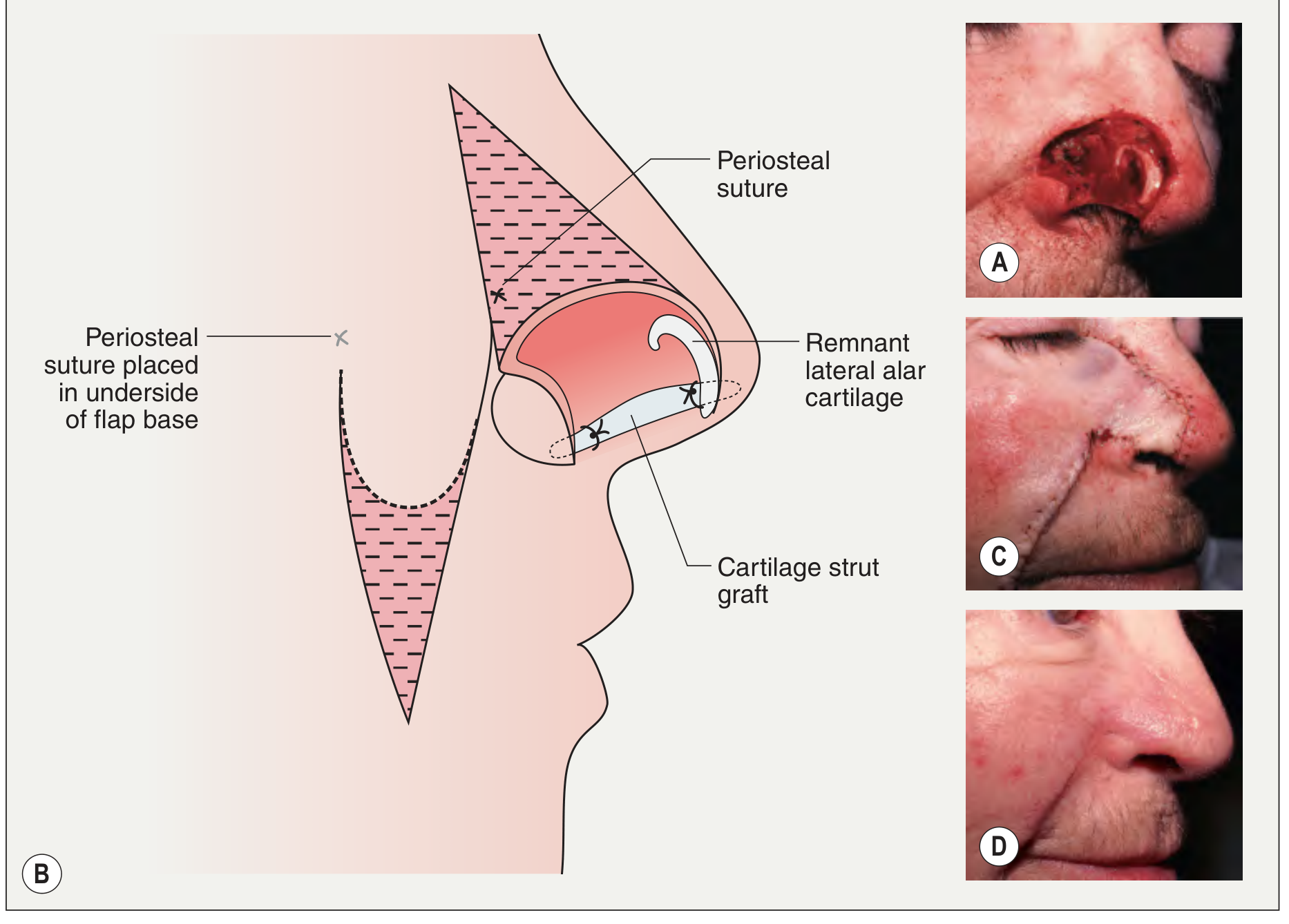
Nasolabial transposition flap with auricular cartilage strut graft for through-and-through alar defect - Dermatology 2-Volume Set 5e
2. Nasolabial (Melolabial) Interpolation Flap
- A two-stage procedure
- Used for large/complex alar defects, alar rim defects, nasal tip defects, and mid upper lip
- Stage 1: Flap is harvested from the nasolabial fold, a foil template of the contralateral ala is made and flipped; the flap is rotated downward, debulked, trimmed, and inset into the defect; the pedicle is left intact and wrapped with Vaseline/Xeroform gauze
- Stage 2 (at ~3 weeks): Pedicle is divided, the wound edges are freshened, tissue is further debulked, and remaining edge sutured; donor defect is closed up to base of pedicle
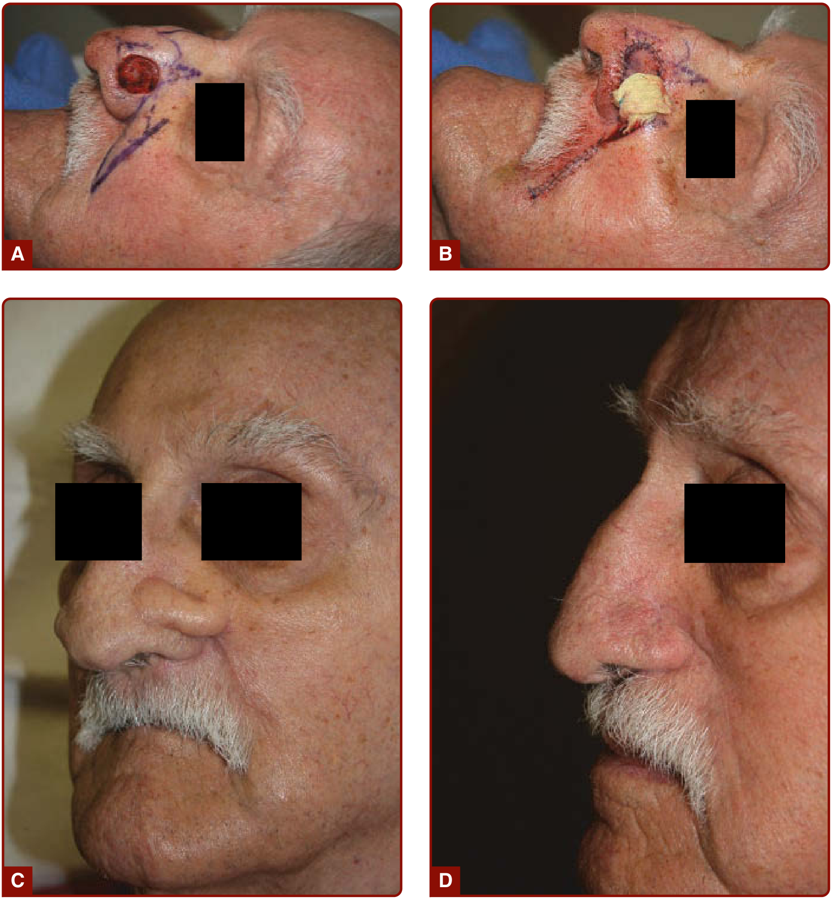
Nasolabial interpolation flap - four stages from lesion to 1-year outcome - Fitzpatrick's Dermatology
3. Island Nasolabial Flap
- Pedicled on perforating vessels; can be rotated 180 degrees to allow primary closure of the donor
- Useful where a skin bridge pedicle would be cosmetically unacceptable or impractical
4. Superiorly Based vs. Inferiorly Based
| Feature | Superiorly Based | Inferiorly Based |
|---|---|---|
| Blood supply | Angular artery | Labial/submental branches |
| Best for | Larger, more medial defects | Smaller, lateral alar defects |
| Hair bearing | Yes (limits use in men) | Finer skin, often hair-free |
| Reach | Greater reach medially | More limited |
| Skin quality | Thicker | Thinner, better color match |
Indications
External (Cutaneous) Uses:
- Nasal ala - the primary and most common indication
- Alar rim - complex defects, especially with cartilage loss
- Nasal tip - moderate size defects when a forehead flap is less optimal
- Nasal sidewall - lateral alar crease
- Upper lip - mid upper lip defects; superiorly based hair-bearing flap for lateral lip cancer reconstruction in men
- Medial cheek defects
Intraoral / Head and Neck Uses:
- Floor of mouth reconstruction (tunnelled submucosally)
- Buccal mucosa defects
- Lower alveolus reconstruction following mandibular surgery
- Used as a lining flap for nasal reconstruction (inner layer of full-thickness alar defects)
- As part of composite nose reconstruction alongside forehead flaps
Surgical Technique - Key Steps
- Defect assessment: Measure the defect; extend to include the entire alar cosmetic subunit for a better aesthetic result
- Template creation: Make a foil/gauze template of the defect (or contralateral ala) and place on the ipsilateral nasolabial skin
- Flap design: Mark the flap ellipse in the nasolabial fold; ensure adequate pedicle width at the base
- Incision and dissection: Incise flap; dissect deep to subcutis to a well-vascularized pedicle; the flap is myocutaneous in its deep plane
- Donor closure: Close the donor site in the nasolabial fold (primary closure, scar hidden in fold)
- Cartilage support (if needed): Insert cartilage strut graft (auricular/septal) at the alar rim before flap insetting
- Flap inset: Twist/rotate flap on pedicle; debulk and trim; suture into defect
- Pedicle management: Wrap pedicle with non-stick gauze
- Stage 2 (3 weeks later for interpolation flap): Divide and inset pedicle; further debulking and suture refinement
The Note Flap
The Note flap is essentially a superiorly based nasolabial flap designed so that the flap and adjoining defect together resemble a musical note. A dog-ear is removed and the flap moved to cover the nasal sidewall defect. Crucially, only one stage is required (unlike standard interpolation flaps). Used for BCC of the nasal sidewall. - Scott-Brown's Otorhinolaryngology Head & Neck Surgery
The Reverse (Spear's) Nasolabial Flap
- Employed when the defect involves the entire ala including the junction of the lateral ala and cheek/upper lip
- The motion of this flap is an upward rotation - opposite to the traditional nasolabial interpolation flap
- Also called the "melolabial flap with a twist"
Advantages
- Excellent color and texture match with nasal and facial skin
- Abundant, redundant tissue in the nasolabial fold
- Inconspicuous donor scar hidden in the nasolabial fold
- Reliable, axial blood supply
- Versatile - can be used as transposition, interpolation, or island flap
- Can provide both outer skin cover and inner lining simultaneously (folded technique)
- Can be used with cartilage grafting for structural support
- Relatively simple technique compared to forehead flap
Disadvantages
- Two stages required for the interpolation flap (3-week interval)
- Hair-bearing skin in men limits use of superiorly based flap (may result in hair growth on the reconstructed ala)
- Limited reach for very large or medially located defects
- Bulky initially - requires secondary debulking in some cases
- Risk of pin-cushioning (trapdoor deformity) if not properly defatted
- Scar contraction may distort the alar margin if not planned carefully
Comparison with Paramedian Forehead Flap
| Feature | Nasolabial Flap | Paramedian Forehead Flap |
|---|---|---|
| Best for | Small-moderate alar/tip defects | Large nasal defects |
| Stages | 1-2 | 2-3 |
| Donor site | Nasolabial fold (concealed) | Forehead (visible scar) |
| Hair issue in men | Significant | Minimal |
| Blood supply | Facial/angular artery | Supratrochlear artery |
| Skin match | Good | Excellent |
| Reach | Limited | Can reach entire nose |
Key Points for Exams
- Named for the nasolabial fold - the skin crease between nose/cheek and upper lip
- Blood supply: facial artery (via angular and labial branches)
- Most common use: alar reconstruction
- Superiorly based = greater reach; inferiorly based = better skin match, less hair
- Two-stage interpolation flap: pedicle divided at 3 weeks
- The Note flap = superiorly based nasolabial flap - one stage only
- Spear's/Reverse flap = for full alar defects - upward rotation
- Can be used intraorally by tunnelling submucosally for floor of mouth/buccal reconstruction
- For full-thickness alar defects: flap folded on itself OR combined with a second lining flap
Sources: Scott-Brown's Otorhinolaryngology Head & Neck Surgery | Fitzpatrick's Dermatology | Dermatology 2-Volume Set 5e
Corticosteroid in net
Your credits are exhausted. You can purchase a top-up or subscribe to a plan to continue using Orris. If you're already subscribed, your daily credits will reset tomorrow.
Intratympanic injection of gentamycine
Your credits are exhausted. You can purchase a top-up or subscribe to a plan to continue using Orris. If you're already subscribed, your daily credits will reset tomorrow.
This is a shared conversation. Sign in to Orris to start your own chat.