
Seizure in depth
Reading File
Finding Sources
Finding Sources
Searching PubMed
"seizure" AND "classification" AND "management"
Now let me read the key sections in parallel — Bradley and Daroff's (classification + types + precipitants), Harrison's, Tintinalli's (disorders + classification), and Rosen's (management):
Reading File
Reading File
Reading File
Reading File
Now let me read the deeper content from Bradley & Daroff block 24 (seizure types, precipitants), the Harrison's seizure/epilepsy treatment section, and search for relevant images in parallel:
Reading File
Reading File
Finding Sources
Searching Images
seizure classification types EEG epilepsy diagram
Excellent — I have the ILAE 2017 classification image. Now let me read the status epilepticus treatment from Tintinalli, the pathophysiology/EEG section, and the antiseizure drugs content:
Reading File
Reading File
Finding Sources
Finding Sources
Reading File
Loading Image
Now let me get the antiseizure drug content from Katzung and Goldman-Cecil, and the status epilepticus treatment flowchart:
Reading File
Reading File
Loading Image
Now let me get the antiseizure drug mechanisms and the Harrison's evaluation flowchart image:
Reading File
Loading Image
I now have comprehensive content. Let me get the specific drug mechanisms table from Katzung and the pathophysiology section:
Reading File
Finding Sources
Finding Sources
Reading File
Reading File
I now have comprehensive, authoritative content from multiple major textbooks. Here is the full in-depth synthesis:
Seizures — A Comprehensive Overview
1. Definition
A seizure is a transient alteration in behavior, sensation, or consciousness caused by an abnormal, synchronized electrical discharge in the brain. The term epilepsy refers to a chronic disorder characterized by the recurrent, unpredictable occurrence of seizures without an identifiable acute provoking cause. Epilepsy affects approximately 1% of the world's population and is the fourth most common neurologic disorder after migraine, stroke, and Alzheimer disease.
2. ILAE 2017 Classification of Seizure Types
The International League Against Epilepsy (ILAE) 2017 framework organizes seizures by onset, awareness, and motor/nonmotor features:
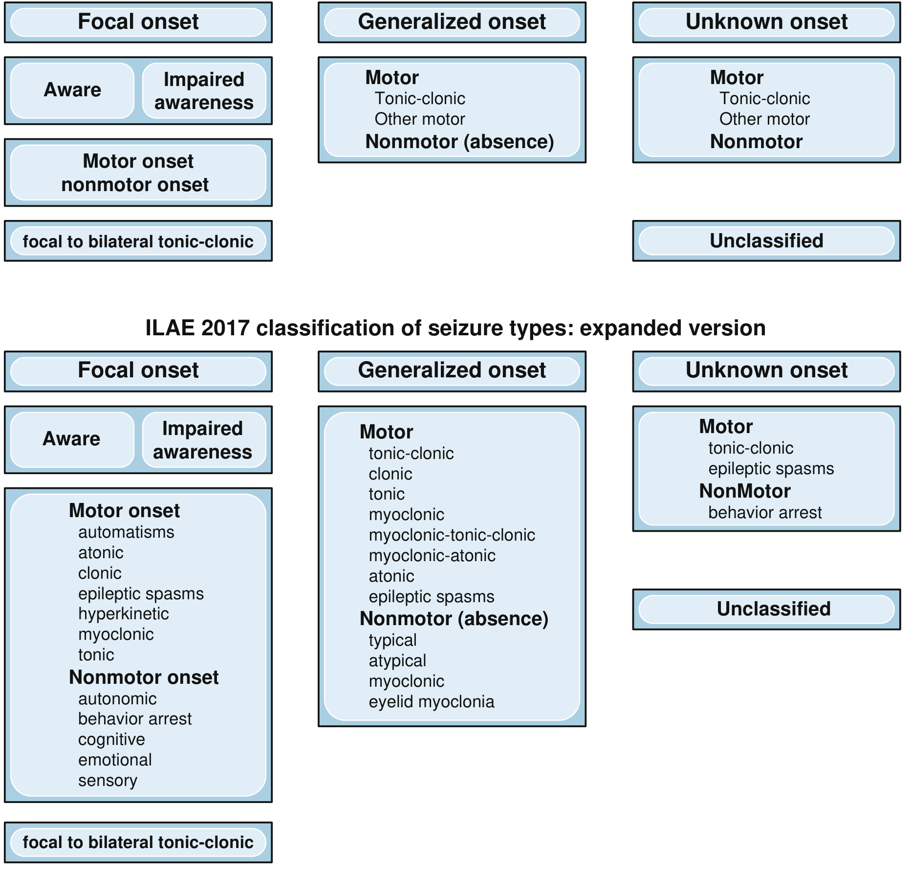
ILAE 2017 Operational Classification of Seizure Types (Bradley and Daroff's Neurology, Fig. 100.3)
Key update from 1981 → 2017 terminology:
| 1981 Term | 2017 Term |
|---|---|
| Simple partial seizure | Focal aware seizure (FAS) |
| Complex partial seizure | Focal impaired awareness seizure (FIAS) |
| Secondarily generalized | Focal to bilateral tonic-clonic |
| Idiopathic generalized epilepsy | Genetic generalized epilepsy (GGE) |
| Symptomatic epilepsy | Structural/metabolic epilepsy |
2A. Focal (Partial) Onset Seizures
Focal Aware Seizures (FAS / Simple Partial)
- Consciousness is fully preserved throughout
- Purely subjective manifestations are often called auras
- Semiology reflects the origin: tonic/clonic activity is contralateral to the hemisphere; somatosensory/visual/auditory auras have strong localizing value
- Auras typically last seconds to minutes; prolonged auras = aura continua (a form of focal nonconvulsive status epilepticus)
Focal Impaired Awareness Seizures (FIAS / Complex Partial)
- Altered awareness — may range from subtle confusion to complete amnesia for the event
- Often begin with an aura or abrupt loss of awareness
- Automatisms (repetitive purposeless movements — lip smacking, picking) are characteristic
- Most commonly arise from the temporal lobe
Focal to Bilateral Tonic-Clonic Seizures (formerly secondary generalization)
- A focal seizure spreads to involve both hemispheres
- Preceded by an aura; key distinction from primary generalized tonic-clonic
2B. Generalized Onset Seizures
Caused by near-simultaneous activation of the entire cerebral cortex — often involving deep subcortical discharge spreading outward. Consciousness is always impaired.
Generalized Tonic-Clonic (GTC / Grand Mal)
- Most dramatic seizure type
- Tonic phase (10–20 sec): sustained muscle contraction, extension of trunk and extremities, apnea, cyanosis
- Clonic phase: rhythmic synchronous muscle contractions
- Autonomic changes: ↑ BP & HR, mydriasis, piloerection, urinary/fecal incontinence, diaphoresis
- Injuries from fall: shoulder dislocation, tongue biting
- Typical duration: 60–90 seconds (bystanders typically overestimate)
- Postictal period: slow return of consciousness, lethargy, confusion, myalgias, fatigue — may last hours; pathologically brisk reflexes may be present
Absence Seizures (Petit Mal)
- Onset: 5–15 years (childhood absence epilepsy typically 4–10 years, usually remits by age 12)
- Brief (usually 10–15 seconds, never >40 seconds), abrupt onset and offset
- Behavioral arrest, staring, eyelid fluttering — no postictal symptoms
- May occur >100 times/day, often mistaken for daydreaming
- Complex absence has additional clonic/tonic/autonomic or automatism components
- Provoked in clinic by hyperventilation for 3–4 minutes
- EEG: classic 3 Hz spike-and-wave discharges
Myoclonic Seizures
- Sudden, brief (<100 ms) involuntary muscle contractions, often bilateral
- Characteristic of juvenile myoclonic epilepsy (JME), Dravet syndrome
- Less rhythmic and less sustained than clonus
Atonic Seizures (Drop Attacks)
- Sudden loss of postural muscle tone → forward fall
- Seen in Lennox-Gastaut syndrome
Tonic Seizures
- Sustained increase in muscle tone; brief impairment of consciousness
Epileptic Spasms
- Sudden flexion, extension, or mixed truncal/proximal limb movements
- Classic in infantile spasms (West syndrome)
3. Etiology and Classification of Epilepsy
| Category | Description | Examples |
|---|---|---|
| Structural | Identifiable structural lesion | Cortical malformations, hippocampal sclerosis, stroke, tumor, TBI |
| Genetic (Idiopathic) | Genetic cause, no structural/metabolic abnormality | Childhood absence epilepsy, JME, GGE |
| Metabolic | Underlying metabolic disorder | Hypoglycemia, hypocalcemia, hyponatremia, uremia |
| Infectious | CNS infection | Meningitis, encephalitis, abscess, neurocysticercosis |
| Immune | Autoimmune encephalitis | Anti-NMDAR, LGI1, CASPR2 antibodies |
| Unknown | No identifiable cause | — |
Developmental and Epileptic Encephalopathies (DEEs):
- Lennox-Gastaut syndrome (multiple etiologies)
- Dravet syndrome (SCN1A mutation ~85%)
- CDKL5 deficiency disorder
- West syndrome (infantile spasms)
- PCDH19 clustering epilepsy, GLUT1 deficiency syndrome
4. Pathophysiology
The core abnormality in a seizure is the development of an abnormal, hypersynchronous electrical discharge in a neuronal population. This involves:
- Failure of GABAergic inhibition — reduced inhibitory tone allows unchecked excitation
- Enhanced glutamatergic excitation — excessive NMDA and AMPA receptor activation
- Voltage-gated sodium channel dysregulation — during normal firing, sodium channels cycle through resting → open → inactivated states. In seizure discharges, repetitive high-frequency depolarization leads to channel accumulation in the inactivated state, but intrinsic channel defects or ion imbalances can impair this regulation
- Abnormal calcium influx via T-type calcium channels (especially relevant in absence seizures — thalamocortical circuits)
- Loss of surround inhibition in the perifocal area that normally contains a discharge
For focal seizures: an epileptogenic zone with a low seizure threshold generates ictal discharges that may remain local (focal aware) or spread (focal impaired awareness → focal to bilateral)
For generalized seizures: thalamocortical resonance circuits generate bilateral synchronous discharges; 3 Hz spike-wave of absence seizures results from abnormal T-type calcium channel oscillations in thalamic relay neurons
5. Precipitants and Risk Factors
Over 50% of patients with epilepsy report at least one precipitant:
| Precipitant | Notes |
|---|---|
| Emotional stress | Most common |
| Sleep deprivation | Most common |
| Fatigue, fever/illness | Common |
| Flickering light | Photosensitive epilepsy |
| Menstruation (catamenial epilepsy) | ~55% of women; perimenstrual pattern most common; related to proconvulsant estradiol vs. anticonvulsant progesterone |
| Missed medication | Carbamazepine and oxcarbazepine cause especially severe withdrawal seizures |
| Medications that lower threshold | TCAs, bupropion, antipsychotics, fluoroquinolones, meperidine, tramadol, CNS stimulants |
| Hyperventilation | Classic trigger for generalized absence seizures |
| Alcohol withdrawal | Major cause of acute symptomatic seizures |
Risk factors for epilepsy:
- Prior febrile seizures
- Family history of seizures
- Prior head trauma, stroke, CNS tumor, CNS infection
- Developmental abnormalities
6. Epidemiology
- Incidence in North America: 16–51 per 100,000/year (higher in developing countries, up to 111/100,000)
- Prevalence: 2.7–7.1 per 1,000 (higher in males, lower socioeconomic status)
- Bimodal distribution: highest incidence in first decade and after age 65
- Cumulative lifetime risk of epilepsy: ~4% by age 80
- 2-year recurrence risk after first unprovoked seizure: ~40%
- Risk doubles after second seizure (61% at 2 years, 73% at 5 years)
- Factors predicting recurrence: abnormal EEG, abnormal neurological examination
7. Clinical Evaluation
Harrison's Evaluation Flowchart
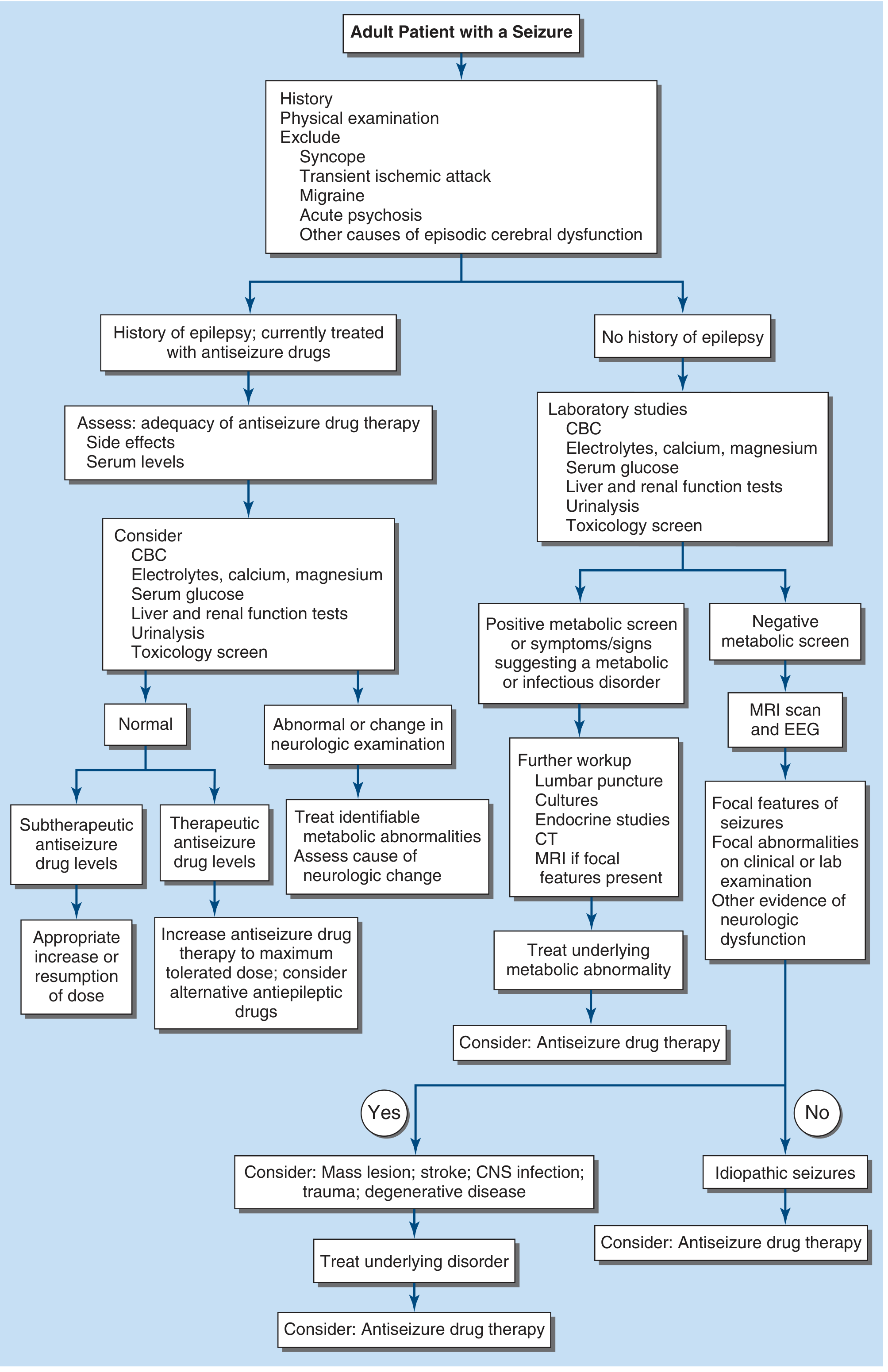
Evaluation of the adult patient with a seizure (Harrison's Principles of Internal Medicine 22e, Fig. 436-2)
History priorities:
- Was this truly a seizure? (exclude syncope, TIA, migraine, acute psychosis, panic attack)
- What was before/during/after: aura, ictal semiology, postictal state
- Witness account is critical (patient often has no recollection of ictal and immediate postictal phases)
- Risk factors: prior febrile seizures, family history, head trauma, stroke, CNS infection, drugs/alcohol
- In known epilepsy: assess medication adherence and current drug levels
Physical examination:
- Signs of systemic illness, infection, trauma
- Skin: tuberous sclerosis, neurofibromatosis, chronic liver/renal disease
- Cardiovascular: auscultation for causes of cerebrovascular disease
- Neurological: cranial nerve examination, motor/sensory exam, mental status (memory, language, abstract thinking), visual fields
Investigations:
| Test | Indication |
|---|---|
| Glucose (bedside) | All cases — exclude hypoglycemia |
| CBC, metabolic panel (electrolytes, Ca, Mg, glucose), LFTs, RFTs | All new-onset seizures |
| Toxicology screen | Suspected drug toxicity |
| Antiseizure drug levels | Known epilepsy |
| EEG | All new-onset seizures; urgent if nonconvulsive status epilepticus suspected |
| MRI (preferred over CT) | First seizure with negative metabolic screen; focal features |
| CT | When MRI unavailable or emergent structural lesion suspected |
| LP | Suspected CNS infection/encephalitis |
EEG findings:
- Absence: 3 Hz bilateral spike-and-wave
- Focal seizure: unilateral sharp waves or spike discharges
- GTC: high-amplitude polyspike and wave
- Interictal epileptiform discharges confirm epileptogenic tendency; normal interictal EEG does not exclude epilepsy
8. Differential Diagnosis
| Condition | Key Distinguishing Features |
|---|---|
| Syncope | Prodrome (lightheadedness, nausea, diaphoresis), brief (seconds), upright posture, rapid full recovery |
| TIA | Negative symptoms (weakness, numbness, vision loss), vascular risk factors, no postictal phase |
| Psychogenic non-epileptic seizures (PNES) | Highly variable semiology, prolonged, eyes often closed, pelvic thrusting, waxing/waning; no postictal changes on EEG |
| Migraine with aura | Aura evolves slowly (minutes), followed by headache |
| Hypoglycemia | Confirmed by glucose level; responds to dextrose |
| Narcolepsy/cataplexy | Triggered by emotion, preserved consciousness in cataplexy |
| Panic attack | Hyperventilation, anxiety, no tonic-clonic activity |
| Acute dystonic reaction | Drug exposure, sustained posturing, no alteration of consciousness |
9. Antiseizure Medications (ASMs) — Mechanisms and Indications
Mechanism Classes
| Mechanism | Drugs | Seizure Types Targeted |
|---|---|---|
| Na⁺ channel blockers (use/voltage-dependent) | Carbamazepine, Oxcarbazepine, Eslicarbazepine, Phenytoin/Fosphenytoin, Lamotrigine, Lacosamide | Focal ± GTC (NOT absence or myoclonic) |
| Broad spectrum (multiple/unknown mechanisms) | Valproate, Levetiracetam, Topiramate, Zonisamide | Focal + generalized |
| T-type Ca²⁺ channel blockers | Ethosuximide | Absence seizures only |
| GABA-A enhancers (BZDs) | Lorazepam, Diazepam, Clonazepam, Midazolam | Acute seizures, status epilepticus |
| GABA-A enhancers (barbiturates) | Phenobarbital | Broad spectrum (chronic + status epilepticus) |
| SV2A ligand | Levetiracetam, Brivaracetam | Focal + generalized |
| AMPA receptor antagonist | Perampanel | Focal ± GTC |
| Cannabidiol | CBD (Epidiolex) | Dravet syndrome, Lennox-Gastaut |
| mTOR inhibitor | Everolimus | Tuberous sclerosis (reduces seizures by ~25%) |
Sodium channel blocker mechanism (Katzung): These drugs bind preferentially to the inactivated state of voltage-gated Na⁺ channels. During high-frequency seizure firing, sodium channels cycle rapidly through the inactivated state — the drug accumulates and produces use-dependent block, selectively suppressing high-frequency ictal discharges while minimally impairing normal low-frequency firing.
Drug Selection by Seizure/Syndrome Type
| Seizure/Syndrome | First-line | Notes |
|---|---|---|
| Focal epilepsy | Lamotrigine > Levetiracetam, Carbamazepine | Lamotrigine superior to LEV/ZNS for new-onset focal; carbamazepine for trigeminal neuralgia overlap |
| Generalized epilepsy (GGE) | Valproate, Lamotrigine, Levetiracetam | Valproate superior to LEV for GGE; avoid Na⁺ channel blockers (can worsen absence/myoclonic) |
| Absence epilepsy | Ethosuximide, Valproate | Ethosuximide is absence-specific; valproate if GTC also present |
| Juvenile myoclonic epilepsy | Valproate, Levetiracetam | Valproate highly effective but teratogenic |
| Lennox-Gastaut | Valproate, Lamotrigine, Clobazam, Cannabidiol | Rufinamide, Topiramate also used |
| Dravet syndrome | Valproate, Clobazam, Stiripentol, Cannabidiol | Avoid Na⁺ channel blockers |
| Tuberous sclerosis | Everolimus (mTOR inhibitor) | Reduces seizure frequency by 25% |
Drug-resistant epilepsy is defined as failure of ≥2 adequate trials of appropriate ASMs. ~35% of patients are drug-resistant; surgical candidacy evaluation (video-EEG, MRI, PET/SPECT, neuropsychological testing) should be initiated promptly.
Key drug interactions:
- Enzyme inducers (carbamazepine, phenytoin, phenobarbital, rufinamide) → reduce levels of oral contraceptives, warfarin, many other drugs
- Enzyme inhibitors (valproate) → increase levels of other ASMs
- Low-interaction drugs (gabapentin, levetiracetam, brivaracetam, lacosamide) → preferred in elderly, polypharmacy, or women on OCP
10. Status Epilepticus (SE)
Definition: Seizure lasting ≥5 minutes OR two or more seizures without return to baseline between them (operational definition per ILAE 2015).
Types:
- Convulsive SE (CSE): GTC activity; medical emergency with significant mortality
- Nonconvulsive SE (NCSE): Ongoing electrical seizures without obvious motor manifestations; requires continuous EEG for diagnosis; commonly missed; mortality increases with delayed recognition especially in age >60
Causes: CNS infection, stroke, TBI, metabolic derangements, drug toxicity/withdrawal, acute encephalitis, medication non-adherence in known epilepsy
Management Protocol
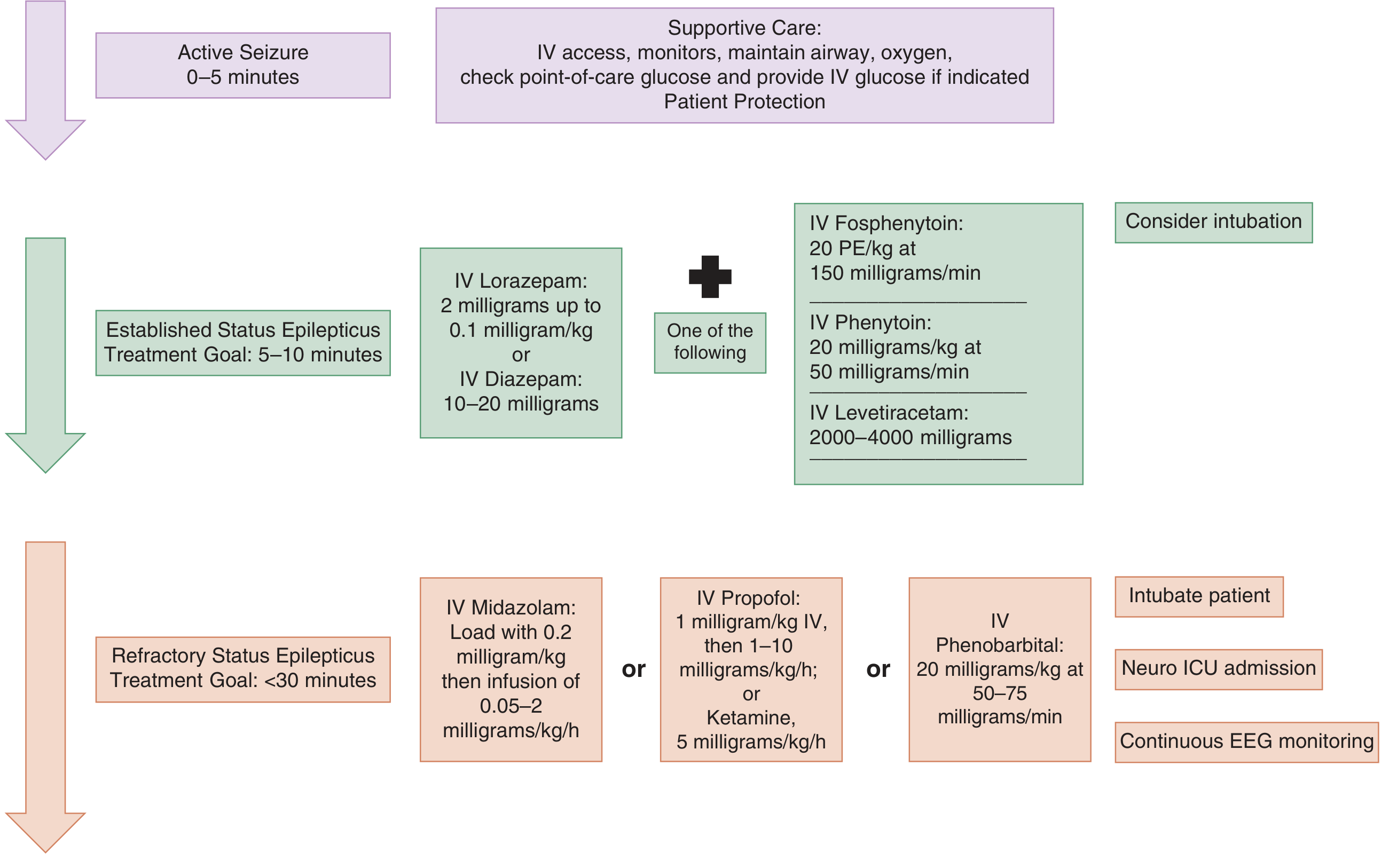
Guidelines for management of active seizures and status epilepticus (Tintinalli's Emergency Medicine, Fig. 171-1)
| Phase | Time Goal | Intervention |
|---|---|---|
| ABCs / Supportive | 0–5 min | IV access, O₂, cardiac monitor, pulse oximetry, bedside glucose; IV normal saline (NOT glucose — incompatible with phenytoin) |
| Established SE | 5–10 min | IV Lorazepam 2 mg (up to 0.1 mg/kg) — first choice OR IV Diazepam 10–20 mg PLUS one of: IV Fosphenytoin 20 PE/kg at 150 mg/min; IV Phenytoin 20 mg/kg at 50 mg/min; IV Levetiracetam 2000–4000 mg |
| Refractory SE | <30 min | IV Midazolam 0.2 mg/kg load then 0.05–2 mg/kg/h OR IV Propofol 1 mg/kg then 1–10 mg/kg/h OR IV Phenobarbital 20 mg/kg at 50–75 mg/min OR Ketamine 5 mg/kg/h; Intubate, Neuro-ICU, continuous EEG |
Key points:
- IV lorazepam has onset in 3 min, duration 12–24 h; preferred over diazepam (15–60 min duration)
- IM midazolam is as safe and effective as IV lorazepam when no IV access; decreased seizure time and fewer ICU admissions in prehospital trials
- Do not attempt LP during SE; if meningitis/encephalitis suspected, start empiric antibiotics/antivirals immediately
- Paralytic agents mask convulsive activity → continuous EEG monitoring mandatory after neuromuscular blockade
11. Special Situations
Febrile Seizures
- Most common seizure type in children (3 months–5 years)
- Simple febrile seizure: generalized, <15 min, single in 24 h — benign prognosis
- Complex febrile seizure: focal, >15 min, or recurs within 24 h — higher risk of epilepsy
Pregnancy and Seizures
- Volume of distribution and plasma clearance of ASMs increase → drug levels may fall
- Most ASMs are teratogenic; valproate, carbamazepine, phenytoin carry highest risk
- Preferred ASMs in pregnancy: levetiracetam or lamotrigine monotherapy
- Fetal bradycardia lasting up to 20 min can follow a single brief maternal seizure
- Status epilepticus in pregnancy carries significant maternal and fetal mortality → aggressive management including early intubation
Catamenial Epilepsy
- ~55% of women with epilepsy have seizure clustering related to menstruation
- Most common pattern: perimenstrual (3 days before to 3 days after onset of menses)
- Mechanism: estradiol (proconvulsant) vs. progesterone (anticonvulsant)
- Treatment: progesterone therapy as adjunct when standard ASMs fail
Post-Surgical Seizure Prophylaxis
- Routine prophylaxis is not indicated for most neurosurgical procedures unless seizure risk is high (traumatic brain injury, supratentorial surgery near cortex)
12. Surgical Treatment
For drug-resistant epilepsy:
- Temporal lobectomy (most evidence): 60–70% seizure freedom in temporal lobe epilepsy with hippocampal sclerosis
- Lesionectomy: removal of epileptogenic lesion (cortical dysplasia, tumor, vascular malformation)
- Corpus callosotomy: palliative for drop attacks in Lennox-Gastaut
- Responsive neurostimulation (RNS): closed-loop device that detects and terminates ictal activity
- Vagal nerve stimulation (VNS): palliative, reduces seizure frequency by ~50% in ~50% of patients
Pre-surgical workup: video-EEG (capture of habitual seizures with ictal EEG), MRI with dedicated epilepsy protocols, FDG-PET, ictal SPECT, neuropsychological evaluation, and in selected cases stereoEEG (SEEG) or subdural grid implantation for Phase 2 evaluation.
13. Prognosis
- ~65% of newly diagnosed epilepsy patients achieve complete remission on ASMs
- ~45–50% remit on the first drug; ~10–15% on the second; ~35% are drug-resistant
- Remission more likely: vascular malformations, stroke, tumors (63–78%)
- Remission less likely: hippocampal sclerosis, cortical malformations (40–50%)
- SUDEP (Sudden Unexpected Death in Epilepsy): rare but real risk (~1/1,000 patient-years in general epilepsy; higher in drug-resistant); mechanism unclear but involves cardiorespiratory dysfunction during/after GTC seizures
- Patients with epilepsy have significantly higher rates of psychiatric comorbidity (depression, anxiety — doubled risk), accidents, and some neurodegenerative conditions
Sources:
- Bradley and Daroff's Neurology in Clinical Practice — Seizure Classification, Types, Precipitants, Epidemiology
- Harrison's Principles of Internal Medicine 22e — Seizure evaluation, history, examination
- Tintinalli's Emergency Medicine — Seizure classification, status epilepticus management
- Katzung's Basic and Clinical Pharmacology 16e — Antiseizure medication mechanisms and pharmacology
- Goldman-Cecil Medicine — Choice of antiseizure medications, drug-resistant epilepsy, surgical therapyThe answer above draws directly from five major textbooks and covers every major domain. Here is a quick orientation to the sections:
What was covered, section by section:
| # | Section | Key Source |
|---|---|---|
| 1 | Definition | Katzung Pharmacology |
| 2 | ILAE 2017 Classification (with diagram) | Bradley & Daroff's Neurology |
| 2A–2B | Focal & Generalized seizure types in depth | Bradley & Daroff, Tintinalli |
| 3 | Etiology (structural/genetic/metabolic/infectious/immune/DEEs) | Katzung, Goldman-Cecil |
| 4 | Pathophysiology (GABAergic failure, Na⁺ channels, T-type Ca²⁺, thalamocortical circuits) | Katzung |
| 5 | Precipitants & risk factors (including catamenial epilepsy) | Bradley & Daroff |
| 6 | Epidemiology | Bradley & Daroff |
| 7 | Clinical evaluation (flowchart + investigations) | Harrison's 22e |
| 8 | Differential diagnosis | Bradley & Daroff, Harrison's |
| 9 | ASMs — mechanisms, drugs, indications, interactions | Katzung, Goldman-Cecil |
| 10 | Status epilepticus — staged protocol (flowchart) | Tintinalli |
| 11 | Special situations (febrile, pregnancy, catamenial) | Tintinalli, Bradley & Daroff |
| 12 | Surgical treatment | Goldman-Cecil |
| 13 | Prognosis & SUDEP | Bradley & Daroff, Goldman-Cecil |
Epileptiform syndrome
Your credits are exhausted. Your daily credits reset tomorrow, or you can purchase a top-up for more.
This is a shared conversation. Sign in to Orris to start your own chat.