
Bacteria basic microbiology in detail based on USMLE
gram positive vs gram negative bacterial cell wall structure peptidoglycan
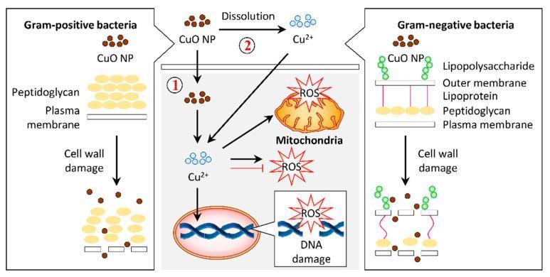
A pathophysiology diagram illustrating the antibacterial mechanisms of copper oxide nanoparticles (CuO NPs) on Gram-positive and Gram-negative bacteria. The left panel shows Gram-positive bacteria with a thick peptidoglycan layer above a plasma membrane. The right panel shows Gram-negative bacteria with a complex cell wall including lipopolysaccharides, an outer membrane, lipoproteins, and a thin peptidoglycan layer. Both panels demonstrate CuO NPs causing cell wall damage and lysis. The central panel details the intracellular bactericidal pathway labeled as mechanism 1 and 2. Mechanism 1 involves the internalization of CuO NPs into the cytosol, while mechanism 2 illustrates the dissolution of CuO NPs into copper ions (Cu2+) which then permeate the cell. Inside the cell, both Cu2+ ions and CuO NPs induce the production of reactive oxygen species (ROS). The resulting oxidative stress leads to mitochondrial disruption and DNA damage, ultimately resulting in cell death. This infographic highlights the role of nanoparticle morphology and ion release in targeting bacterial structural and metabolic integrity.
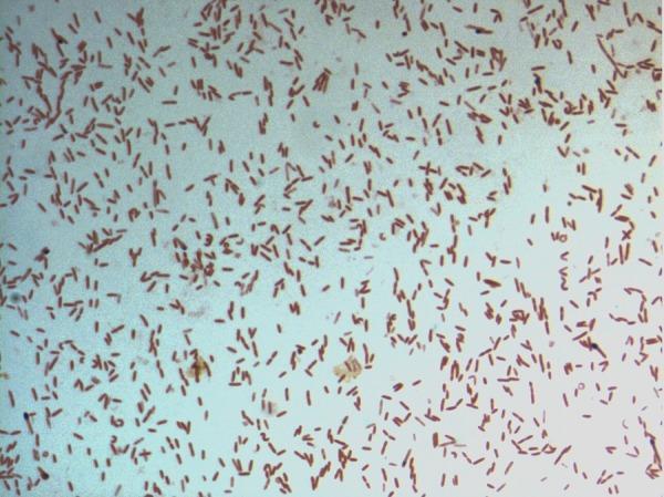
This diagnostic image shows a light microscopy view of a bacterial culture following Gram staining. The specimen displays numerous small, rod-shaped (bacilli) bacteria distributed across the field. The microorganisms exhibit a consistent pink-to-red hue, identifying them as Gram-negative. This staining characteristic indicates a cell wall structure with a thin peptidoglycan layer and an outer membrane that does not retain the crystal violet stain. The arrangement of the bacilli is predominantly individual or scattered, with occasional small clusters and pairs. Specifically identified as D. fastidiosa strain JC13T, this image serves as a primary microbiological reference for the morphological and staining properties of this novel genus within the family Erysipelotrichaceae. The visual demonstrates key diagnostic features including cell morphology, arrangement, and Gram reaction, which are essential for clinical microbiology classification and initial pathogen identification.
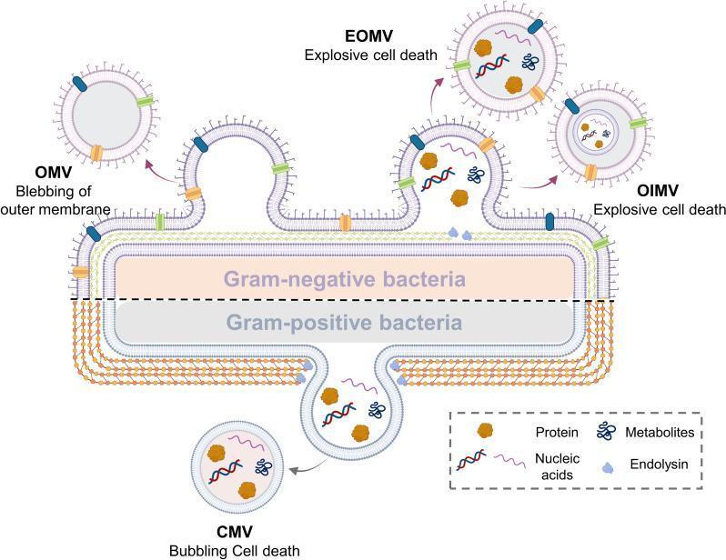
A medical microbiology diagram illustrating the biogenesis and classification of bacterial extracellular vesicles (BEVs) from Gram-negative and Gram-positive bacteria. The top half depicts a Gram-negative bacterium with its triple-layered cell envelope (outer membrane, periplasmic space, and cytoplasmic membrane). Three vesicle formation mechanisms are shown: blebbing of the outer membrane creating Outer Membrane Vesicles (OMV), and explosive cell death leading to Explosive Outer Membrane Vesicles (EOMV) and Outer-Inner Membrane Vesicles (OIMV). The bottom half represents a Gram-positive bacterium characterized by a thick peptidoglycan cell wall and a single cytoplasmic membrane. This section illustrates 'bubbling cell death' resulting in Cytoplasmic Membrane Vesicles (CMV). A legend identifies the internal cargo within these vesicles, including proteins (brown shapes), nucleic acids (wavy lines), metabolites (blue branched icons), and endolysin (light blue shapes). This pathophysiology diagram highlights the structural differences in bacterial envelopes and the various pathways—blebbing, explosive death, and bubbling—that contribute to BEV diversity and composition.
bacterial growth curve lag log stationary death phase
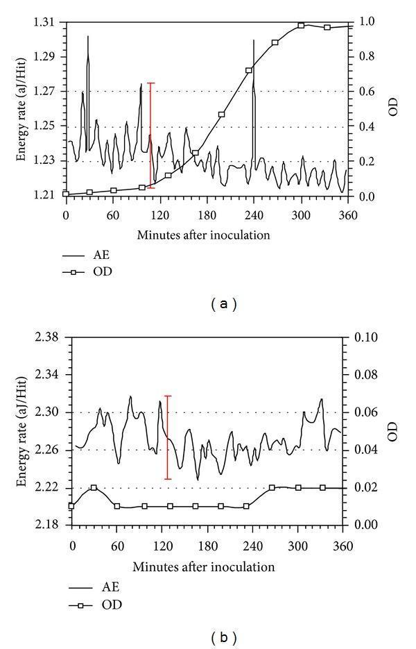
This figure presents two comparison charts, (a) and (b), depicting the growth kinetics and metabolic activity of Lactococcus lactis over 360 minutes. The primary metrics are Optical Density (OD), indicating bacterial concentration, and Energy Rate measured via Acoustic Emission (AE) in attojoules per hit (aJ/Hit). Graph (a) shows normal bacterial growth: the OD curve follows a characteristic sigmoidal pattern with lag, log, and stationary phases, while the AE data exhibits significant fluctuations and distinct peaks (e.g., around 108 and 240 minutes) representing metabolic energy release. Graph (b) demonstrates the effect of the metabolic inhibitor Sodium Azide (NaN3). In this panel, the OD remains near-baseline (indicating total inhibition of growth), and the AE signal shows only low-level background fluctuations without significant metabolic peaks. Red bars indicate ±3σ deviation standards for noise assessment. These graphs are utilized in microbiology and biomedical engineering to evaluate non-invasive methods for monitoring microbial activity and metabolic inhibition.
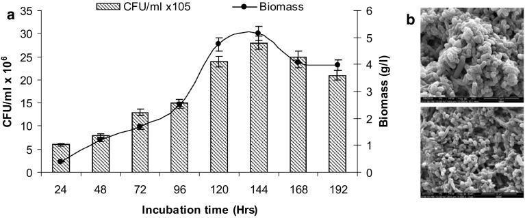
Educational microbiology graphic consisting of a growth curve plot (1a) and scanning electron micrographs (1b) documenting a bacterial consortium's development. Panel (a) presents a dual-axis line and bar graph showing the relationship between incubation time (24–192 hours), bacterial colony-forming units (CFU/mL x 10^6), and biomass concentration (g/L). The growth kinetics demonstrate a logarithmic increase in both parameters, peaking at 144 hours with a biomass of approximately 5.15 g/L and a CFU count of 28 x 10^6, followed by a slight decline indicating the stationary or death phase. Panel (b) features two scanning electron micrographs (SEM) at different magnifications, visualizing the bacterial morphology. The microbes are characterized as densely packed, rod-shaped bacilli forming a thick, interconnected biofilm matrix. The rough surface texture suggests the presence of an extracellular polymeric substance (EPS) coating. This figure illustrates bacterial growth dynamics and structural arrangement relevant to microbial degradation processes in biomedical or environmental health contexts.
bacterial genetics conjugation transformation transduction plasmid
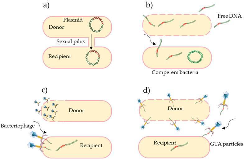
A multi-panel microbiological diagram illustrating the four primary mechanisms of horizontal gene transfer (HGT) in bacteria, which facilitate the spread of antibiotic resistance. (a) Conjugation: Shows a donor and recipient cell physically connected by a sexual pilus, mediating the direct transfer of a circular plasmid. (b) Transformation: Depicts a competent bacterial cell actively taking up naked, 'free DNA' fragments from the extracellular environment across its membrane. (c) Transduction: Illustrates viral-mediated transfer where a bacteriophage infects a donor, carrying genetic material to a recipient cell. (d) Gene Transfer Agents (GTAs): Shows the release of phage-like GTA particles from a donor cell (indicated by a disrupted membrane) to a recipient cell. The diagram effectively contrasts these pathways by highlighting their unique vectors: direct contact (pilus), environmental uptake, viral infection (bacteriophage), and specialized particles (GTAs). This educational resource is designed for microbiology and infectious disease curricula, emphasizing the mechanisms of genetic recombination and bacterial evolution.
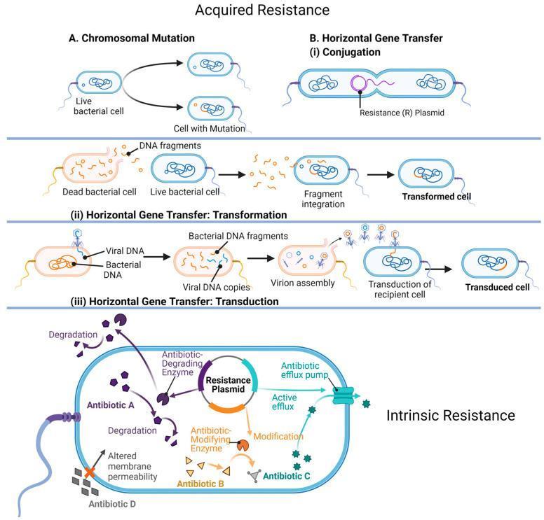
A multi-panel medical illustration detailing the mechanisms of bacterial antibiotic resistance, categorized into 'Acquired Resistance' and 'Intrinsic Resistance'. Acquired Resistance (upper panels) depicts four pathways: 1. Chromosomal Mutation: Shows a live bacterial cell undergoing spontaneous genetic changes. 2. Horizontal Gene Transfer (HGT) via Conjugation: Illustrates the direct transfer of a Resistance (R) Plasmid between two bacteria through a pilus. 3. HGT via Transformation: Shows a live cell uptaking DNA fragments released from a dead bacterial cell, leading to genetic integration. 4. HGT via Transduction: Demonstrates viral DNA (bacteriophage) incorporating bacterial DNA fragments and assembling into virions to infect a recipient cell. Intrinsic Resistance (lower panel) highlights structural and enzymatic mechanisms within a single bacterium: - Antibiotic Degradation: An enzyme chemically breaks down 'Antibiotic A'. - Antibiotic Modification: An enzyme alters 'Antibiotic B' to neutralize it. - Active Efflux: A membrane efflux pump actively transports 'Antibiotic C' out of the cytoplasm. - Altered Membrane Permeability: Structural changes block 'Antibiotic D' from entering the cell. This educational diagram is designed for microbiology and pharmacology studies to explain how pathogens evade antimicrobial therapy.
bacterial toxin exotoxin endotoxin mechanism virulence
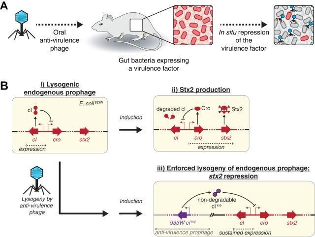
Educational diagram illustrating an anti-virulence phage strategy to neutralize Shiga toxin (Stx2) production in E. coli 933W. Panel A depicts a mouse model receiving oral anti-virulence phages that target gut bacteria, resulting in the in situ repression of virulence factors without killing the bacterial host. Panel B shows the molecular mechanism across three states: (i) Normal lysogeny where the cI repressor protein prevents expression of cro and stx2 genes. (ii) Induction leading to cI degradation, triggering the expression of Cro and the virulence factor Stx2 (indicated by a skull-and-crossbones icon). (iii) Engineered enforced lysogeny where an anti-virulence prophage delivers a non-degradable cI variant (cI ind-). This engineered repressor constitutively inhibits cro and stx2 expression even under inducing conditions, effectively neutralizing toxin production. This represents a precision microbiology approach to treating enteric infections by modifying bacterial function rather than employing traditional bacteriolytic or antibiotic methods.
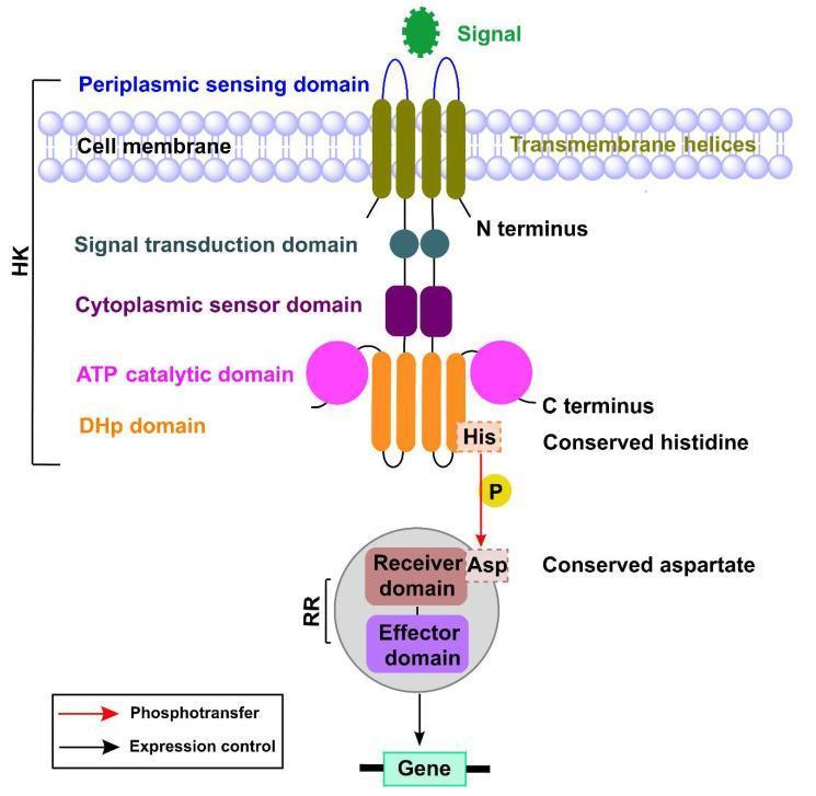
A pathophysiology diagram illustrating the basic mechanism of a bacterial Two-Component Signaling (TCS) system, commonly involved in virulence and environmental adaptation. The diagram depicts two primary protein components: a Sensor Histidine Kinase (HK) and a Response Regulator (RR). The HK is an integral membrane protein featuring a periplasmic sensing domain for extracellular signals, transmembrane helices, and a cytoplasmic C-terminus. The cytoplasmic portion of the HK contains the signal transduction domain, a cytoplasmic sensor domain, an ATP catalytic domain, and a dimerization histidine phosphotransfer (DHp) domain. A phosphotransfer event is illustrated where a phosphate group (P) moves from a conserved histidine (His) residue on the HK to a conserved aspartate (Asp) residue on the receiver domain of the RR. The RR consists of a receiver domain and an effector domain; upon phosphorylation, the effector domain exerts expression control by binding to a target gene. This schematic demonstrates the fundamental biochemical architecture of signal transduction from the extracellular environment to the bacterial genome.
Bacteria: Basic Microbiology (USMLE High-Yield)
Source: Medical Microbiology 9e | Jawetz, Melnick & Adelberg's Medical Microbiology 28E | Sherris & Ryan's Medical Microbiology, 8th Ed
1. PROKARYOTE vs EUKARYOTE - The Foundation
| Feature | Prokaryote (Bacteria) | Eukaryote (Human/Fungi) |
|---|---|---|
| Nuclear membrane | Absent | Present |
| Ribosomes | 70S (50S + 30S) | 80S (60S + 40S) |
| Cell wall | Peptidoglycan (most) | Absent (humans); Ergosterol (fungi) |
| Mitochondria | Absent | Present |
| Plasmids | Yes | Rarely |
| Reproduction | Binary fission | Mitosis/meiosis |
2. BACTERIAL MORPHOLOGY
Shape (USMLE mnemonics)
- Cocci (spherical): Staph, Strep, Neisseria, Enterococcus
- Bacilli (rods): E. coli, Klebsiella, Pseudomonas, Clostridium
- Spirochetes (spiral): Treponema, Borrelia, Leptospira
- Coccobacilli: Haemophilus, Bordetella, Brucella, Pasteurella ("HiBBP")
- Vibrio (comma-shaped): Vibrio cholerae
- Pleomorphic: Mycoplasma, Bartonella
Arrangement
- Clusters (Staphylo-): Staphylococcus aureus
- Chains (Strepto-): Streptococcus pyogenes
- Pairs (Diplo-): S. pneumoniae, Neisseria
- Tetrads: Micrococcus
- Cuboidal packets of 8: Sarcina
3. THE GRAM STAIN - Most Tested Lab Concept
| Gram-Positive | Gram-Negative | |
|---|---|---|
| Color | Purple/violet | Pink/red |
| Peptidoglycan layer | Thick (20-80 nm) | Thin (2-7 nm) |
| Outer membrane | Absent | Present |
| Teichoic acids | Present | Absent |
| LPS (endotoxin) | Absent | Present (Lipid A) |
| Periplasmic space | Absent | Present |
| Lysozyme sensitivity | High | Low (outer membrane blocks) |
| Penicillin sensitivity | Generally higher | Lower (porin-dependent) |
- Mycobacteria - too much mycolic acid (use Acid-Fast/Ziehl-Neelsen)
- Mycoplasma - no cell wall at all
- Legionella, Rickettsia, Chlamydia - intracellular; Legionella stains poorly (use silver stain)
- Treponema - too thin (use dark-field microscopy)
4. BACTERIAL CELL WALL STRUCTURE (High-Yield)
Peptidoglycan
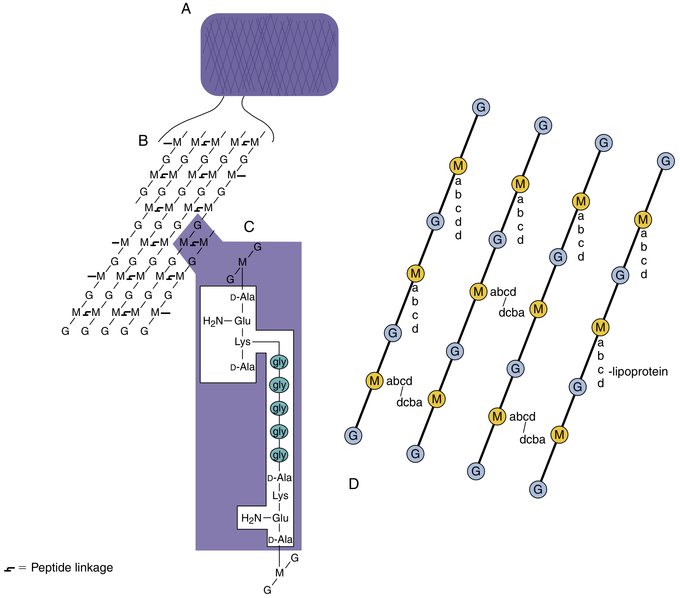
Gram-Positive Unique Components
- Teichoic acids: Polymers of ribitol/glycerol phosphate; anchored to peptidoglycan. Define bacterial serotype (antigenic determinants). Lipoteichoic acid is anchored in the membrane.
- Thick peptidoglycan: Acts as physical scaffold; lysozyme cleaves NAG-NAM bond → protoplast formation → osmotic lysis
Gram-Negative Unique Components
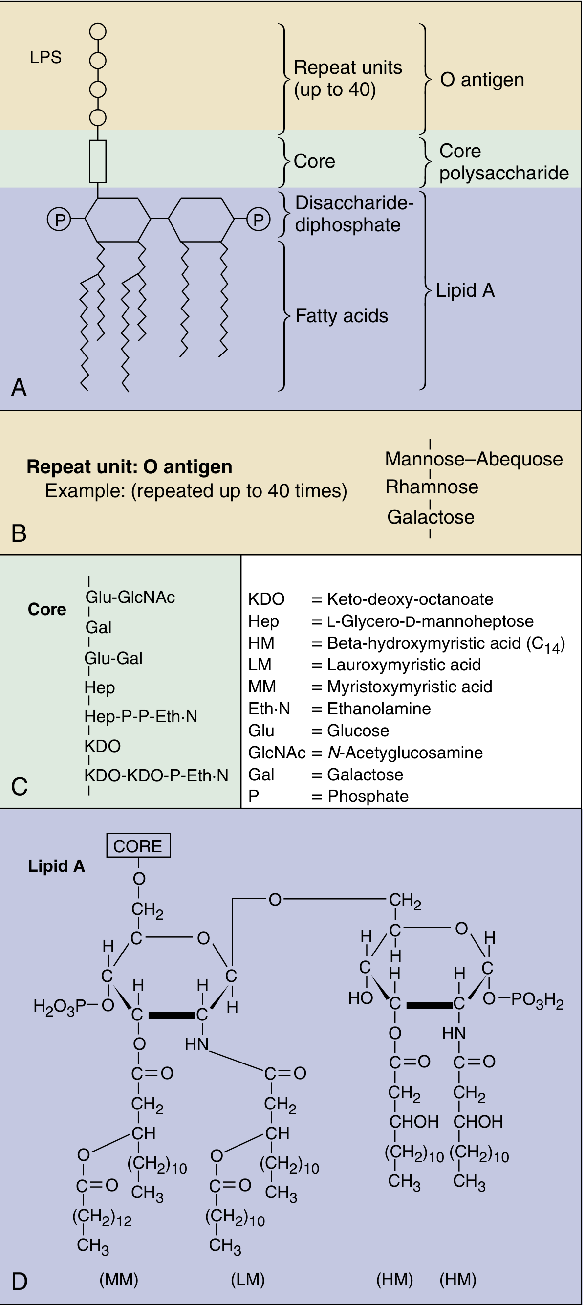
| LPS Component | Role |
|---|---|
| Lipid A | Endotoxin activity → fever, hypotension, DIC, septic shock |
| Core polysaccharide | Contains KDO; structural integrity |
| O antigen | Surface antigen; serotype determinant; antiphagocytic |
5. EXTERNAL STRUCTURES (Virulence Factors)
Capsule
- Composition: Polysaccharide (exception: B. anthracis - polypeptide D-glutamate capsule)
- Function: Antiphagocytic (major virulence factor), poorly antigenic
- Visualized by India ink (negative stain - capsule appears as clear halo)
- Key encapsulated organisms: "SHiNE SKiS" - S. pneumoniae, H. influenzae (type b), Neisseria meningitidis, E. coli (K1), Salmonella typhi, Klebsiella, Streptococcus agalactiae (GBS)
- Asplenic patients are particularly susceptible to these encapsulated organisms
Flagella
- Ropelike propellers of flagellin protein; responsible for motility
- H antigen (flagellar antigen) - used in serotyping (e.g., E. coli O157:H7)
- Arrangement: monotrichous, lophotrichous, peritrichous, amphitrichous
Pili (Fimbriae)
- Type I pili: Mediate adherence to uroepithelium (E. coli UTI)
- Type IV pili: Twitching motility; virulence in Neisseria, Pseudomonas
- Sex pilus (F pilus): Conjugation (gene transfer)
Biofilm
- Produced by P. aeruginosa, S. aureus, and others when quorum reached
- Protects bacteria from antibiotics and host defenses
- S. mutans dextran/levan biofilm → tooth plaque
6. BACTERIAL GROWTH
Growth Phases
| Phase | Features |
|---|---|
| Lag | Metabolic activity; no cell division; RNA/protein synthesis ramps up |
| Log (exponential) | Rapid binary fission; most susceptible to antibiotics (especially cell-wall agents) |
| Stationary | Nutrient depletion; growth = death rate; endospore formation begins |
| Death | Cell death exceeds growth; toxin production may peak |
Oxygen Requirements
| Type | O₂ Requirement | Examples |
|---|---|---|
| Obligate aerobe | Requires O₂ | Mycobacteria ("TB loves apex"), Nocardia, Pseudomonas, Bordetella |
| Obligate anaerobe | Killed by O₂ | Clostridium, Bacteroides, Fusobacterium, Actinomyces |
| Facultative anaerobe | Prefers O₂, tolerates absence | E. coli, Staph, most Enterobacteriaceae |
| Microaerophile | Low O₂ | Campylobacter, H. pylori |
| Aerotolerant anaerobe | Ignores O₂ | Streptococcus (no catalase) |
Metabolism Types
- Autotrophs: Use CO₂ as carbon source (rare pathogens)
- Heterotrophs: Use organic molecules (most pathogens)
- Fermentation: Anaerobic; pyruvate → acids/alcohols; 2 ATP per glucose
- Aerobic respiration: TCA cycle + electron transport chain; 38 ATP per glucose
- Key metabolic tests: Oxidase test, catalase test, fermentation patterns (used for ID)
7. ENDOSPORES (High-Yield USMLE)
| Feature | Details |
|---|---|
| Composition | Dipicolinic acid (calcium chelated) + dehydrated core |
| Resistance | Heat, UV, desiccation, chemicals, autoclaving (121°C/15 min kills them) |
| Stain | Schaeffer-Fulton stain (malachite green + safranin) |
| Key pathogens | B. anthracis (anthrax), B. cereus (food poisoning), C. tetani, C. botulinum, C. perfringens, C. difficile |
8. BACTERIAL GENETICS - Gene Transfer (Highly Tested)
Three Mechanisms of Horizontal Gene Transfer
| Mechanism | How | Requires | Key Example |
|---|---|---|---|
| Transformation | Cell takes up naked DNA from environment | DNA in medium; competent cell | S. pneumoniae (Griffith's experiment); H. influenzae |
| Transduction | Bacteriophage transfers DNA | Bacteriophage (virus) | S. aureus toxins (TSST-1, exfoliatin), Shigella toxin, V. cholerae toxin |
| Conjugation | Direct cell-to-cell contact via sex pilus; plasmid transfer | F plasmid (F+ → F-) | Antibiotic resistance transfer (R-factors); E. coli |
- Generalized transduction: Any bacterial gene can be transferred (lytic phage accidentally packages host DNA)
- Specialized (lysogenic) transduction: Only specific genes transferred (phage integrates near specific chromosomal genes)
- Many toxins are phage-encoded: Cholera toxin (V. cholerae), Shiga toxin (STEC), Diphtheria toxin (C. diphtheriae), TSST-1 (S. aureus), Erythrogenic toxin (S. pyogenes)
Plasmids
- Extrachromosomal circular dsDNA; replicate autonomously
- Encode: resistance genes (R-plasmids), toxins, metabolic enzymes, virulence factors
- R-factors: Transmissible via conjugation → spread of multi-drug resistance (USMLE loves this)
9. BACTERIAL TOXINS
Exotoxins vs Endotoxin
| Feature | Exotoxin | Endotoxin (LPS) |
|---|---|---|
| Source | Gram+ and Gram- | Gram-negative ONLY |
| Chemistry | Protein | Lipopolysaccharide (Lipid A) |
| Heat stability | Labile (60°C destroys most) | Stable |
| Toxicity | Very high (ng range) | Lower (mg range) |
| Antigenicity | High → toxoid vaccines | Poor |
| Fever | Yes (if absorbed) | Yes (pyrogenic) |
| Action | Specific mechanism | Activates macrophages/complement |
| Examples | Diphtheria, botulinum, tetanus, cholera | Neisseria, Salmonella, E. coli (gram-neg sepsis) |
Key Exotoxin Mechanisms (USMLE Must-Know)
| Toxin | Organism | Mechanism | Disease |
|---|---|---|---|
| Cholera toxin | V. cholerae (phage-encoded) | ADP-ribosylates Gs → ↑cAMP | Watery diarrhea ("rice water") |
| Pertussis toxin | B. pertussis | ADP-ribosylates Gi → ↑cAMP | Whooping cough |
| Diphtheria toxin | C. diphtheriae (phage) | ADP-ribosylates EF-2 → stops protein synthesis | Pseudomembrane, myocarditis |
| Botulinum toxin | C. botulinum (spore) | Blocks ACh release (SNARE cleavage) | Descending flaccid paralysis |
| Tetanus toxin | C. tetani (spore) | Blocks glycine/GABA release in spinal cord | Spastic paralysis, trismus |
| TSST-1 | S. aureus (phage) | Superantigen → massive T-cell activation | Toxic shock syndrome |
| Shiga toxin | E. coli O157:H7 / Shigella (phage) | Cleaves 28S rRNA → stops protein synthesis | HUS (hemolytic uremic syndrome) |
| Anthrax toxin | B. anthracis | EF = adenylate cyclase (↑cAMP); LF = protease | Anthrax, edema |
10. BACTERIAL METABOLISM - Lab Identification (USMLE-Relevant Tests)
| Test | Principle | Positive Organisms |
|---|---|---|
| Catalase | H₂O₂ → H₂O + O₂ (bubbles) | Staph (+ vs Strep -) |
| Coagulase | Fibrinogen → fibrin clot | S. aureus (+) vs CoNS (-) |
| Oxidase | Cytochrome c oxidase present | Neisseria, Pseudomonas, Campylobacter, Vibrio |
| Urease | Urea → NH₃ + CO₂ | H. pylori, Proteus, Klebsiella, Ureaplasma |
| Indole | Tryptophan → indole | E. coli (+); Klebsiella (-) |
| Optochin | Inhibits S. pneumoniae | Strep pneumo (sensitive) vs viridans (resistant) |
| Bile solubility | Lyses S. pneumoniae | Strep pneumo (+) |
| Bacitracin | Inhibits S. pyogenes | GAS sensitive; GBS resistant |
| Novobiocin | Coagulase-neg Staph | S. saprophyticus resistant; S. epidermidis sensitive |
11. SPECIAL CELL WALL TYPES
| Organism | Cell Wall Feature | Significance |
|---|---|---|
| Mycobacteria | Mycolic acid + arabinogalactan; "Acid-fast" | Resist gram stain; need Ziehl-Neelsen; resistant to drying/disinfectants |
| Mycoplasma | No cell wall | Not gram-stainable; penicillin ineffective; only bacteria with cholesterol in membrane |
| Chlamydia | Minimal peptidoglycan; obligate intracellular | Cannot be gram-stained; use DIF or PCR |
| Rickettsia | Gram-negative-like; obligate intracellular | Transmitted by arthropod vectors |
| L-forms | Lost cell wall (under antibiotic pressure) | Become penicillin resistant; osmotically fragile |
12. NORMAL FLORA BY BODY SITE (USMLE-Tested)
| Site | Dominant Flora | Clinical Significance |
|---|---|---|
| Skin | S. epidermidis (CoNS), Propionibacterium | IV catheter infections; acne |
| Oropharynx | Viridans streptococci | Subacute bacterial endocarditis (dental procedures) |
| GI tract | Bacteroides fragilis (most common anaerobe), E. coli | Post-op peritonitis; C. diff after antibiotics |
| Vagina | Lactobacillus | BV when disrupted (Gardnerella replaces it) |
| Urogenital | S. saprophyticus (young women) | Second most common UTI in young women |
13. QUICK-REFERENCE CLASSIFICATION TABLE
| Property | Gram+ | Gram- |
|---|---|---|
| Cell wall | Thick peptidoglycan + teichoic acids | Thin PG + outer membrane + LPS |
| Endotoxin | No | Yes (Lipid A) |
| Exotoxin | Common (Staph, Strep, Clostridium) | Less common (E. coli, V. cholerae) |
| Beta-lactam effect | Very susceptible | Variable (outer membrane barrier) |
| Vancomycin effect | Susceptible (targets D-Ala-D-Ala) | Intrinsically resistant (outer membrane) |
| Examples | S. aureus, S. pyogenes, S. pneumoniae, Enterococcus, Clostridium, Bacillus, Listeria, Corynebacterium | E. coli, Klebsiella, Pseudomonas, Neisseria, H. influenzae, Salmonella, Shigella, V. cholerae |
Top USMLE High-Yield Summary Points
- 70S ribosome = antibiotic target (30S: aminoglycosides, tetracyclines; 50S: macrolides, chloramphenicol, clindamycin, linezolid)
- Peptidoglycan synthesis is blocked by beta-lactams (transpeptidase), vancomycin (D-Ala-D-Ala), and bacitracin (bactoprenol)
- Endotoxin = Lipid A of LPS → septic shock, DIC, fever; gram-negative organisms ONLY
- Exotoxins are protein-based; many are ADP-ribosylating enzymes (cholera, pertussis, diphtheria, anthrax EF)
- Capsule = antiphagocytic virulence factor; absent spleen → susceptibility to encapsulated bacteria
- Spores (Bacillus + Clostridium) resist autoclaving, UV, alcohol; killed at 121°C/15 min under pressure
- Conjugation is the primary mechanism of antibiotic resistance transfer between bacteria (R-plasmids)
- Toxin-encoded phages (lysogenic): cholera, diphtheria, TSST-1, erythrogenic toxin, Shiga toxin
- Mycoplasma has no cell wall → penicillin useless; treat with macrolides or doxycycline
- Mycobacteria are acid-fast due to mycolic acid; gram stain fails on them