
Give me detail about Posterior cerebral artery
Reading File
Now let me search the medical library for information on the posterior cerebral artery in parallel.
Finding Sources
Finding Sources
Searching the Web
posterior cerebral artery anatomy branches territory clinical
Excellent results. Now let me read the key sections in parallel from multiple authoritative textbooks.
Reading File
Reading File
Reading File
Reading File
Now let me read the most detailed section from Adams & Victor and the clinical syndromes section:
Reading File
Reading File
Reading File
Excellent - I now have rich, comprehensive information from multiple authoritative sources. Let me also fetch a key anatomical diagram and read the proximal syndromes section in detail:
Loading Image
Reading File
I now have comprehensive information from multiple authoritative neurological textbooks. Let me compile the detailed answer.
Posterior Cerebral Artery (PCA)
Origin and Anatomy
The posterior cerebral artery (PCA) is one of a pair of terminal branches of the basilar artery, arising at its bifurcation at the superior border of the pons, posterior to the dorsum sellae.
- In approximately 70% of individuals, both PCAs arise from the basilar bifurcation in the standard configuration, connected to the ipsilateral internal carotid arteries (ICAs) via thin posterior communicating arteries (PComA).
- In 20-25%, one PCA arises from the basilar normally, while the other arises from the ICA - a "fetal PCA" (persistent fetal circulation pattern).
- Fewer than 5% have bilateral fetal PCAs, where both arise from the respective ICAs.
- Adams and Victor's Principles of Neurology, 12th Edition, p. 810
- Localization in Clinical Neurology, 8e, p. 1350
Segments
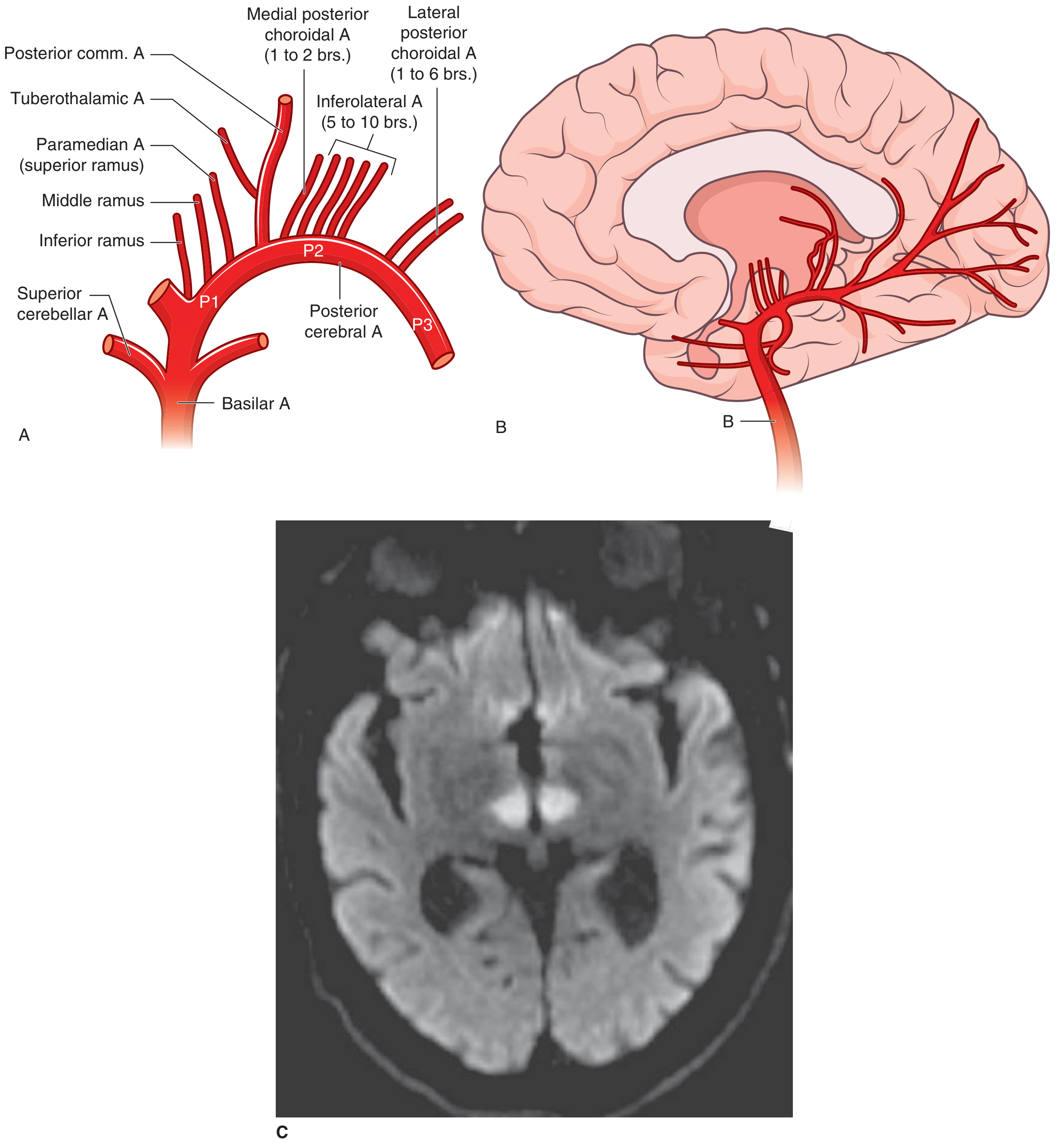
Four named segments are recognized (P1-P4):
| Segment | Also Known As | Description |
|---|---|---|
| P1 | Precommunal / Mesencephalic segment | From basilar bifurcation to junction with PComA |
| P2 | Ambient segment | From PComA junction, curves around midbrain in the ambient cistern; thalamogeniculate branches arise here |
| P3 | Quadrigeminal segment | Posterior to lateral aspect of midbrain, to origin of parieto-occipital and calcarine arteries |
| P4 | Calcarine segment | Terminal cortical division; the calcarine artery |
- Localization in Clinical Neurology, 8e, p. 1350-1351
Branches and Their Territories
PCA branches are divided into three functional groups:
1. Penetrating (Central) Branches
These supply the brainstem and thalamus:
-
Interpeduncular (paramedian mesencephalic) branches - arise just above the basilar bifurcation; supply:
- Red nuclei and substantia nigra (bilaterally)
- Medial cerebral peduncles
- Oculomotor (CN III) and trochlear (CN IV) nuclei and nerves
- Reticular substance of upper brainstem
- Decussation of superior cerebellar peduncles
- Medial longitudinal fasciculi
- Medial lemnisci
- (The artery of Percheron is an anatomical variant where a single unpaired artery from one P1 supplies both paramedian thalami - occlusion causes a bilateral "butterfly" thalamic infarct)
-
Thalamoperforant (paramedian thalamic) branches - arise near P1-P2 junction; supply inferior, medial, and anterior thalamus
-
Thalamogeniculate branches - arise from P2 opposite the lateral geniculate body; supply the geniculate body and central/posterior thalamus
-
Medial branches (from the PCA as it encircles the midbrain) - supply the lateral cerebral peduncle, lateral tegmentum, corpora quadrigemina, and pineal gland
-
Posterior choroidal branches - supply the posterosuperior thalamus, choroid plexus, posterior hippocampus, and psalterium (fornix decussation)
2. Dorsal Callosal (Splenial) Artery
Anastomoses with distal ACA branches; supplies the splenium of the corpus callosum.
3. Cortical Branches (4 main branches)
The distal PCA divides into anterior and posterior divisions, giving rise to:
| Branch | Territory |
|---|---|
| Anterior temporal artery | Inferior and medial temporal lobe (middle cranial fossa) |
| Posterior temporal artery | Posterior temporal lobe |
| Parieto-occipital artery | Parieto-occipital region |
| Calcarine artery | Primary visual cortex (calcarine/striate cortex, Brodmann areas 17, 18, 19); lingula, cuneus, precuneus |
Terminal branches anastomose with those of both the MCA and ACA.
- Adams and Victor's Principles of Neurology, 12th Edition, p. 811-812
- Localization in Clinical Neurology, 8e, p. 1350
Overall Territories Supplied
The PCA supplies:
- Occipital lobes (including primary visual cortex)
- Inferomedial temporal lobe
- Thalamus (most of it, via multiple branches)
- Midbrain (parts of peduncles, tegmentum, quadrigeminal plate)
- Choroid plexus of lateral and third ventricles
- Splenium of corpus callosum
- Posterior limb of the internal capsule (via penetrating vessels)
PCA Anatomy Diagram
Below is a diagram of the posterior cerebral artery segments and branches from Adams & Victor's Principles of Neurology:
Clinical Syndromes of PCA Occlusion
Etiology of PCA strokes: Most are embolic (cardioembolic or artery-to-artery); atherosclerosis of the proximal PCA is less common.
PCA syndromes are divided into three groups:
1. Proximal PCA Syndromes (Thalamic and Midbrain)
Thalamic Syndrome of Dejerine-Roussy
- Caused by occlusion of thalamogeniculate branches
- Features:
- Contralateral hemisensory loss (deep and cutaneous), often severe
- May include transient hemiparesis
- Possible homonymous hemianopia
- After recovery, develops thalamic pain (burning/aching hemibody pain, hyperpathia, dysesthesia) - may persist for years
- Occasionally: distortion of taste, athetotic hand posturing, mood alteration (mania/depression)
Central Midbrain and Subthalamic Syndromes
- Due to occlusion of interpeduncular (Percheron) branches
- Features: vertical gaze palsy, stupor or coma
Paramedian Midbrain Syndromes (proximal PCA/P1)
- Weber syndrome: CN III palsy (ipsilateral) + contralateral hemiplegia
- Claude syndrome: CN III palsy + contralateral cerebellar ataxia and tremor
- Benedikt syndrome: CN III palsy + contralateral ataxia, tremor, and corticospinal signs
Anteromedial-Inferior Thalamic Syndrome
- Due to thalamoperforant branch occlusion
- Features: extrapyramidal movement disorders (hemiballismus, hemichoreoathetosis, asterixis); deep sensory loss, hemiataxia, or tremor in combinations
2. Cortical PCA Syndromes (Temporal and Occipital)
Visual Field Defects
- Contralateral homonymous hemianopia - most characteristic; due to involvement of calcarine cortex (Brodmann 17, 18, 19) or the geniculocalcarine fibers
- Hemianopia may be incomplete, often more severe in upper quadrants
- Macular sparing is common due to collateral supply of the occipital pole from MCA
Left (Dominant) PCA Occlusion
- Alexia without agraphia - when the left occipital cortex AND the splenium of the corpus callosum are both involved (visual info from right hemisphere cannot cross to left language areas)
- Visual agnosia - inability to recognize objects by sight
- Anomia / thalamic aphasia - if left thalamus involved
- Color agnosia
Right (Non-dominant) PCA Occlusion
- Prosopagnosia (failure to recognize faces)
- Topographic disorientation
- Visual hallucinations (release hallucinations in the hemianopic field)
3. Bilateral PCA Occlusion
- Cortical blindness (Anton's syndrome) - patient is blind but denies it; can occur with sequential bilateral occipital infarctions
- Bilateral thalamic infarction (artery of Percheron variant)
- Severe memory impairment (bilateral hippocampal and temporal involvement)
Key Clinical Notes
| Feature | Detail |
|---|---|
| Most common symptom | Contralateral homonymous hemianopia |
| Macular sparing | Present due to MCA collaterals to occipital pole |
| Fetal PCA | 20-25%; PCA territory stroke may appear as carotid rather than vertebrobasilar disease |
| Proximal occlusion | May be asymptomatic if collateral flow from Circle of Willis is adequate |
| Artery of Percheron | Variant; bilateral "butterfly" paramedian thalamic infarction from a single artery occlusion |
Sources: Adams and Victor's Principles of Neurology, 12th Edition, pp. 810-814 | Localization in Clinical Neurology, 8e, pp. 1350-1351 | Neuroanatomy through Clinical Cases, 3rd Edition, p. 426 | Goldman-Cecil Medicine, p. 3924
This is a shared conversation. Sign in to Orris to start your own chat.