
Mechanism of hearing Sembulingam
Reading File
Finding Sources
Finding Sources
Searching Images
organ of Corti cochlea structure diagram hearing mechanism
Finding Sources
Finding Sources
Reading File
Reading File
Mechanism of Hearing
Note: Sembulingam's Essentials of Medical Physiology isn't in this textbook library, but the mechanism of hearing is standard physiology covered identically in Guyton and Hall and Costanzo Physiology, which I've used below. The steps and terminology match what Sembulingam presents.
Hearing is a sequence of mechanical, hydraulic, and electrical events that convert sound waves in air into nerve impulses.
1. Conduction through the external and middle ear (impedance matching)
Sound waves travel down the external auditory canal and strike the tympanic membrane, setting it into vibration. The membrane's vibrations are transmitted through the ossicular chain (malleus → incus → stapes), which acts as a lever system attached to the oval window.
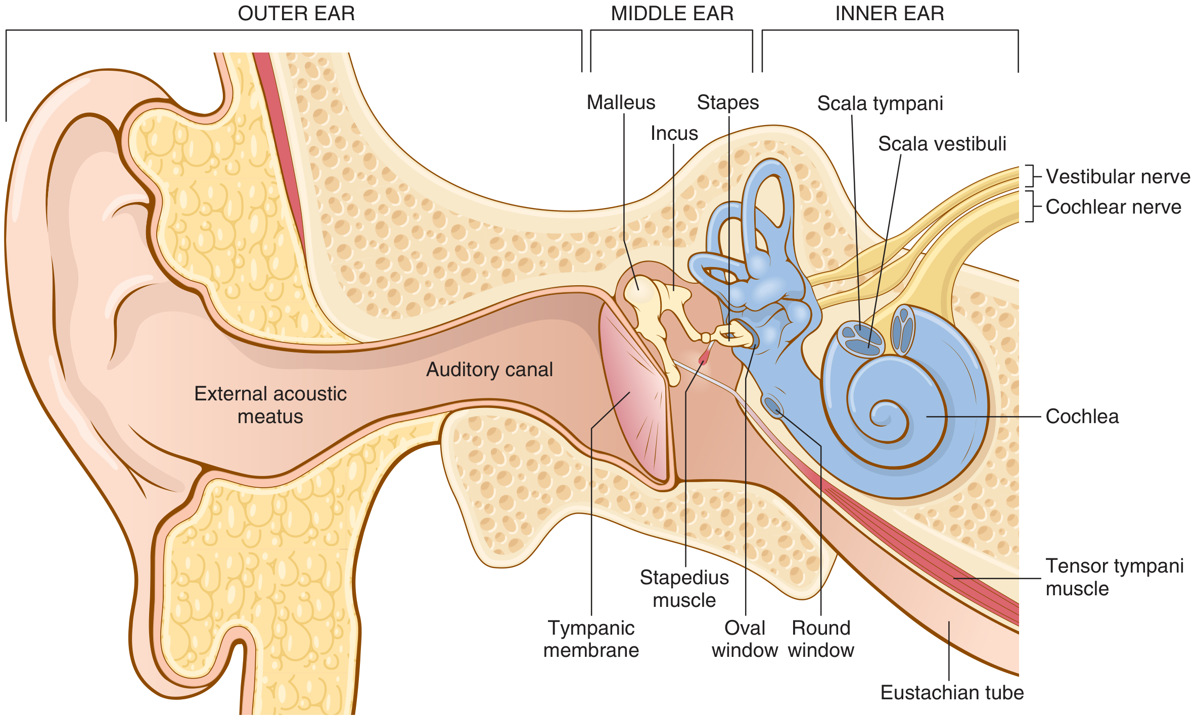
Two mechanisms boost the force delivered to the cochlear fluid, which is necessary because fluid has much greater inertia than air:
- Lever action of the malleus-incus system increases force about 1.3 times (while slightly reducing amplitude).
- Area ratio: the tympanic membrane (~55 mm²) is about 17 times larger than the stapes footplate (~3.2 mm²), concentrating the same total force onto a smaller area.
Together these give roughly a 22-fold increase in force at the oval window compared to the force on the eardrum, achieving about 50-75% efficient impedance matching between air and cochlear fluid (Guyton and Hall, p. 656).
2. Transmission into the cochlea
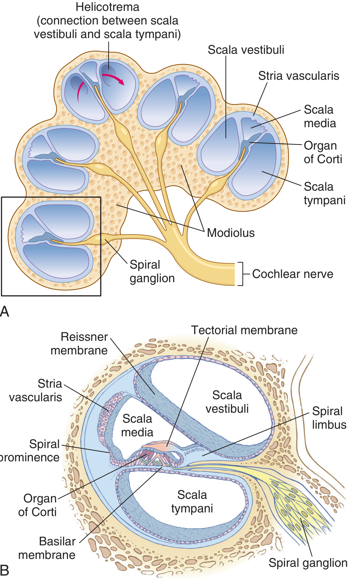
The stapes footplate rocks in the oval window: inward movement pushes fluid forward in the scala vestibuli, and outward movement pulls it back. The cochlea is a coiled tube of three compartments:
- Scala vestibuli and scala media, separated by Reissner's membrane
- Scala media and scala tympani, separated by the basilar membrane
The organ of Corti, containing the hair cells, sits on the basilar membrane.
3. Traveling wave and frequency (place) discrimination
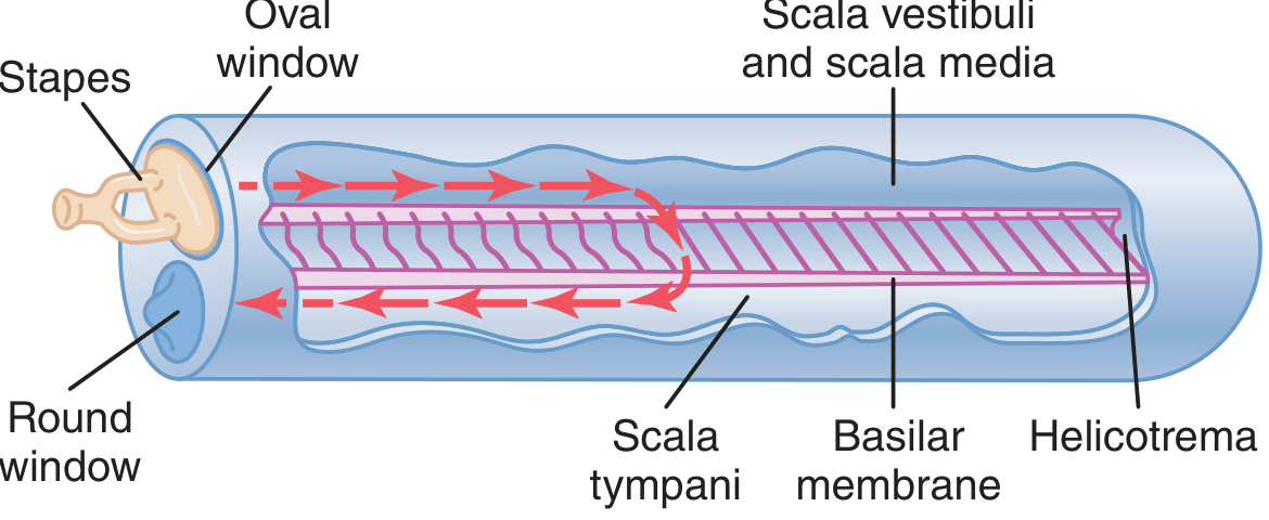
Pressure changes in the scala vestibuli are transmitted across the flexible basilar membrane into the scala tympani, producing a traveling wave that moves from the base toward the apex of the cochlea. The basilar membrane's fibers are short and stiff near the base (oval window) and progressively longer and more flexible toward the apex.
- High-frequency sounds cause maximal vibration near the base of the cochlea (short, stiff fibers).
- Low-frequency sounds cause maximal vibration near the apex (long, flexible fibers).
This is the basis of the place principle of frequency discrimination: the position along the basilar membrane where vibration amplitude peaks determines the pitch perceived, and this "tonotopic" map is preserved all the way up the auditory pathway to the cortex (Guyton and Hall, p. 657-658).
4. Transduction by the organ of Corti (hair cells)
The organ of Corti contains:
- Inner hair cells (the primary sensory receptors, ~3500 in a single row)
- Outer hair cells (~12,000-20,000 in 3-4 rows, mainly involved in mechanical amplification/tuning)
The tips of the hair cell stereocilia are embedded in or touch the overlying tectorial membrane. As the basilar membrane vibrates up and down, a shearing motion develops between the basilar membrane (carrying the hair cells) and the tectorial membrane, bending the stereocilia.
- Bending toward the tallest stereocilia opens mechanically-gated K+/cation channels, depolarizing the hair cell (K+ influx occurs because endolymph, unlike normal extracellular fluid, is high in K+).
- Bending in the opposite direction closes the channels and hyperpolarizes the cell.
Depolarization triggers Ca2+ influx at the base of the hair cell, causing release of neurotransmitter (glutamate) onto the afferent fibers of the cochlear (auditory) nerve, generating action potentials that carry auditory information centrally through the cochlear nuclei, superior olivary complex, inferior colliculus, and medial geniculate body to the auditory cortex in the temporal lobe.
Summary of the pathway
External ear → tympanic membrane → ossicles (impedance matching) → oval window → cochlear fluid (traveling wave along basilar membrane) → organ of Corti hair cell stereocilia bending → mechanotransduction and neurotransmitter release → cochlear nerve → central auditory pathways → auditory cortex.
Recent evidence check: This is settled classical physiology; a quick literature check found no recent systematic reviews or meta-analyses that revise this core mechanism, so no contradiction warning applies.
If you want, I can go deeper into any one part - the impedance matching math, the outer hair cell "cochlear amplifier" mechanism, or the central auditory pathway.
Process of urine formation Sembulingam
Finding Sources
Finding Sources
Searching Images
nephron structure glomerulus tubule diagram urine formation
Reading File
Process of Urine Formation
Note: Sembulingam's Essentials of Medical Physiology isn't indexed in this textbook library, but urine formation is core renal physiology covered in identical fashion by Guyton and Hall, which I've drawn on below. The three-process framework matches what Sembulingam presents.
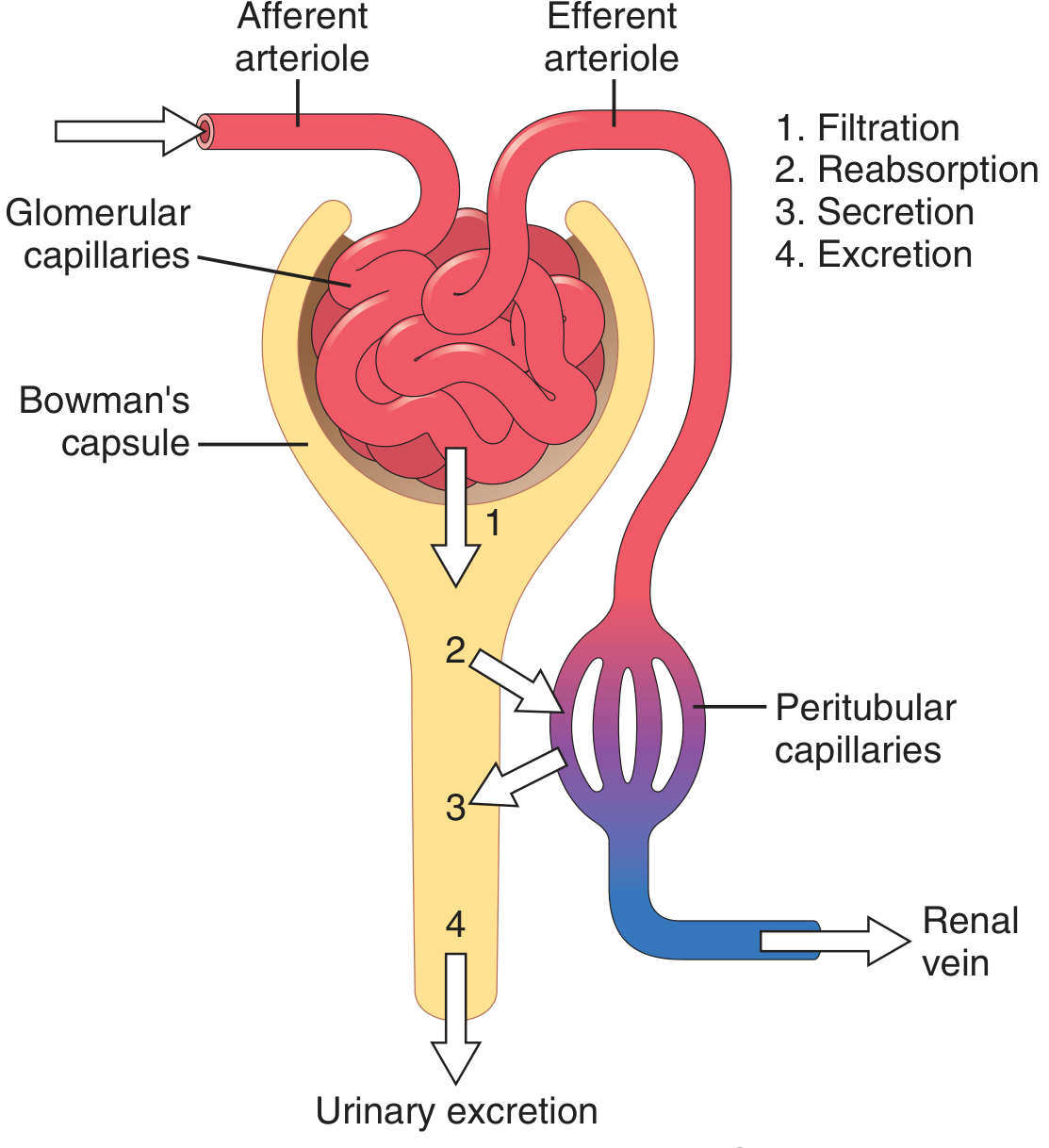
Urine formation results from three sequential renal processes occurring along the nephron:
Urinary excretion rate = Filtration rate - Reabsorption rate + Secretion rate
1. Glomerular Filtration
Urine formation begins when a large volume of plasma, virtually free of protein, is filtered from the glomerular capillaries into Bowman's capsule. Because plasma proteins are largely retained, the composition of the filtrate (glomerular filtrate) is essentially identical to plasma for all small solutes (electrolytes, glucose, urea, creatinine).
- Normal GFR is about 125 mL/min (~180 L/day) in a healthy adult.
- Filtration depends on the balance of Starling forces across the glomerular capillary: glomerular hydrostatic pressure (favoring filtration) versus plasma colloid osmotic pressure and Bowman's capsule hydrostatic pressure (opposing filtration).
- GFR is regulated by afferent and efferent arteriolar resistance, renal blood flow, and autoregulatory mechanisms (myogenic response and tubuloglomerular feedback).
2. Tubular Reabsorption
As the filtrate passes through the proximal tubule, loop of Henle, distal tubule, and collecting duct, water and needed solutes (glucose, amino acids, most electrolytes, and the bulk of filtered water) are reabsorbed from the tubular lumen back into the peritubular capillary blood. About 99% of the filtered water and most filtered solutes are normally reabsorbed, so only a small fraction of the original filtrate is excreted as urine.
- The proximal tubule reabsorbs the bulk (~65%) of filtered Na+, water, glucose, and amino acids.
- The loop of Henle (especially the thick ascending limb) reabsorbs Na+, K+, Cl- and is central to the countercurrent mechanism that concentrates urine.
- The distal tubule and collecting duct provide fine control of water, Na+, K+, and acid-base balance, under hormonal regulation (ADH, aldosterone).
3. Tubular Secretion
Certain substances are actively or passively transported from the peritubular capillary blood into the tubular fluid, adding to what is filtered. This includes H+, K+, and various organic acids/bases (e.g., drugs, urea, creatinine to a small degree), which supplements filtration in eliminating certain solutes and helps regulate acid-base balance.
Net Handling Patterns
Guyton illustrates four patterns of renal handling for different substances (Guyton and Hall, p. 328-329):
- Filtered only, not reabsorbed or secreted: excretion rate equals filtration rate (e.g., creatinine, used clinically to estimate GFR).
- Filtered and partly reabsorbed: excretion is less than filtration (most electrolytes, urea).
- Filtered and completely reabsorbed: substance does not appear in urine at all under normal conditions (e.g., glucose, amino acids, at normal plasma levels).
- Filtered and secreted: excretion exceeds filtration (some organic acids and drugs, e.g., PAH, used to estimate renal plasma flow).
Summary
Renal artery → afferent arteriole → glomerulus (ultrafiltration into Bowman's capsule) → proximal tubule (bulk reabsorption) → loop of Henle (countercurrent concentration/dilution) → distal tubule and collecting duct (fine regulation of water, electrolytes, acid-base, aided by ADH and aldosterone) → renal pelvis → ureter as final urine.
Recent evidence check: This is well-established, textbook-level renal physiology; no recent systematic reviews or meta-analyses were sought since this is not an evolving evidence topic, and no contradiction is expected.
If you'd like, I can go deeper into any one step - Starling forces and GFR determinants, the countercurrent multiplier mechanism in the loop of Henle, or hormonal regulation of reabsorption (ADH, aldosterone, ANP).
This is a shared conversation. Sign in to Orris to start your own chat.