
Acute pancreatitis management
"acute pancreatitis"[MeSH Terms] AND management
acute pancreatitis CT scan necrosis management
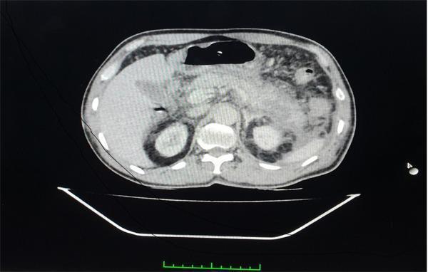
This diagnostic image is a contrast-enhanced computed tomography (CECT) scan of the abdomen in the axial plane, demonstrating acute necrotizing pancreatitis. The pancreas shows significant morphological changes, including a swollen neck and tail with indistinct margins and heterogeneous parenchymal enhancement. Specific areas of non-enhancement are visible, consistent with approximately 30% pancreatic necrosis. Extensive peripancreatic inflammation and fat stranding are evident, accompanied by fluid collections within the peritoneal cavity. These fluid collections are notably observed surrounding the kidneys and extending into the pararenal spaces. The stomach, partially visualized with an air-fluid level, and the liver are also present within the frame. This scan illustrates key radiological features used to calculate the modified CT severity index (CTSI) in the management of severe acute pancreatitis, specifically highlighting parenchymal necrosis and extra-pancreatic inflammatory involvement.
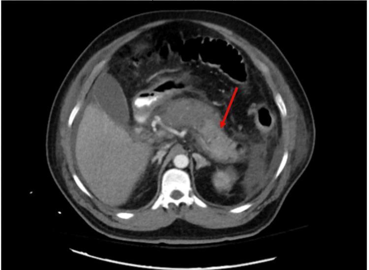
This diagnostic image is an axial contrast-enhanced Computed Tomography (CT) scan of the upper abdomen, primarily demonstrating severe acute necrotizing pancreatitis. The pancreas is significantly enlarged and edematous, showing heterogeneous enhancement. A red arrow points to non-enhancing, hypodense regions within the pancreatic parenchyma, particularly involving the body and tail, which are consistent with pancreatic necrosis. There is also evidence of peripancreatic fat stranding and fluid accumulation, indicating a severe inflammatory process. Surrounding anatomical structures are visible, including the liver (right), spleen (left), and cross-section of the spine and aorta. The image serves as a clinical illustration of advanced acute pancreatitis with parenchymal destruction, highlighting the importance of contrast-enhanced imaging in evaluating the extent of glandular necrosis for clinical staging and management decisions.
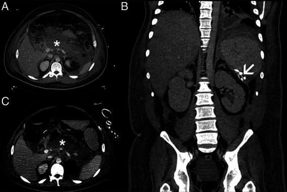
This composite diagnostic image features three panels (A, B, C) of a computerized tomography (CT) scan illustrating necrotizing pancreatitis and its management. Panels A and C are axial views showing extensive peripancreatic and retroperitoneal necrosis, marked by asterisks (*). The necrotic tissue appears as heterogeneous, mottled areas of low attenuation (density) surrounding the pancreatic head and body, extending into the retroperitoneal space anterior to the vertebral column and major vessels. Panel B is a coronal reformatted image demonstrating a transcutaneous drainage catheter (indicated by a white arrowhead) reaching the left retroperitoneum near the splenic hilum and the superior pole of the left kidney. This radiological series highlights a typical diagnostic and interventional approach for infected or symptomatic walled-off necrosis (WON) and acute necrotic collections. Key educational concepts include the recognition of pancreatic parenchymal destruction, retroperitoneal extension of inflammation, and the anatomical placement of minimally invasive percutaneous drains in the management of acute pancreatitis complications.
Acute Pancreatitis: Management
1. Overview & Epidemiology
- 85-90% of cases are self-limited, resolving within 3-7 days
- Mortality: <1% mild; ~10% moderate; 20-40% severe; >50% critical
- Most admissions are for interstitial pancreatitis (90-95%); necrotizing pancreatitis accounts for 5-10%
2. Severity Classification (Revised Atlanta Criteria)
| Class | Features | Mortality |
|---|---|---|
| Mild | No organ failure (OF), no local complications (LC) | <1% |
| Moderately Severe | Transient OF (<48 h) and/or LC without persistent OF | Low |
| Severe | Persistent OF (>48 h), 1+ organ systems | 20-40% |
| Critical (DBC) | Persistent OF + infected necrosis | >50% |
- SIRS (≥2 criteria): temp <36 or >38°C; HR >90; RR >20 or PCO₂ <32; WBC >12,000 or <4,000 or >10% bands
- BISAP score (≥3 predicts severity): BUN >25, impaired mental status, SIRS, age >60, pleural effusion
- APACHE II ≥8 at 24 h indicates severe disease
- Hemoconcentration (Hct >44%) and admission BUN >20 mg/dL are poor prognostic markers
3. Initial Management (First 4-6 Hours)
A. Fluid Resuscitation (Most Important Intervention)
- Lactated Ringer's solution is preferred over normal saline - shown to reduce systemic inflammation (lower CRP) and decrease SIRS
- Historical aggressive strategy: bolus 15-20 mL/kg, then 2-3 mL/kg/h
- Recent evidence (Evans et al., 2024 meta-analysis, PMID 38872377): A recent meta-analysis found aggressive vs. non-aggressive fluid therapy shows no clear outcome benefit from aggressive hydration; a less aggressive strategy (10 mL/kg bolus → 1.5 mL/kg/h) avoids fluid overload without worsening outcomes
- Target: urine output >0.5 mL/kg/h, normalization of HR, MAP, hematocrit, and BUN
- Monitor response every 6-8 hours with BUN and hematocrit every 8-12 h
B. Analgesia
- IV narcotic analgesics for pain control
- Nonopioid strategies preferred for maintenance, especially in pediatric and chronic cases
C. NPO & Oxygen
- Patient made NPO initially to minimize pancreatic stimulation
- Supplemental oxygen as needed; monitor O₂ saturation
D. Monitoring
- Vital signs, CVP, urine output, blood gases (in severe cases)
- Labs: LFTs, renal function, CBC, clotting, serum calcium (hypocalcemia), blood glucose, triglycerides
- Patients not responding to initial resuscitation → ICU/step-down unit
4. Determine Etiology
| Cause | Investigation |
|---|---|
| Gallstones (commonest) | Ultrasound; more likely if female, >50 yrs, high ALP/ALT/amylase |
| Alcohol | History + blood ethanol |
| Hypertriglyceridemia | Serum triglycerides |
| Hypercalcemia | Serum calcium, PTH |
| Drugs | ~30 causative agents confirmed by rechallenge (Class 1A); stop suspected drug |
| Post-ERCP | Pancreatic duct stenting and/or rectal indomethacin for prevention |
| Idiopathic | MRCP; consider genetic testing |
5. Imaging
- CT scan with IV contrast is best performed 3-5 days into hospitalization (not in first 72 h unless clinical deterioration)
- Recent data show CT is overutilized within 72 h, especially in mild disease
- CT indicated if: organ failure, clinical deterioration, signs of sepsis, suspected necrosis/complications, abdominal compartment syndrome
- Revised Atlanta Criteria outline CT terminology for local complications:
| Local Complication | Timing | Content |
|---|---|---|
| Acute Pancreatic Fluid Collection (APFC) | <4 weeks | Fluid only |
| Acute Necrotic Collection (ANC) | <4 weeks | Solid ± fluid |
| Pseudocyst | >4 weeks | Fluid with defined wall |
| Walled-off Necrosis (WON) | >4 weeks | Solid ± fluid, defined wall |
6. Nutritional Support
- Mild pancreatitis: resume oral low-fat solid diet early once hungry with normal bowel function and no nausea/vomiting
- Moderate-Severe pancreatitis: enteral nutrition (EN) preferred over total parenteral nutrition (TPN), started 2-3 days after admission
- Nasogastric (NG) tube feeding is acceptable; jejunal (NJ) feeding offers theoretical gut-barrier advantages but proven superiority is not established
- EN maintains gut barrier integrity, limits bacterial translocation, is cheaper, and has fewer complications than TPN
- "Resting the pancreas" is no longer supported - enteral feeding is the standard of care
- Consider supplemental parenteral nutrition by day 4 if enteral route is not tolerated
7. Antibiotics
- Prophylactic antibiotics are NOT recommended for severe or necrotizing pancreatitis - they do not improve survival and may promote opportunistic fungal infections
- Empiric antibiotics should be started if clinical decompensation occurs or infected necrosis is suspected
- Specific indications: confirmed cholangitis, concomitant respiratory/urinary tract infection
- Once infection confirmed and organism identified: targeted antibiotics
- Regimens used: IV cefuroxime, imipenem, or ciprofloxacin + metronidazole
- Duration of prophylaxis (if used): should not exceed 14 days
8. Gallstone-Specific Management
- ERCP within 72 hours is indicated for gallstone pancreatitis with cholangitis or obstructive jaundice
- ERCP is NOT indicated solely for cholestasis or predicted severe disease without cholangitis
- Cholecystectomy: should be performed during the same admission in mild disease; delayed in severe/complicated disease until recovery
9. Management of Local Complications
Necrosis
- Sterile necrosis: managed conservatively in most cases
- Infected necrosis: empiric antibiotics + drainage/debridement
- Fine-needle aspiration (FNA) of necrosis has fallen out of favor due to risk of contaminating sterile collections
- Nearly two-thirds of infected necrosis patients respond to antibiotics ± drainage without surgery
Step-Up Approach (Current Standard)
- Percutaneous or endoscopic transgastric/transduodenal drainage (first-line)
- If failure: minimally invasive debridement (video-assisted retroperitoneal debridement, endoscopic transluminal debridement, percutaneous nephroscopic debridement)
- Laparotomy reserved for: failed step-up approach, acute abdomen (perforation/ischemia), severe abdominal compartment syndrome
Pseudocysts
- Asymptomatic pseudocysts: observe
- Symptomatic/enlarging pseudocysts: endoscopic cystgastrostomy or percutaneous drainage
10. Summary Table: Early Management of Severe Acute Pancreatitis
| Action | Details |
|---|---|
| Admission | HDU/ICU |
| Analgesia | IV opioid analgesics |
| Fluid resuscitation | Lactated Ringer's, goal-directed; target UO >0.5 mL/kg/h |
| Supplemental O₂ | As needed; monitor SpO₂ |
| Invasive monitoring | Vitals, CVP, UO, ABG |
| Labs | FBC, LFTs, RFTs, clotting, Ca²⁺, glucose - frequent |
| Nutrition | NG enteral feeding once normovolemic (do not "rest" pancreas) |
| Antibiotics | Only for cholangitis/confirmed infection; NOT prophylactic |
| CT scan | If deterioration, organ failure, or signs of sepsis (not routine <72 h) |
| ERCP | Within 72 h if cholangitis present |
| Organ failure support | Inotropes, ventilation, hemofiltration as needed |
| Interventional | Step-up approach; delay ≥3-4 weeks if possible |
Recent Evidence Updates
- Fluid therapy (PMID 38872377): 2024 meta-analysis confirms aggressive IV fluid therapy does not improve outcomes vs. non-aggressive strategy; LR preferred
- Pharmacological interventions (PMID 40035297): 2025 systematic review of pharmacological interventions in adults - supports role of antioxidants and omega-3 fatty acids; LMWH also studied
- LMWH (PMID 39256790): 2024 systematic review and meta-analysis suggests low-molecular-weight heparin may reduce severity and complications in acute pancreatitis, though not yet standard of care