
Retinal Detachments
Reading File
Reading File
Reading File
Reading File
“Retinal detachments” is a broad ophthalmology topic. Key areas are:
- Classification: rhegmatogenous, tractional, and exudative/serous detachment
- Risk factors and pathophysiology: retinal breaks, posterior vitreous detachment, myopia, trauma, diabetic retinopathy, inflammation, tumors
- Clinical features: flashes, floaters, visual-field curtain/shadow, reduced vision
- Examination and diagnosis: dilated fundus examination, ocular ultrasound when the fundus cannot be visualized
- Urgency and initial actions: macula-on versus macula-off detachment, referral and activity advice
- Treatment: laser retinopexy, cryotherapy, pneumatic retinopexy, scleral buckling, pars plana vitrectomy
- Complications and prognosis: proliferative vitreoretinopathy, redetachment, macular involvement, visual outcomes
- Differentials: vitreous hemorrhage, posterior vitreous detachment, migraine aura, retinal tear without detachment
A retinal detachment is an ophthalmic emergency. New flashes or floaters, particularly with a peripheral “curtain,” shadow, or sudden visual-field loss, require same-day urgent ophthalmic assessment. Do not wait for symptoms to settle.
Which section do you want in detail: classification, diagnosis, management, or an exam-oriented note?
Puri btana details mey intro dif bera sab kuchh with imagr
Reading File
Reading File
Searching the Web
retinal detachment diagnosis management AAO patient information retinal tears detachment
Searching PubMed
retinal detachment treatment
Finding Sources
Finding Sources
Searching Images
retinal detachment types rhegmatogenous tractional exudative fundus images diagram
Ye topic kaafi broad hai. Main Hindi-English mixed, exam-oriented notes bana sakta hoon with diagrams/images. Pehle batao kis depth mein chahiye.
Asking for Preferences
Aapka preferred format kya hai? · Images kis type ki chahiye?
“Retinal detachments” is a broad ophthalmology topic. Key areas are:
- Classification: rhegmatogenous, tractional, and exudative/serous detachment
- Risk factors and pathophysiology: retinal breaks, posterior vitreous detachment, myopia, trauma, diabetic retinopathy, inflammation, tumors
- Clinical features: flashes, floaters, visual-field curtain/shadow, reduced vision
- Examination and diagnosis: dilated fundus examination, ocular ultrasound when the fundus cannot be visualized
- Urgency and initial actions: macula-on versus macula-off detachment, referral and activity advice
- Treatment: laser retinopexy, cryotherapy, pneumatic retinopexy, scleral buckling, pars plana vitrectomy
- Complications and prognosis: proliferative vitreoretinopathy, redetachment, macular involvement, visual outcomes
- Differentials: vitreous hemorrhage, posterior vitreous detachment, migraine aura, retinal tear without detachment
A retinal detachment is an ophthalmic emergency. New flashes or floaters, particularly with a peripheral “curtain,” shadow, or sudden visual-field loss, require same-day urgent ophthalmic assessment. Do not wait for symptoms to settle.
Which section do you want in detail: classification, diagnosis, management, or an exam-oriented note?Ye topic kaafi broad hai. Main Hindi-English mixed, exam-oriented notes bana sakta hoon with diagrams/images. Pehle batao kis depth mein chahiye.
Bsc nursing
Reading File
Reading File
Finding Sources
Finding Sources
query_images_db
Reading Web Page
https://www.aao.org/young-ophthalmologists/yo-info/article/y…
Reading File
Reading File
Reading File
Loading Image
Loading Image
Finding Sources
Retinal Detachment (RD) - BSc Nursing Notes
1. Introduction / Definition
Retinal detachment (RD) mein eye ki neurosensory retina apni underlying layer, retinal pigment epithelium (RPE), se separate ho jaati hai. Is separation ke beech subretinal fluid (SRF) collect hota hai. Retina light ko nerve signals mein convert karti hai, isliye detachment untreated rehne par permanent visual loss ho sakta hai.
- Retina: eye ki inner light-sensitive layer
- RPE: retina ko support, nutrition aur attachment provide karne wali layer
- Macula: central sharp vision ka area
- Fovea: macula ka central part, finest vision ke liye responsible
Kanski's Clinical Ophthalmology, p. 682.
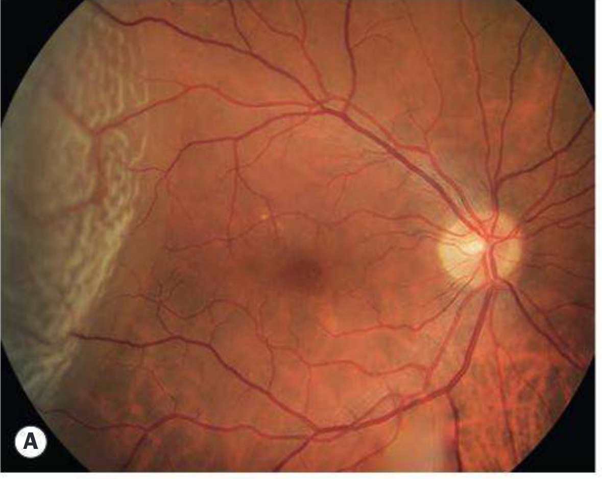
Fresh retinal detachment ka fundus appearance. Detached retina elevated, greyish/opaque aur corrugated lag sakti hai.
2. Why is it important?
Retinal detachment is a vision-threatening ophthalmic emergency.
Agar detachment macula tak nahi pahuncha hai (macula-on), to urgent surgery se central vision save hone ka chance zyada hota hai. Agar macula detach ho jaaye (macula-off), visual prognosis comparatively poorer ho sakta hai, though repair remains urgent.
The American Academy of Ophthalmology states that macula-on RD requires emergent retinal referral, while macula-off RD also needs urgent assessment and repair planning. AAO retinal detachment guidance
3. Types of Retinal Detachment
There are three main types:
| Type | Basic mechanism | Retinal break? | Common causes |
|---|---|---|---|
| Rhegmatogenous RD | Retinal tear/hole se liquefied vitreous retina ke neeche chala jata hai | Present | PVD, high myopia, trauma, lattice degeneration, post-cataract surgery |
| Tractional RD | Fibrous membranes contract karke retina ko pull karte hain | Usually absent | Proliferative diabetic retinopathy, retinopathy of prematurity |
| Exudative/serous RD | Retina/choroid vessels se fluid leakage | Absent | Inflammation, tumors, severe hypertension, Coats disease, VKH |
Kanski's Clinical Ophthalmology, p. 682.
A. Rhegmatogenous Retinal Detachment - Most common surgical type
Meaning
“Rhegma” means break/tear. Is type mein retina mein full-thickness tear, hole, or dialysis hota hai. Liquefied vitreous fluid is break ke through subretinal space mein chala jata hai and retina RPE se separate ho jaati hai.
Pathogenesis
Posterior vitreous detachment (PVD) ke time vitreous retina ko pull kar sakta hai. Strong adhesion wale point par traction retinal tear bana sakta hai. Tear ke through fluid retina ke neeche jaakar detachment spread karta hai.
Risk factors
- Increasing age and vitreous degeneration
- High myopia
- Previous retinal detachment in other eye
- Family history of RD
- Lattice degeneration
- Previous cataract surgery, especially complicated surgery
- YAG laser capsulotomy in susceptible patients
- Blunt or penetrating trauma
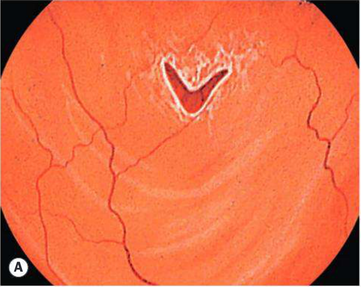
- Retinal tear or retinal hole
- Previous intraocular surgery
More than 40% of rhegmatogenous RDs occur in myopic eyes, and risk increases with degree of myopia. Kanski's Clinical Ophthalmology, p. 695.
Features
- Sudden flashes of light: photopsia
- Sudden onset/increase in floaters
- “Cobwebs” or black dots in vision
- Peripheral visual-field defect
- “Curtain/veil/shadow coming over the eye”
- Central visual acuity may remain normal initially if macula is still attached
- If macula detaches: decreased central vision, distortion, and poor reading vision
Fundus findings
- Elevated, mobile, convex, wrinkled or corrugated retina
- Retinal tear may appear red
- Retinal vessels may look dark and tortuous
- Pigment cells in anterior vitreous called tobacco dust or Shafer sign may indicate a retinal break
- Vitreous hemorrhage may occur
Kanski's Clinical Ophthalmology, pp. 695-696.
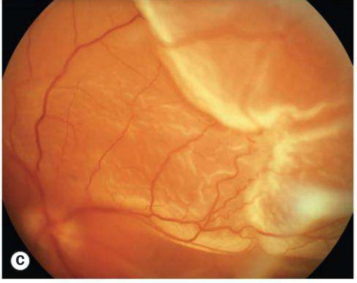
Fresh RD may appear as an elevated, opaque and corrugated retina. Kanski describes the fresh detachment as convex and slightly opaque due to retinal edema. Kanski's Clinical Ophthalmology, p. 696.
B. Tractional Retinal Detachment (TRD)
Meaning
Is type mein retina mein tear usually nahi hota. Fibrovascular membranes retina aur vitreous ke surface par form hote hain. In membranes ke contraction se retina RPE se pull away ho jaati hai.
Common causes
- Proliferative diabetic retinopathy: most important cause
- Retinopathy of prematurity (ROP)
- Sickle-cell retinopathy
- Retinal vein occlusion with neovascularization
- Ocular trauma
- Previous retinal surgery or inflammation
Clinical features
- Vision loss is usually gradual
- Flashes and floaters may be absent because traction slowly develops
- Progressive visual-field defect
- May remain stable for months or years
- If a retinal break develops, it becomes a combined tractional-rhegmatogenous RD, which may progress rapidly.
Fundus features
- Detached retina has a concave configuration
- Retina is relatively immobile
- Retinal breaks are absent in pure TRD
- Shifting fluid absent
- Fibrovascular membranes may be seen
Kanski's Clinical Ophthalmology, p. 705.
Tractional RD is caused by contraction of preretinal/fibrovascular tissue pulling the retina away from the RPE.
C. Exudative / Serous Retinal Detachment
Meaning
Is type mein retinal tear nahi hota. Retina ke vessels or choroidal vessels se fluid leak hoke retina ke neeche accumulate hota hai.
Causes
- Inflammatory eye disease: posterior uveitis, VKH disease
- Severe hypertension, including pre-eclampsia/eclampsia
- Choroidal tumors: melanoma, hemangioma, metastasis
- Coats disease
- Central serous chorioretinopathy
- Scleritis
- Renal disease or systemic inflammatory conditions
Features
- Blurred vision and visual-field defect
- Usually no flashes and floaters unless another retinal problem is present
- Retina smooth, elevated and often mobile
- Shifting subretinal fluid may occur with change in posture
- No retinal break seen
Treatment principle
Underlying cause treat karna zaroori hai. For example:
- Inflammation: corticosteroids/immunosuppression as prescribed
- Hypertension/pre-eclampsia: urgent medical and obstetric management
- Tumor: oncology/ophthalmology-directed treatment
4. Macula-On vs Macula-Off RD
| Feature | Macula-on RD | Macula-off RD |
|---|---|---|
| Macula | Attached | Detached |
| Central vision | Often preserved | Reduced |
| Urgency | Emergency | Urgent |
| Visual prognosis | Better if repaired before macular detachment | More guarded |
| Main nursing action | Immediate ophthalmology referral | Prompt ophthalmology referral and preparation for surgery |
Exam point:
Macula-on RD mein vision central area mein relatively good ho sakta hai, but peripheral field shadow may already be present. It should be treated as an emergency because macular involvement can cause permanent central visual loss.
Macula-on RD mein vision central area mein relatively good ho sakta hai, but peripheral field shadow may already be present. It should be treated as an emergency because macular involvement can cause permanent central visual loss.
5. Clinical Manifestations
Subjective symptoms
- Floaters: black dots, spots, cobwebs
- Photopsia: flashes of light, commonly peripheral visual field mein
- Curtain/veil effect: peripheral side se shadow spreading over vision
- Painless loss of vision
- Blurring of vision
- Peripheral visual-field loss
- Distorted vision if macula is involved
Important: RD is generally painless. Painful red eye suggests other conditions such as acute angle-closure glaucoma, uveitis, or trauma.
A sudden increase in floaters, new flashes, or a veil/curtain over vision may signify retinal detachment. Textbook of Family Medicine, p. 279.
6. Assessment and Diagnosis
History taking
Nurse/clinician should ask:
- Symptoms kab start hue?
- Flashes of light hain?
- New floaters hain?
- Curtain, shadow, or field loss hai?
- Vision suddenly reduced hui?
- Eye trauma hua?
- High myopia hai?
- Diabetes mellitus hai?
- Previous cataract or eye surgery?
- Retinal detachment in family or other eye?
- Any previous retinal tear or laser treatment?
Eye examination
- Visual acuity in both eyes
- Pupil examination, including relative afferent pupillary defect in significant macular/optic involvement
- Visual-field assessment by confrontation
- Slit-lamp examination
- Dilated fundus examination with indirect ophthalmoscopy
- Scleral depression to locate peripheral tear
- Intraocular pressure may be low in RD
Investigations
- Dilated fundus examination: most important test
- B-scan ultrasonography: useful when media are opaque due to dense cataract, vitreous hemorrhage, or severe inflammation
- Optical coherence tomography (OCT): useful for macular involvement and shallow RD
- Fundus photography, fluorescein angiography, or OCT angiography when indicated for underlying disease
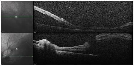
OCT can demonstrate separation of the neurosensory retina from the underlying RPE and subretinal fluid.
7. Differential Diagnosis
| Condition | Difference from RD |
|---|---|
| Posterior vitreous detachment (PVD) | Floaters/flashes can occur, but retina remains attached |
| Retinal tear without detachment | Tear present but no extensive SRF/detachment |
| Vitreous hemorrhage | Sudden floaters and haze; fundus view may be obscured |
| Migraine aura | Usually bilateral visual phenomenon, transient, often zig-zag lights |
| Retinoschisis | Splitting of retinal layers, usually smooth and less mobile |
| Choroidal detachment | Often associated with hypotony/eye surgery; different fundus configuration |
| Central retinal artery occlusion | Sudden profound painless loss of vision but no curtain-like progressive field loss |
8. Treatment
A. Treatment of retinal tear before detachment
If retinal tear is detected before RD develops, treatment can prevent detachment:
- Laser photocoagulation
- Cryotherapy (cryopexy)
These produce chorioretinal adhesion around the break and seal it.
The AAO notes that acute horseshoe tears and traumatic retinal breaks usually require treatment, while many asymptomatic atrophic/operculated breaks do not. AAO 2024 PPP
B. Surgical repair of established RD
1. Pneumatic retinopexy
- Gas bubble injected into vitreous cavity
- Bubble presses against retinal tear
- Laser or cryopexy seals tear
- Suitable for selected uncomplicated RRD, especially superior breaks
- Patient must maintain prescribed head position
2. Scleral buckle
- Silicone band/sponge is placed around outer wall of eye
- Indents sclera inward to support retinal break
- Usually combined with cryotherapy or laser
- Commonly used in selected young or phakic patients and some uncomplicated RRDs
3. Pars plana vitrectomy (PPV)
- Vitreous gel removed
- Traction relieved
- Retinal break treated with laser/cryopexy
- Gas or silicone oil may be injected as internal tamponade
- Often used for complex RD, vitreous hemorrhage, PVR, giant tear, pseudophakia, or tractional RD
4. Drainage of subretinal fluid
May be performed during surgery where needed.
5. Treatment of underlying cause
Particularly essential in tractional and exudative RD:
- Diabetic retinopathy: glycemic control, panretinal photocoagulation, anti-VEGF treatment, vitrectomy when indicated
- Uveitis: anti-inflammatory therapy
- Hypertension/preeclampsia: urgent medical treatment
- Tumor: specialist treatment
9. Complications
- Permanent visual impairment or blindness
- Macular involvement
- Recurrent retinal detachment
- Proliferative vitreoretinopathy (PVR)
- Cataract, especially after vitrectomy
- Raised intraocular pressure or glaucoma
- Infection, though uncommon after surgery
- Endophthalmitis, rare but serious
- Subretinal fibrosis/scarring
- Diplopia or refractive change after scleral buckle
Proliferative Vitreoretinopathy (PVR)
PVR is a major cause of failure after RD surgery. Scar-like membranes form and contract on or under the retina, pulling it away again. It may require repeat vitrectomy and membrane removal.
10. Nursing Management
A. Immediate nursing priorities
-
Treat as urgent
- Sudden flashes, floaters, curtain/shadow, or painless field loss needs immediate ophthalmology evaluation.
- Do not delay for routine appointment.
-
Assess vision
- Record visual acuity in both eyes.
- Ask about onset and progression of vision loss.
- Note peripheral field loss and central vision status.
-
Protect the affected eye
- Avoid eye rubbing or pressure.
- Use protective shield if instructed.
- Do not apply pressure dressing unless prescribed.
-
Positioning
- Follow ophthalmologist’s instructions.
- In some patients, position may help keep fluid away from macula before surgery.
- After gas tamponade, strict posturing is often prescribed.
-
Prevent falls/injury
- Reduced vision causes fall risk.
- Assist with ambulation, toileting, and unfamiliar environments.
- Keep call bell within reach.
- Ensure adequate lighting and remove hazards.
-
Emotional support
- Sudden visual loss causes fear and anxiety.
- Explain that urgent treatment is intended to preserve vision.
- Encourage patient and family questions.
B. Preoperative Nursing Care
- Confirm consent and identity according to policy.
- Record baseline vital signs, visual status, allergies, comorbidities, medicines, and anticoagulant use.
- Ensure fasting status if surgery under anesthesia is planned.
- Administer prescribed preoperative eye drops or systemic medicines.
- Teach not to rub or press the eye.
- Explain procedure, possible positioning, eye shield, follow-up, and activity restrictions.
- For diabetic patients: monitor blood glucose as ordered.
- For hypertensive patients: monitor BP and report severe elevation.
- Maintain infection-control measures.
C. Postoperative Nursing Care
Eye care
- Keep eye pad/shield clean and intact as prescribed.
- Do not allow patient to rub or press eye.
- Administer antibiotic, steroid, cycloplegic, and pressure-lowering eye drops exactly as ordered.
- Teach correct eye-drop technique:
- Hand hygiene
- Avoid touching bottle tip to eye/eyelashes
- Leave 5 minutes between different drops
- Apply gentle pressure over inner canthus only if advised
Monitor for warning signs
Report immediately:
- Sudden decrease in vision
- Increasing severe eye pain
- Marked redness/swelling
- Purulent discharge
- Severe headache, nausea, vomiting
- New curtain, flash, or many floaters
- Fever
- Injury to operated eye
Activity restriction
Usually avoid:
- Heavy lifting
- Straining
- Vigorous exercise
- Bending with head down
- Rubbing the eye
- Swimming until surgeon permits
Follow-up
Stress that postoperative reviews are essential to detect:
- Raised intraocular pressure
- Cataract
- Re-detachment
- Infection
- PVR
11. Special Instruction After Intraocular Gas Injection
If a gas bubble is placed during surgery:
- Maintain prescribed head position exactly.
- No air travel until gas completely absorbs.
- Avoid high-altitude travel.
- Inform every doctor, dentist, and anesthetist that an intraocular gas bubble is present.
- Nitrous oxide anesthesia must not be used while gas remains in the eye because gas expansion can dangerously raise intraocular pressure.
- Vision may remain blurred while bubble is present.
12. Health Education and Prevention
- Seek urgent eye review for sudden flashes, floaters, or a curtain over vision.
- Use protective eyewear during sports, industrial work, and trauma-risk activities.
- Diabetic patients should maintain glycemic control and attend retinal screening.
- High-myopia patients need regular dilated retinal examination.
- Attend follow-up after cataract surgery or retinal laser treatment.
- Patients with RD in one eye should report symptoms in the other eye immediately.
- Avoid self-medication and delaying specialist consultation.
13. Short Exam Answer
Retinal detachment is separation of neurosensory retina from retinal pigment epithelium due to accumulation of subretinal fluid. It is of three types: rhegmatogenous, tractional, and exudative. The patient may present with sudden floaters, flashes of light, painless peripheral field loss, and curtain-like shadow over vision. Diagnosis is by dilated fundus examination and B-scan ultrasonography if the fundus cannot be seen. Treatment includes laser photocoagulation or cryotherapy for tears, and pneumatic retinopexy, scleral buckle, or pars plana vitrectomy for RD. Nursing care includes urgent referral, visual assessment, eye protection, fall prevention, pre/postoperative care, prescribed posturing, eye-drop administration, and patient education.
14. One-Line Memory Trick
“Flashes + Floaters + Curtain = Retinal Detachment until proven otherwise.”
References
- Kanski's Clinical Ophthalmology: A Systematic Approach, 10th ed., pp. 682, 695-696, 705.
- The AAO retinal detachment triage guide emphasizes urgent referral, particularly for macula-on detachments.
- The AAO retinal-break guideline discusses retinal tears, lattice degeneration, and early prevention of rhegmatogenous RD.
This is a shared conversation. Sign in to Orris to start your own chat.