
Brachial artery for 5 marks
Reading File
The brachial artery is the main artery of the arm, a continuation of the axillary artery, running from the lower border of teres major to the cubital fossa. Now searching the textbook library for detailed anatomical content.
Searching sources...
Finding Sources
Reading File
Reading File
Reading File
Finding Sources
Loading Image
Loading Image
Finding Sources
Reading File
Reading File
Reading File
I now have comprehensive information from multiple authoritative textbooks. Here is the full 5-mark answer:
Brachial Artery
1. Origin and Course
The brachial artery is the major artery of the arm and a direct continuation of the axillary artery at the lower (inferior) border of teres major. It runs down the anterior compartment of the arm and terminates in the cubital fossa at the level of the neck of the radius, where it divides into the radial and ulnar arteries.
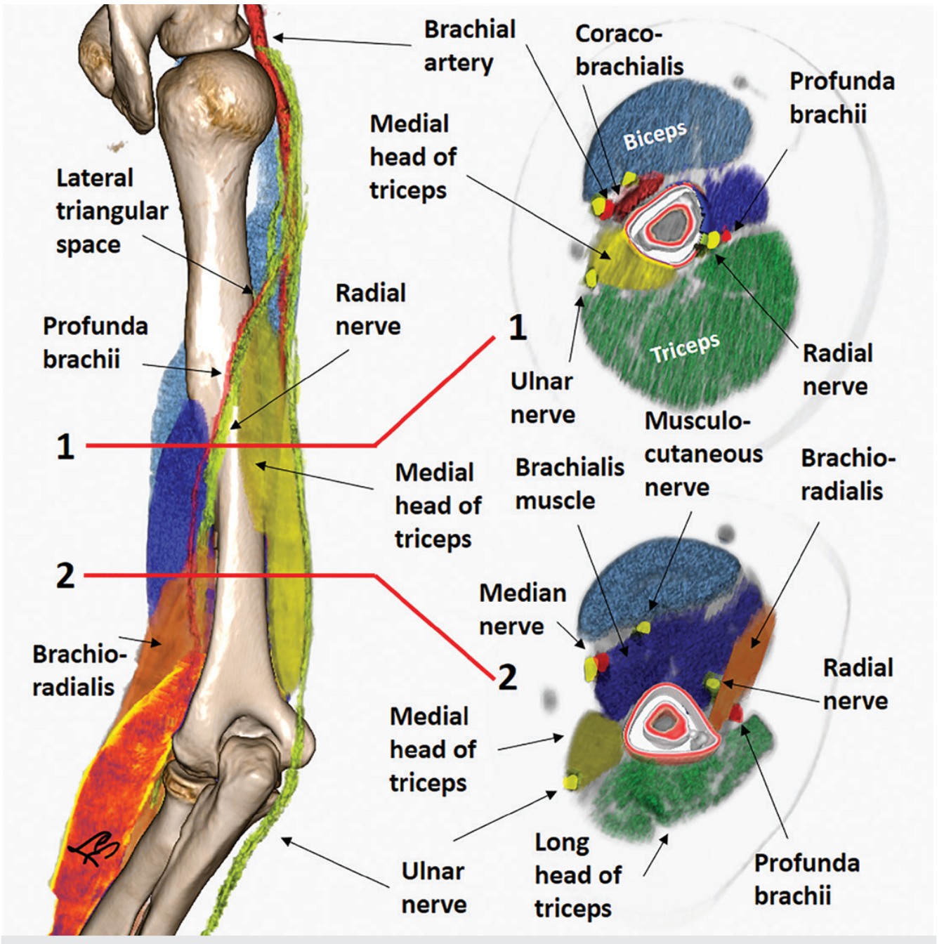
- In the proximal arm: lies on the medial side of the humerus in the medial bicipital groove, closely related to the ulnar and median nerves.
- In the distal arm: moves anteriorly and laterally to lie midway between the medial and lateral epicondyles.
- Throughout its course, it is palpable along its length and can be compressed against the humerus proximally.
(Gray's Anatomy for Students)
2. Relations
| Level | Medial | Lateral | Anterior | Posterior |
|---|---|---|---|---|
| Proximal arm | Ulnar nerve | - | Skin/fascia | Medial head of triceps |
| Mid-arm | Median nerve (crosses from lateral to medial) | - | Median nerve (crosses over it) | Brachialis |
| Cubital fossa | Median nerve | Biceps tendon | Bicipital aponeurosis | Brachialis |
The median nerve crosses superficial to the brachial artery from lateral to medial as it descends.
3. Branches
| Branch | Notes |
|---|---|
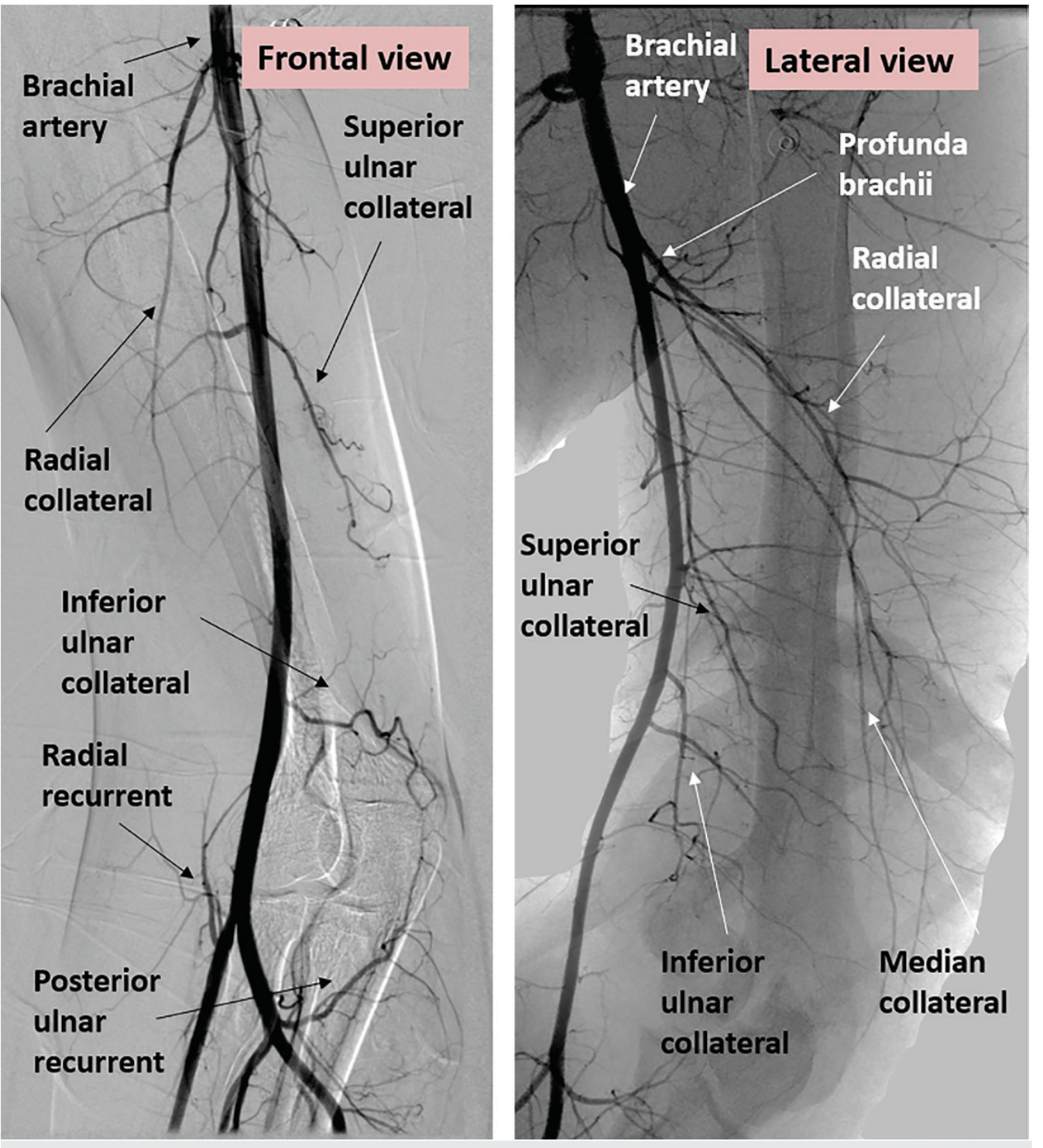
| Profunda brachii (deep brachial artery) | Largest branch; arises near teres major; accompanies the radial nerve through the lateral triangular interval and along the radial groove; divides into radial collateral and middle (medial) collateral arteries |
| Superior ulnar collateral artery | Runs with the ulnar nerve; anastomoses with posterior ulnar recurrent artery |
| Inferior ulnar collateral artery | Arises above the elbow; contributes to elbow anastomosis |
| Muscular branches | To anterior compartment muscles |
| Nutrient artery to humerus | Enters anteromedial surface of humeral shaft |
| Terminal branches: Radial & Ulnar arteries | At the apex of the cubital fossa |
(Fischer's Mastery of Surgery, Imaging Anatomy Vol. 3, Gray's Anatomy for Students)
4. In the Cubital Fossa
The cubital fossa contents, from lateral to medial: Biceps tendon → Brachial artery → Median nerve (covered anteriorly by the bicipital aponeurosis). The brachial artery bifurcates here into radial and ulnar arteries. The bicipital aponeurosis protects the brachial artery during venepuncture of the median cubital vein.
5. Anatomical Diagrams
Cross-sectional relations and course of brachial artery with neurovascular structures:
Angiographic view showing branches - profunda brachii, superior and inferior ulnar collateral arteries:
6. Clinical Importance
- Blood pressure measurement: The cuff compresses the brachial artery against the humerus; the Korotkoff sounds are auscultated in the cubital fossa over the brachial artery.
- Arterial line / ABG: Brachial artery access is used in catheterization and blood gas sampling.
- Supracondylar fracture in children: The brachial artery is vulnerable and injury causes Volkmann's ischemic contracture.
- Superficial brachial artery variant (~4-12%): Runs superficial to the median nerve; more prone to iatrogenic injury.
Sources: Gray's Anatomy for Students | Imaging Anatomy Vol. 3 | Fischer's Mastery of Surgery 8e | THIEME Atlas of Anatomy
This is a shared conversation. Sign in to Orris to start your own chat.