Proper Notes of all upper limbs bones, arteries,veins with tricks and proper explanation
Reading File
Finding Sources
Finding Sources
Finding Sources
Finding Sources
Finding Sources
Reading File
Reading File
Reading File
Reading File
Reading File
Reading File
Loading Image
Loading Image
Loading Image
Loading Image
Excellent! I have all the content and images needed. Now let me compile the complete, well-structured notes.
Complete Notes: Upper Limb Bones, Arteries & Veins
Source: THIEME Atlas of Anatomy - General Anatomy and Musculoskeletal System; Gray's Anatomy for Students
PART 1: BONES OF THE UPPER LIMB
The upper limb skeleton has 4 main segments: shoulder girdle, arm (brachium), forearm (antebrachium), and hand.
1. SHOULDER GIRDLE
Two bones connect the upper limb to the axial skeleton:
| Bone | Key Features | Clinical Note |
|---|---|---|
| Clavicle | S-shaped; medial 2/3 convex forward, lateral 1/3 concave forward | Most commonly fractured bone; fractures at junction of medial 2/3 and lateral 1/3 |
| Scapula | Flat triangular bone; has spine, acromion, coracoid process, glenoid cavity | Winged scapula = long thoracic nerve injury |
Memory Trick - Clavicle "S" shape: "Starts Medially Convex (like a C-shape turned in), ends Laterally Concave" - think "S for Shoulder"
2. HUMERUS (Arm Bone)
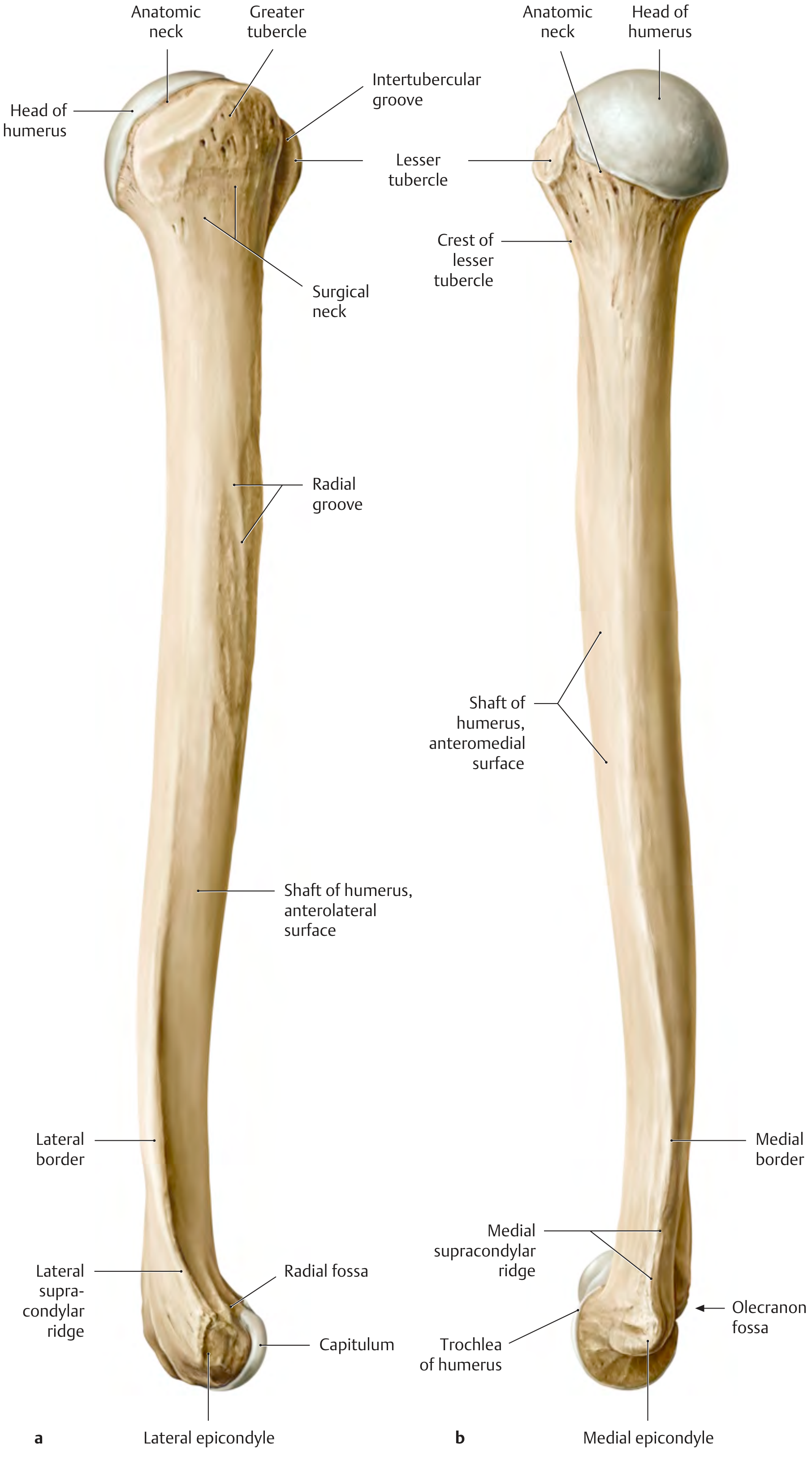
Proximal end features:
- Head - articulates with glenoid cavity of scapula (glenohumeral joint)
- Anatomic neck - narrow constriction at margin of head (articular cartilage boundary)
- Surgical neck - below tubercles; most commonly fractured part (axillary nerve at risk)
- Greater tubercle (lateral) - insertion of supraspinatus, infraspinatus, teres minor
- Lesser tubercle (anterior/medial) - insertion of subscapularis
- Intertubercular (bicipital) groove - between tubercles; long head of biceps tendon runs here
Shaft features:
- Deltoid tuberosity - lateral surface, mid-shaft; deltoid muscle insertion
- Radial (spiral) groove - winds around posterior shaft; radial nerve + profunda brachii artery run here
Distal end features:
- Capitulum - lateral; articulates with head of radius
- Trochlea - medial; articulates with trochlear notch of ulna
- Medial epicondyle - ulnar nerve passes behind it ("funny bone")
- Lateral epicondyle - extensor muscle origin
- Olecranon fossa (posterior) - receives olecranon during extension
- Coronoid fossa (anterior) - receives coronoid process during flexion
- Radial fossa (anterior, lateral) - receives radial head during flexion
Memory Trick for humerus distal features: "My Caddy Takes Long Rounds"
- Medial epicondyle, Capitulum, Trochlea, Lateral epicondyle, Radial fossa + olecranon fossa
Clinical Trick - Which nerve is injured?
- Surgical neck fracture → Axillary nerve (deltoid paralysis, loss of shoulder abduction)
- Midshaft fracture → Radial nerve (wrist drop)
- Medial epicondyle fracture → Ulnar nerve (claw hand - ring + little finger)
- Supracondylar fracture (kids) → Median nerve or anterior interosseous nerve (can't make "OK" sign)
3. RADIUS & ULNA (Forearm Bones)
| Feature | Radius | Ulna |
|---|---|---|
| Position | Lateral (thumb side) | Medial (little finger side) |
| Proximal end | Head (disc-shaped), neck, radial tuberosity | Olecranon, coronoid process, trochlear notch, radial notch |
| Shaft | Wider distally | Wider proximally |
| Distal end | Wider; articular surface for wrist joint, styloid process | Head (small), styloid process |
| Key surface | Radial tuberosity = biceps brachii insertion | Olecranon = triceps insertion |
Interosseous membrane connects radius and ulna, transmitting forces between them.
Radioulnar joints:
- Proximal radioulnar joint - head of radius rotates in radial notch of ulna
- Distal radioulnar joint - head of ulna rotates on ulnar notch of radius
- Together they allow pronation and supination
Memory Trick - Radius vs Ulna: "RULE: Radius is Lateral, Ulna is Medial" (when anatomical position) Or: "The THUMB side = Radius" (both start with the same position - lateral)
Trick for forearm fractures:
- Colles' fracture - distal radius fracture, dinner fork deformity (fall on outstretched hand, elderly)
- Smith's fracture - reverse Colles (fall on back of hand, garden spade deformity)
- Monteggia = proximal ulna fracture + radial head dislocation → "MR" = Monteggia-Radial
- Galeazzi = distal radius fracture + distal radioulnar dislocation → "GU" = Galeazzi-Ulnar
4. HAND BONES
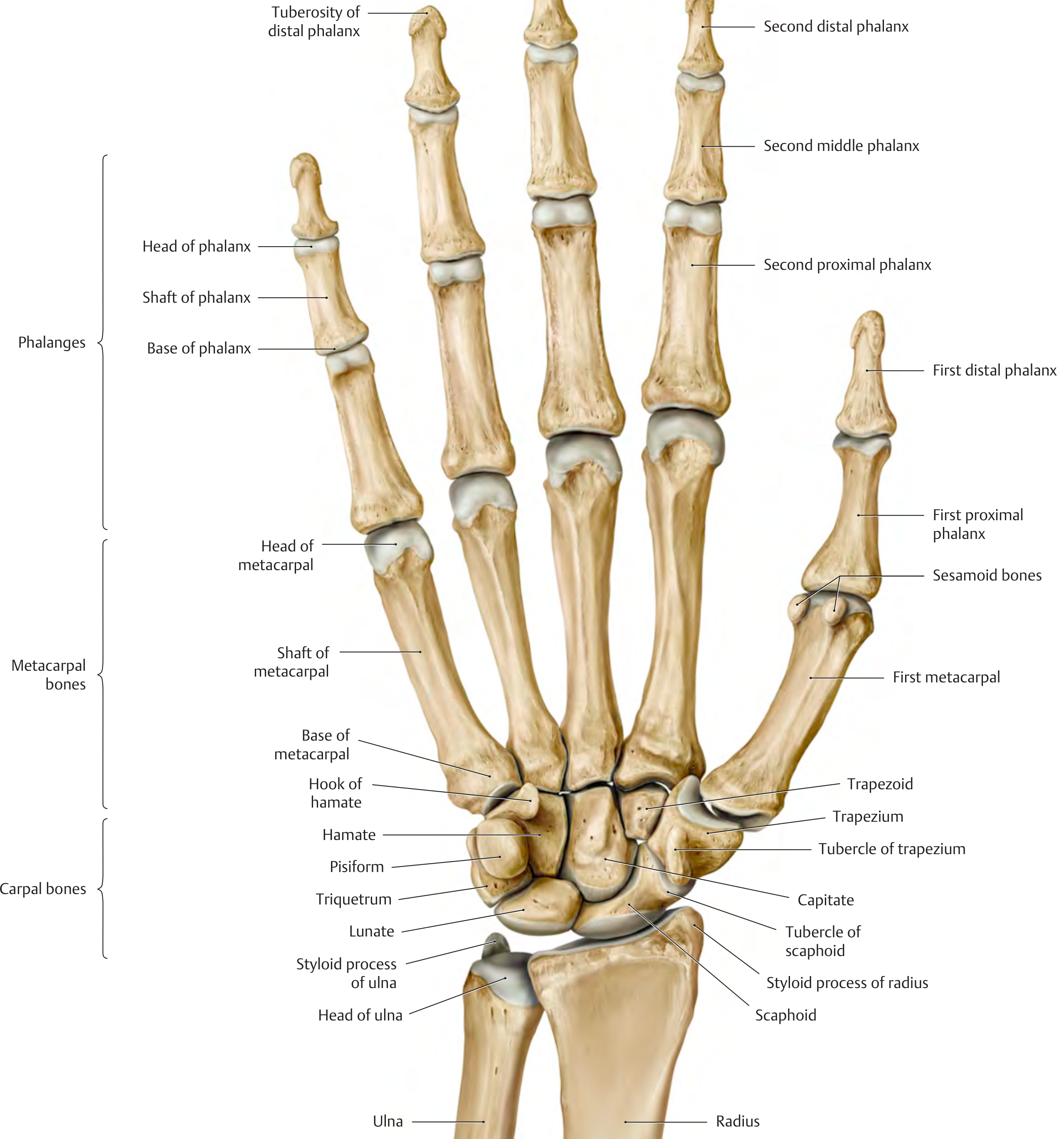
The hand skeleton has three groups:
A. Carpal Bones (8 bones, 2 rows)
Proximal row (lateral to medial): Scaphoid - Lunate - Triquetrum - Pisiform
Distal row (lateral to medial): Trapezium - Trapezoid - Capitate - Hamate
Classic Memory Trick: "Some Lovers Try Positions That They Can't Handle"
- Scaphoid, Lunate, Triquetrum, Pisiform | Trapezium, Trapezoid, Capitate, Hamate
Alternative (more fun): "She Looks Too Pretty, Try To Catch Her"
Key carpal facts:
| Bone | Clinical Importance |
|---|---|
| Scaphoid | Most commonly fractured carpal; in anatomical snuffbox; avascular necrosis risk (blood supply enters distally) |
| Lunate | Most commonly dislocated carpal; can compress median nerve |
| Pisiform | Sesamoid bone in flexor carpi ulnaris tendon |
| Hamate | Hook of hamate fracture in golfers/cyclists; ulnar nerve at risk |
| Trapezium | At base of thumb; CMC joint of thumb formed here |
| Capitate | Largest carpal bone |
Trick - Scaphoid fracture: Tender in anatomical snuffbox (between APL/EPB and EPL tendons). X-ray may be normal initially - treat as fracture if tender. Blood supply enters from distal end, so proximal pole avascular necrosis is a complication of proximal scaphoid fractures.
B. Metacarpal Bones (5 bones)
- Numbered 1-5 (thumb to little finger)
- Each has a base (proximal), shaft, and head (distal - forms the "knuckle")
- 1st metacarpal (thumb) is shortest and most mobile
Trick: "Boxer's fracture" = fracture of neck of 5th metacarpal (from punching with a closed fist)
C. Phalanges (14 bones)
- Thumb has 2 phalanges: proximal + distal
- Fingers 2-5 have 3 each: proximal + middle + distal
- Total: 2 + (3 × 4) = 14 phalanges
Memory Trick: "The thumb has 2, rest have 3 - total 14 is the key"
PART 2: ARTERIES OF THE UPPER LIMB
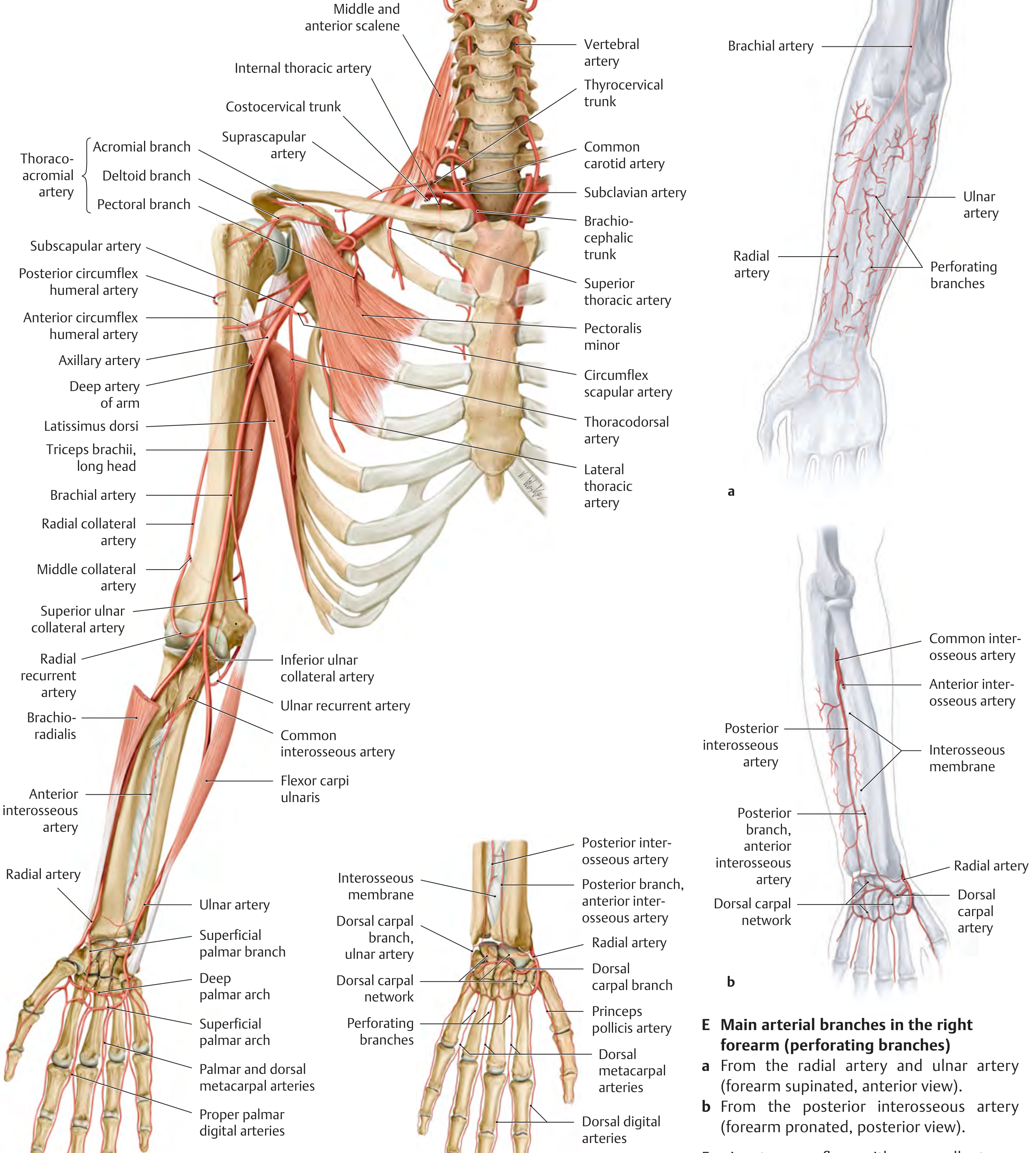
The main arterial trunk flows: Subclavian → Axillary → Brachial → Radial + Ulnar → Palmar arches → Digital arteries
1. SUBCLAVIAN ARTERY
Becomes axillary artery as it crosses the lateral border of the 1st rib.
Key branches (divided into 3 parts by scalenus anterior):
| Branch | Origin | Supply |
|---|---|---|
| Vertebral artery | Part 1 | Brain via foramen magnum |
| Internal thoracic artery | Part 1 | Anterior chest wall, breast |
| Thyrocervical trunk | Part 1 | Thyroid, scapular region, neck |
| - Suprascapular artery | from thyrocervical trunk | Scapular muscles |
| - Transverse cervical artery | from thyrocervical trunk | Trapezius, neck muscles |
| Costocervical trunk | Part 2 | Deep neck, upper intercostals |
Memory Trick for subclavian branches: "VITamins Come From Scot's Country"
- Vertebral, Internal thoracic, Thyrocervical trunk (with Suprascapular + Transverse cervical), Costocervical trunk
2. AXILLARY ARTERY
Runs from lateral border of 1st rib to lower border of teres major (where it becomes brachial).
Divided into 3 parts by pectoralis minor muscle.
Branches by part:
| Part | # of Branches | Branches |
|---|---|---|
| Part 1 (medial to pec minor) | 1 | Superior thoracic artery |
| Part 2 (behind pec minor) | 2 | Thoracoacromial artery, Lateral thoracic artery |
| Part 3 (lateral to pec minor) | 3 | Subscapular artery, Anterior circumflex humeral artery, Posterior circumflex humeral artery |
Memory Trick: "1 branch, 2 branches, 3 branches" (Parts 1, 2, 3 give 1, 2, 3 branches)
Trick for thoracoacromial branches: "CAMP"
- Clavicular, Acromial, M (deltoid), Pectoral
Clinically important: Posterior circumflex humeral artery travels with the axillary nerve through the quadrangular space.
Axillary artery relations:
- Medially: axillary vein
- Surrounded by cords of brachial plexus
- Enclosed in axillary sheath (fascial investment)
3. BRACHIAL ARTERY
Runs from lower border of teres major to the cubital fossa (where it bifurcates at the neck of the radius).
Lies medial to biceps tendon in cubital fossa.
Key branches:
| Branch | Supply |
|---|---|
| Deep artery of arm (profunda brachii) | Runs in radial groove with radial nerve; gives radial + middle collateral arteries |
| Superior ulnar collateral artery | Elbow collateral network (medial side) |
| Inferior ulnar collateral artery | Elbow collateral network (medial side) |
Bifurcation: In cubital fossa → Radial artery (lateral) + Ulnar artery (medial)
Clinical Trick:
- Blood pressure is measured using the brachial artery in the cubital fossa
- Brachial artery is the only artery in the arm - no major branches until it bifurcates
- Brachial artery pulse palpated medial to biceps tendon
4. RADIAL ARTERY
Continues from brachial artery along the lateral (radial) side of the forearm.
Passes deep to abductor pollicis longus and extensor pollicis brevis at the wrist → enters the anatomical snuffbox → passes between the 1st and 2nd metacarpals into the palm.
Key branches:
| Branch | Location | Supply |
|---|---|---|
| Radial recurrent artery | Immediately after origin | Anastomoses with radial collateral (elbow network) |
| Palmar carpal branch | Wrist | Palmar carpal network |
| Superficial palmar branch | Wrist | Superficial palmar arch (minor contribution) |
| Dorsal carpal branch | Wrist | Dorsal carpal network → dorsal metacarpal arteries |
| Princeps pollicis artery | Palm | Thumb (both sides) |
| Radialis indicis | Palm | Lateral side of index finger |
| Deep palmar arch | Palm | Main contribution (anastomoses with deep branch of ulnar) |
| Palmar metacarpal arteries | From deep arch | Deep finger supply |
Trick: Radial artery pulse felt at the radial side of wrist, between FCR tendon and radial styloid - used for pulse, Allen test, arterial blood gas sampling
5. ULNAR ARTERY
Larger of the two terminal branches. Runs along the medial (ulnar) side of the forearm.
Enters hand superficial to flexor retinaculum via Guyon's canal (ulnar tunnel).
Key branches:
| Branch | Supply |
|---|---|
| Ulnar recurrent artery (anterior + posterior) | Elbow network |
| Common interosseous artery | Bifurcates into anterior and posterior interosseous arteries |
| - Anterior interosseous | Runs on anterior interosseous membrane |
| - Posterior interosseous | Runs on posterior aspect; also gives recurrent interosseous |
| Palmar carpal branch | Palmar carpal network |
| Deep palmar branch | Deep palmar arch (minor contribution) |
| Superficial palmar arch | Main contribution to superficial arch |
| Common/proper palmar digital arteries | Fingers |
6. PALMAR ARCHES
| Arch | Formed mainly by | Location | Gives |
|---|---|---|---|
| Superficial palmar arch | Ulnar artery (+ superficial branch of radial) | Superficial to flexor tendons; at level of fully extended thumb | Common palmar digital arteries → proper palmar digital arteries |
| Deep palmar arch | Radial artery (+ deep branch of ulnar) | Deep to flexor tendons; 1 cm proximal to superficial arch | Palmar metacarpal arteries + perforating branches |
Memory Trick: "SUDA" = Superficial = Ulnar; Deep = rAdial Or: "SU-DR" - Superficial-Ulnar, Deep-Radial
Allen's Test: Compress both radial and ulnar arteries at wrist. Release one. If hand flushes pink, that artery is patent and contributes to palmar arch. Used before radial artery cannulation.
PART 3: VEINS OF THE UPPER LIMB
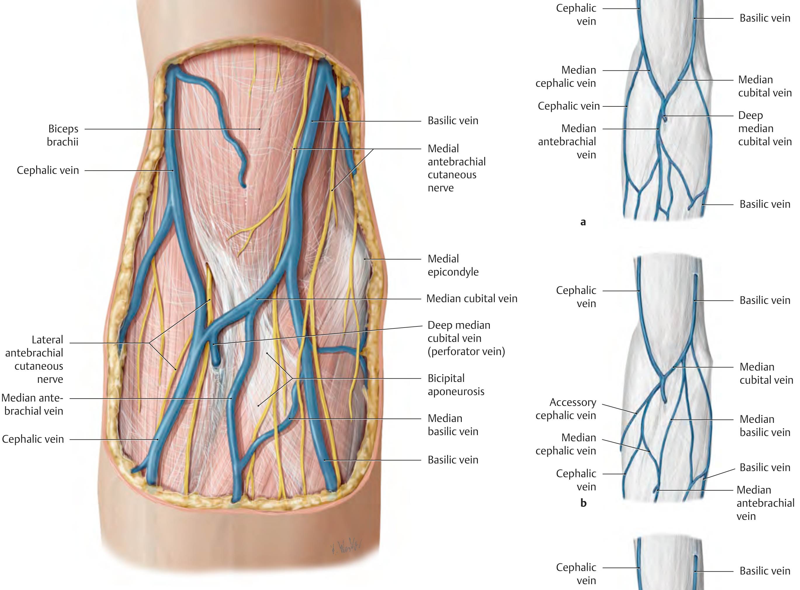
Veins of the upper limb are divided into superficial (subcutaneous, clinically accessible) and deep (accompanying arteries as venae comitantes).
DEEP VEINS (Venae Comitantes)
These travel alongside their corresponding arteries (usually paired):
Deep palmar venous arch
        ↓
Palmar metacarpal veins
        ↓
Radial veins + Ulnar veins  ←  (from superficial + deep palmar arch drainage)
        ↓
Brachial veins (2, paired around brachial artery)
        ↓
Axillary vein
        ↓
Subclavian vein
| Deep Vein | Accompanies |
|---|---|
| Radial veins | Radial artery |
| Ulnar veins | Ulnar artery |
| Anterior/posterior interosseous veins | Interosseous arteries |
| Brachial veins | Brachial artery |
| Axillary vein | Axillary artery |
| Subclavian vein | Subclavian artery |
Note: The axillary vein lies medial to the axillary artery in the axilla.
SUPERFICIAL VEINS (Clinically Most Important)
These travel in the subcutaneous tissue, independent of arteries.
A. Cephalic Vein
- Origin: Dorsal venous network of hand (radial/lateral side)
- Course: Ascends along lateral forearm → lateral aspect of biceps brachii → deltopectoral groove between deltoid and pectoralis major → pierces clavipectoral fascia → drains into axillary vein in clavipectoral triangle
- Clinical use: IV access, peripherally inserted central catheter (PICC), venous cutdown site
Trick: "Cephalic = Clateral (lateral side) = Clavipectoral (drains there)"
B. Basilic Vein
- Origin: Dorsal venous network of hand (ulnar/medial side)
- Course: Ascends along medial forearm → medial bicipital groove → at mid-arm level pierces the basilic hiatus (deep fascia) → runs subfascially → joins brachial vein to form axillary vein
- Lies close to medial antebrachial cutaneous nerve at elbow (injection risk)
Trick: "Basilic = Big, Basement (deep fascia), and Brachial (joins it to form axillary)"
C. Median Antebrachial Vein
- Runs up the anterior (flexor) surface of the forearm
- Drains mainly from dorsum of hand cutaneous veins, not from palmar surface
- Divides at elbow into median cephalic (joins cephalic) and median basilic (joins basilic)
D. Median Cubital Vein
- Connects cephalic and basilic veins across the cubital fossa
- Most common site for venipuncture and blood collection
- Bicipital aponeurosis separates it from the brachial artery beneath
Trick: "Median Cubital = Most Common for cannulation"
Summary Table: Superficial Veins
| Vein | Side | Drains into |
|---|---|---|
| Cephalic | Lateral (radial) | Axillary vein (via deltopectoral groove) |
| Basilic | Medial (ulnar) | Axillary vein (via basilic hiatus at mid-arm) |
| Median antebrachial | Anterior forearm | Median cephalic + median basilic |
| Median cubital | Cubital fossa | Connects cephalic to basilic |
| Accessory cephalic | Extensor forearm | Cephalic vein |
| Dorsal venous network | Dorsum of hand | Source of cephalic and basilic |
PERFORATOR VEINS
- Connect superficial to deep veins through the deep fascia
- Contain valves that direct blood from superficial → deep systems
- The deep median cubital vein is an important perforator at the elbow
MASTER TRICK SUMMARY
| Topic | Trick |
|---|---|
| Carpal bones | "Some Lovers Try Positions That They Can't Handle" |
| Axillary artery branches | Parts 1,2,3 give 1,2,3 branches |
| Thoracoacromial branches | "CAMP" (Clavicular, Acromial, deltoid, Pectoral) |
| Palmar arches | "SUDR" - Superficial=Ulnar, Deep=Radial |
| Nerve injured at surgical neck | Axillary nerve |
| Nerve injured at midshaft | Radial nerve (wrist drop) |
| Nerve injured at medial epicondyle | Ulnar nerve (claw hand) |
| Cephalic vein drainage | Axillary vein via deltopectoral groove |
| Basilic vein drainage | Axillary vein via basilic hiatus |
| Best IV access site | Median cubital vein |
| Most commonly fractured carpal | Scaphoid |
| Most commonly dislocated carpal | Lunate |
| Monteggia vs Galeazzi | MR (Monteggia-Radius head dislocated), GU (Galeazzi-Ulna DRUJ dislocated) |
CLINICAL PEARLS
- Allen's Test - before radial artery cannulation; tests patency of palmar arch collateral supply via ulnar
- IV cannulation - median cubital vein is safest; watch for superficial ulnar artery variant (~3% of people) that may mimic a vein
- Anatomical snuffbox - bounded by APL/EPB (radial) and EPL (ulnar); floor = scaphoid + trapezium; radial artery passes through; tender in scaphoid fracture
- Cephalic vein PICC - sometimes difficult to advance past deltopectoral groove due to angle; basilic vein route preferred for central access
- Quadrangular space - bounded by teres minor, teres major, long head of triceps, humerus; contains axillary nerve + posterior circumflex humeral artery - damaged in shoulder dislocation or humerus fracture
Sources: THIEME Atlas of Anatomy - General Anatomy and Musculoskeletal System, Gray's Anatomy for Students
This is a shared conversation. Sign in to Orris to start your own chat.