
A patient presents with difficulty in Catidiplopia,and reading,automobile accidents peripheral vision e. Whi loss). He also has headaches relieved by painkillers. What is the most likely diagnosis? Open-angle glaucoma Closed-angle glaucoma Optic chiasm lesion Occipital cortex lesion
- Bilateral peripheral vision loss (tunnel vision) - gradual, often unnoticed until advanced
- Diplopia and reading difficulty - from arcuate scotomas encroaching on the central field
- Headaches - from elevated intraocular pressure (IOP), typically dull and relieved by analgesics
- Automobile accidents - a hallmark symptom of peripheral visual field loss, patients clip mirrors or hit objects at the sides
open angle glaucoma visual field defect peripheral vision loss tunnel vision
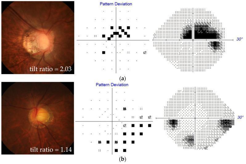
A comparison of optic disc morphology and visual field defects in two patients with myopia and open-angle glaucoma (OAG). The image is organized into two rows, (a) and (b), each featuring three components: stereo disc photography, a Humphrey perimetry pattern deviation plot, and a grayscale visual field map. Patient (a) displays high optic disc tilt (tilt ratio 2.03) with significant elongation and a shifted central retinal vessel trunk. This correlates with a paracentral scotoma, visible as a cluster of significant defects within the central 10 degrees on the pattern deviation plot and a dense superior/central defect on the grayscale map. Patient (b) shows a less tilted, more circular optic disc (tilt ratio 1.14) associated with peripheral scotoma. The perimetry for patient (b) illustrates defects primarily in the inferior and superior peripheral regions, with relative sparing of the central field. This comparison illustrates the relationship between myopic optic disc deformation (tilt) and the localization of glaucomatous visual field loss, highlighting the difference between paracentral and peripheral scotoma patterns in highly myopic eyes.
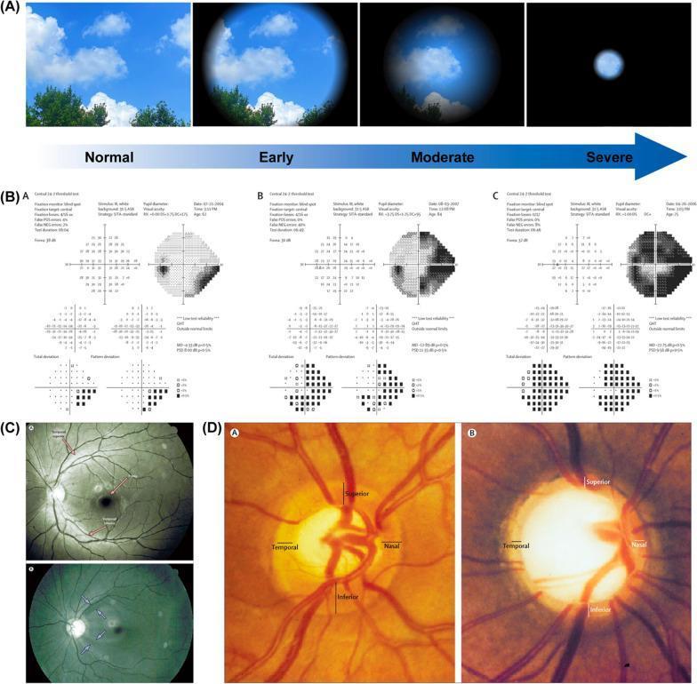
This composite educational graphic illustrates the progression and diagnostic findings of glaucoma across four panels (A-D). Panel A presents a visual simulation of disease progression, showing normal vision, peripheral blurring in early stages, and advanced 'tunnel vision.' Panel B displays Automated Static Perimetry (visual field tests) categorized as early, moderate, and severe functional loss, demonstrating increasing scotoma size and pattern deviation. Panel C contains two ophthalmoscopic photographs highlighting the retinal nerve fiber layer (RNFL); the healthy eye (A) shows intact reflexes (red arrows), whereas the glaucomatous eye (B) exhibits localized defects and reduced reflexes (light blue arrows). Panel D provides a detailed comparison of the optic disc: the healthy disc (A) shows a normal neuroretinal rim with characteristic inferior thickening, while the glaucomatous disc (B) demonstrates advanced pathological cupping, characterized by a significantly larger, deeper cup and thinned neuroretinal rim. This visual summarizes the correlation between structural damage (RNFL and optic disc) and functional vision loss in glaucoma.
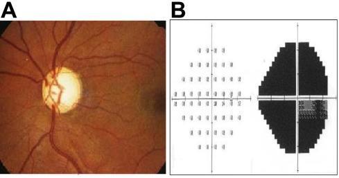
This composite educational graphic illustrates clinical findings of primary open-angle glaucoma (POAG). Panel A is a fundus photograph of the right eye displaying characteristic glaucomatous optic disc atrophy. Visible pathological features include significant enlargement of the optic cup (increased cup-to-disc ratio), thinning of the neuroretinal rim, and generalized disc pallor, suggesting advanced optic nerve damage. Panel B presents the corresponding visual field test (automated perimetry) results. The greyscale plot reveals extensive scotomas or visual field defects in the right eye, characterized by a large darkened area indicative of profound sensitivity loss. The pattern demonstrates significant functional impairment, such as quadrantanopia or arcuate-style defects, consistent with severe glaucomatous neuropathy. This material serves to demonstrate the clinical correlation between anatomical structural damage to the optic nerve head and subsequent functional vision loss in ophthalmic patients.
Answer: A. Open-angle Glaucoma
Symptom-by-Symptom Analysis
| Symptom | Explanation in Open-Angle Glaucoma |
|---|---|
| Peripheral vision loss | Hallmark feature - arcuate/nasal step scotomas that start peripherally and progress inward (tunnel vision) |
| Automobile accidents | Classic functional consequence of peripheral field loss - patients clip objects at the sides of the road without awareness |
| Difficulty reading | Arcuate scotomas eventually encroach on the central field in advanced disease |
| Diplopia (catidiplopia) | Due to progressive visual field defects disrupting binocular fusion |
| Headaches relieved by painkillers | Dull ache from chronically elevated IOP; mild enough to respond to analgesics (unlike closed-angle, which causes severe, unrelenting headache) |
Why NOT the Other Options?
Key Distinguishing Features of Open-Angle Glaucoma
- Bilateral, gradual, painless peripheral visual field loss (or mild dull ache)
- Elevated IOP (usually >21 mmHg) with anatomically open anterior chamber angle
- Progressive optic disc cupping (increased cup-to-disc ratio)
- Chronic, insidious course - patients often unaware until very late
- Common in older adults, with risk factors: family history, African descent, myopia, diabetes
Visual Field Progression in Glaucoma