
Now I am making notes which can be prepared for viva:- This is for bones of upper limb which include clavicle, scapula, humerus, radius, ulna, and bones of hand. go this way, • introduce the bone along with which side i am holding • then anatomical position • side determination • features or parts • important landmarks, muscle attachments, ligament attachments • Articulations • important clinical points and clinical anatomy. Let’s start with clavicle.
clavicle bone anatomy landmarks muscle attachments
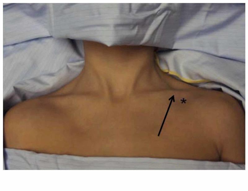
This clinical photograph illustrates the surface anatomy and landmarks for infraclavicular subclavian vein catheterization. The image shows the upper chest and neck of a patient in a supine position, prepped and draped for a sterile procedure. Key anatomical landmarks include the clavicle, sternocleidomastoid muscle, and suprasternal notch. An asterisk marks the needle insertion site, located approximately 1-2 cm inferior and lateral to the junction of the medial one-third and lateral two-thirds of the clavicle. A black arrow indicates the planned trajectory of the needle, aiming medially toward the suprasternal notch and passing immediately beneath the clavicle to access the subclavian vein. This visual guide demonstrates the standard landmark-based approach for central venous access, emphasizing the relationship between bony structures and deep vascular anatomy to minimize risks such as pneumothorax or arterial puncture.
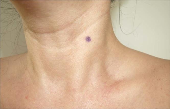
This clinical photograph displays the surface anatomy of the human neck and upper shoulder region in an anterior-lateral view. The primary anatomical landmark visible is the sternocleidomastoid (SCM) muscle, whose contour is clearly delineated as it courses from the mastoid process toward the sternum and clavicle. A circular, purplish skin marking is present on the mid-substance of the SCM muscle belly. This mark identifies the standardized measurement site for assessing myofascial tissue properties, specifically located midway between the muscle's insertion at the manubrium sterni and its origin at the mastoid process, where the sternal and clavicular heads converge. Other visible landmarks include the clavicle, the suprasternal notch, and the lateral cervical triangle. This image serves as a reference for clinical assessments of muscle stiffness and elasticity using devices such as the MyotonPro, demonstrating the appropriate anatomical positioning for perpendicular probe placement in physical therapy and musculoskeletal research.
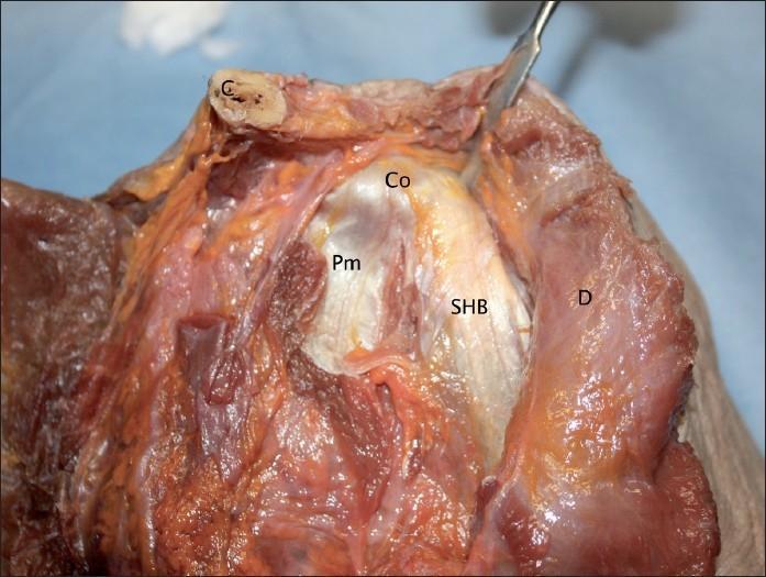
Anatomical dissection of a human cadaveric shoulder specimen focusing on the coracoid process (Co) and its associated muscular attachments. The image demonstrates the origin of the short head of the biceps brachii muscle (SHB) and the insertion of the pectoralis minor muscle (Pm). The short head of the biceps is shown with a broad, direct muscular attachment to the apex of the coracoid process, covered by a distinct white, fibrous tendinous aponeurosis on its anterior surface. The pectoralis minor tendon inserts onto the medial aspect of the coracoid process, adjacent but distinct from the SHB origin. Surrounding anatomical landmarks include the clavicle (C) positioned superiorly and the deltoid muscle (D) visible laterally. This clinical photograph provides high-resolution detail of the soft tissue relationships and aponeurotic structures critical for understanding shoulder anatomy and surgical approaches like the Latarjet procedure.
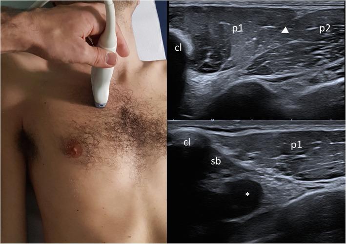
This composite educational resource consists of a clinical photograph showing ultrasound probe placement and two corresponding musculoskeletal ultrasound images of the infraclavicular region. The clinical photograph demonstrates a sagittal parasternal probe orientation with the cranial edge on the clavicle. The ultrasound images (top and bottom right) provide a sagittal view of the chest wall anatomy. Key landmarks include the hyperechoic clavicle (cl) with posterior acoustic shadowing. Originating from the clavicle is the clavicular head of the pectoralis major muscle (p1). A distinct hyperechoic cleavage plane (arrowhead) separates p1 from the sterno-costal head of the pectoralis major (p2). Deep to the clavicle, the subclavius muscle (sb) is visualized. Further deep to these muscular layers, the axillary artery (*) appears as an anechoic circular structure, adjacent to the axillary vein and components of the brachial plexus. This visual sequence is used in medical education to teach standard ultrasound screening protocols for assessing pectoralis muscle integrity, particularly in diagnosing congenital anomalies such as Poland Syndrome.
🦴 CLAVICLE - Viva Notes
1. Introduction & Side Identification
How to identify the side:
| Feature | What to look for |
|---|---|
| Shape | Medial 2/3 - convex anteriorly; Lateral 1/3 - concave anteriorly (S-shape) |
| Sternal end | Larger, quadrangular, bulky |
| Acromial end | Flat, smaller |
| Inferior surface | Rougher than superior surface |
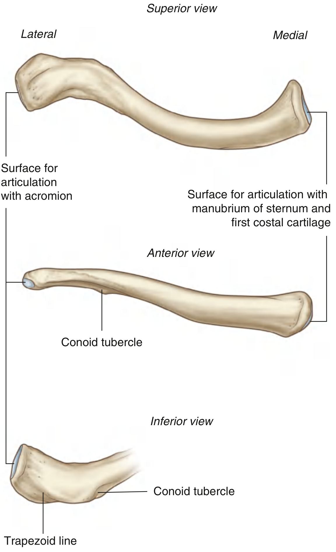
| Conoid tubercle | Prominent projection on inferior surface of lateral 1/3 |
| Trapezoid line | Roughened ridge extending laterally from conoid tubercle |
2. Anatomical Position
- Place the sternal (medial) end medially and the acromial (lateral) end laterally
- The superior surface faces upward and is relatively smooth
- The inferior surface faces downward and is rougher
- The medial 2/3 is rounded/cylindrical in cross-section; the lateral 1/3 is flattened
3. Parts / Features
| Part | Description |
|---|---|
| Shaft | S-shaped; medial 2/3 convex forward, lateral 1/3 concave forward |
| Sternal (medial) end | Bulky, quadrangular; has a large articular facet |
| Acromial (lateral) end | Flat; has a small oval articular facet |
| Superior surface | Smooth; no major bony markings |
| Inferior surface | Rough; has conoid tubercle and trapezoid line laterally; has a groove for subclavius medially |
4. Important Landmarks
Inferior Surface (most important for viva):
| Landmark | Location | Significance |
|---|---|---|
| Conoid tubercle | Inferior surface, at the bend (junction of middle and lateral thirds) | Attachment of conoid ligament (part of coracoclavicular ligament) |
| Trapezoid line | Roughened ridge running laterally from conoid tubercle toward acromial end | Attachment of trapezoid ligament (part of coracoclavicular ligament) |
| Subclavian groove | Inferior surface, medial portion | Lodges the subclavius muscle |
| Costoclavicular impression | Inferior surface, near sternal end | Attachment of costoclavicular ligament |
5. Muscle Attachments
On the Clavicle:
| Muscle | Attachment Site | Action Relevance |
|---|---|---|
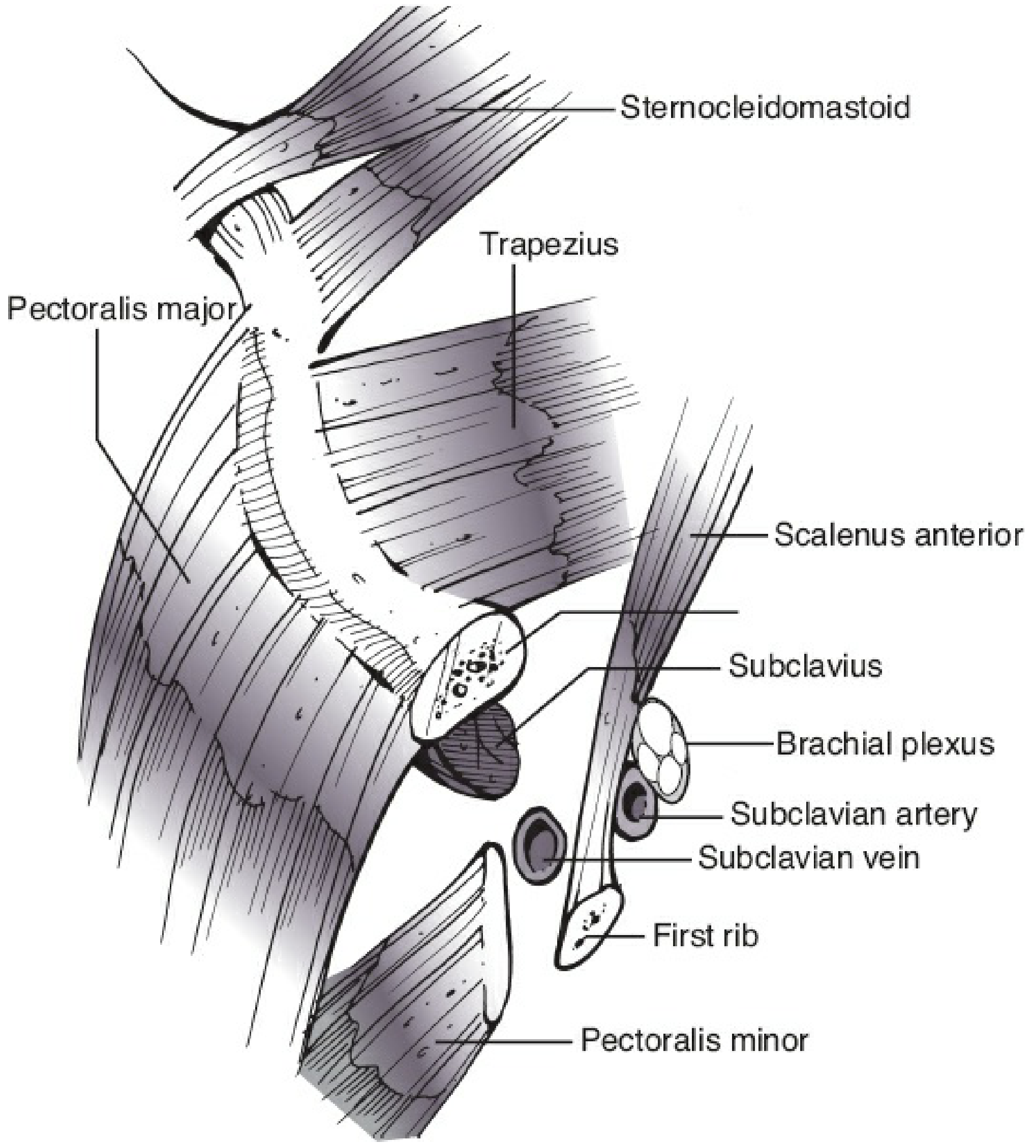
| Sternocleidomastoid (SCM) | Superiorly on medial 1/3 (origin) | Elevates medial fragment after fracture |
| Pectoralis major (clavicular head) | Anteroinferior surface of medial 2/3 (origin) | Pulls distal fragment medially and downward after fracture |
| Deltoid (anterior fibers) | Anteroinferior surface of lateral 1/3 (origin) | Pulls distal fragment downward after fracture |
| Trapezius | Posterosuperior surface of lateral 1/3 (insertion) | Blends with deltoid at lateral margin |
| Subclavius | Inferior (subclavian groove) of middle 1/3 (insertion) | Acts as a soft tissue buffer protecting subclavian vessels and brachial plexus |
6. Ligament Attachments
| Ligament | Attachment Site on Clavicle | Notes |
|---|---|---|
| Costoclavicular ligament | Costoclavicular impression, inferior surface near sternal end | Binds clavicle to 1st rib; limits elevation and protraction at SCJ |
| Coracoclavicular ligament (composed of 2 parts): | Responsible for vertically suspending scapula from clavicle | |
| - Conoid ligament | Conoid tubercle (inferior surface, lateral 1/3) | More medial, cone-shaped |
| - Trapezoid ligament | Trapezoid line (lateral to conoid tubercle) | More lateral, quadrangular |
| Acromioclavicular ligament | Superior surface near acromial end | Reinforces AC joint capsule |
| Anterior/Posterior SCJ ligaments | Around sternal end | Reinforce the sternoclavicular joint capsule |
| Interclavicular ligament | Superior aspect of sternal end | Connects the two clavicles across the jugular notch |
7. Articulations
a) Sternoclavicular Joint (SCJ) - Medial end
- Type: Synovial, saddle (sellar) joint - the only true synovial joint between the upper limb and axial skeleton
- Articular surfaces: Sternal end of clavicle + Manubrium sterni + 1st costal cartilage
- Has an intra-articular disc (fibrocartilaginous)
- Movements: elevation, depression, protraction, retraction, circumduction, and a small amount of rotation
b) Acromioclavicular Joint (ACJ) - Lateral end
- Type: Synovial, plane (gliding) joint
- Articular surfaces: Acromial end of clavicle + Medial surface of acromion of scapula
- May have a partial intra-articular disc
- Stabilized primarily by the coracoclavicular ligament (for vertical stability) and the AC ligament (for horizontal stability)
8. Clinical Points & Clinical Anatomy
Clavicle Fracture - Most Common Fracture of the Shoulder Girdle
- The clavicle is the most commonly fractured bone in the shoulder region
- Middle 1/3 is the most common site (~80%) - weakest point at the junction of the two curves
- Typical deformity: Medial fragment elevated (SCM pull) + distal fragment displaced downward and medially (deltoid + pectoralis major)
- Mechanism: Fall on outstretched hand (FOOSH) or direct blow
The "Strut" Function
- The clavicle acts as a strut (brace) that holds the shoulder away from the chest wall. Loss of this strut (fracture) causes the shoulder to fall medially and forward.
Neurovascular Relations (Important!)
- Inferior to the clavicle lie - subclavian vein (most anterior), subclavian artery, and brachial plexus (most posterior)
- The subclavius muscle acts as a protective buffer between the clavicle and these structures - in clavicle fractures, the subclavius prevents injury to the subclavian vessels in most cases
- Subclavian vein is accessed just below the medial 1/3 of the clavicle for central venous catheterization
Supraclavicular Nerves (Surgical Relevance)
- Arise from C3 and C4; cross the clavicle from medial to lateral
- At risk during surgical plating of clavicle fractures
- Injury causes numbness over the upper chest wall (below incision)
Ossification
- The clavicle is the first bone to ossify in the body (5th-6th week of intrauterine life) - by membranous ossification (intramembranous, not endochondral - the only long bone to ossify this way)
- It is also one of the last bones to fuse - the medial epiphysis fuses at ~22-25 years of age (important for age estimation in forensics and birth injury cases)
Birth Injury
- Fracture of the clavicle is the most common birth injury during difficult deliveries (shoulder dystocia)
ACJ Dislocation
- Grade I-III managed conservatively; Grade IV-VI may require surgical fixation
- In Grade III+, the coracoclavicular ligament is torn - the lateral clavicle appears elevated ("piano key sign")
Surface Anatomy
- The medial end of the clavicle is the most prominent bony point at the root of the neck
- The clavicle demarcates: anteriorly - clavipectoral fascia; posteriorly - posterior triangle of neck
Use Bd chaurasia and gray’s anatomy only for data. And for images you can use from anywhere which is very simple to understand. Now this is just a instruction, just say yes and nothing else.
Umm my credits are about to over in yours so i want you to make a complete explanation of how to teach and complete this such that i can make other ai to do the exact same as yours. Everything you use to generate your response. Only for this chat
Complete Prompt for Viva Notes on Bones of Upper Limb
Role & Task
Source Restriction
- BD Chaurasia's Human Anatomy (Handbook of General Anatomy or the Upper Limb volume)
- Gray's Anatomy for Students
Format for Each Bone (Strict Order)
1. Introduction + Side Identification (while holding the bone)
- State what bone it is
- State which side (right or left) you are holding
- Give 2-3 quick identifying clues that distinguish it from the opposite side
2. Anatomical Position
- How to orient the bone correctly in space
- Which surface faces anteriorly, posteriorly, superiorly, inferiorly
3. Side Determination
- Step-by-step method to identify right vs left
- Key asymmetric features to look for
- Present as a table or bullet list
4. Features / Parts
- All named parts of the bone (ends, surfaces, borders, angles, processes)
- Brief description of each
- Use a table format where possible
5. Important Landmarks + Muscle Attachments + Ligament Attachments
- List every named landmark (tubercle, tuberosity, groove, line, ridge, fossa, etc.)
- For each: location, what attaches there (muscle or ligament), and functional note
- Use a table with columns: Landmark | Location | Muscle/Ligament | Note
- Include fracture deformity logic where relevant (which muscle pulls which fragment where)
6. Articulations
- Every joint the bone participates in
- Joint type (synovial/fibrous/cartilaginous + subtype)
- Articular surfaces on both sides
- Stabilizing ligaments
- Movements permitted
7. Important Clinical Points & Clinical Anatomy
- Most common fracture site and mechanism
- Nerve/vessel at risk in fracture or dislocation
- Ossification (first to ossify? membranous vs endochondral? when does epiphysis fuse?)
- Any eponymous signs, clinical tests, or conditions (e.g. piano key sign, carrying angle, etc.)
- Surface anatomy / palpation points
- Surgical relevance
Formatting Rules
- Use markdown with headers, tables, and bullet points
- Keep language simple and direct - suitable for viva speech
- Bold all key terms
- After each section, include a relevant anatomical diagram (simple and labeled)
- At the end, cite the sources used (BD Chaurasia / Gray's)
- End each bone with: "Ready to move on to [next bone] whenever you are!"
Sequence of Bones
- Clavicle
- Scapula
- Humerus
- Radius
- Ulna
- Bones of the Hand
Example of the Viva Introduction Style
"Sir, this is the clavicle - the collar bone. It is a long bone with a gentle S-shaped curve. This one I am holding appears to be the RIGHT clavicle."
Key Content Points to Cover Per Bone (Quick Reference)
Clavicle
- Only bone connecting upper limb to axial skeleton
- S-shaped; medial 2/3 convex forward, lateral 1/3 concave forward
- First bone to ossify (5th-6th week IU) - intramembranous; last to fuse (medial epiphysis at 22-25 yrs)
- Middle 1/3 most common fracture site
- Fracture deformity: SCM elevates medial fragment; deltoid + pectoralis major pull distal fragment down and medially
- Subclavius protects subclavian vessels from fracture spikes
- Landmarks: conoid tubercle, trapezoid line, costoclavicular impression, subclavian groove
- Muscles: SCM, pectoralis major, deltoid, trapezius, subclavius
- Ligaments: costoclavicular, conoid, trapezoid, acromioclavicular, interclavicular
- Joints: Sternoclavicular (saddle, with intra-articular disc) + Acromioclavicular (plane)
Scapula
- Flat triangular bone; 3 angles, 3 borders, 2 surfaces, 3 processes
- Side ID: glenoid cavity faces laterally, costal (subscapular) fossa faces anteriorly, spine on posterior surface
- Supraspinous, infraspinous, subscapular fossae
- Coracoid process - "lighthouse of the shoulder" - many attachments
- Notch: suprascapular notch (nerve passes through, artery passes over - "generals go over the bridge, soldiers go through")
- Scapulothoracic joint (not a true joint - functional joint)
- Winging of scapula - long thoracic nerve (serratus anterior)
Humerus
- Longest bone of upper limb
- Head, anatomical neck, surgical neck, greater and lesser tubercles, intertubercular sulcus (bicipital groove), deltoid tuberosity, radial groove (spiral groove), medial and lateral epicondyles, capitulum, trochlea, coronoid fossa, olecranon fossa, radial fossa
- Surgical neck - axillary nerve at risk
- Radial groove - radial nerve at risk (Holstein-Lewis fracture)
- Medial epicondyle - ulnar nerve at risk
- Carrying angle formed at elbow (cubitus valgus/varus)
Radius
- Lateral bone of forearm
- Head (proximal), neck, radial tuberosity, styloid process, ulnar notch, dorsal tubercle (Lister's tubercle)
- Interosseous membrane attachment
- Colles' fracture (distal radius, most common - dinner fork deformity)
- Smith's fracture (reverse Colles')
- Head of radius - FOOSH injury in children (pulled elbow / nursemaid's elbow)
Ulna
- Medial bone of forearm
- Olecranon, coronoid process, trochlear notch, radial notch, styloid process, head (distal)
- Olecranon - triceps attachment; pulled off in avulsion
- Monteggia fracture - proximal ulna fracture + radial head dislocation
- Ulnar nerve runs behind medial epicondyle; lies between olecranon and medial epicondyle
Bones of the Hand
- 8 carpal bones (proximal row: Scaphoid, Lunate, Triquetrum, Pisiform; distal row: Trapezium, Trapezoid, Capitate, Hamate)
- Mnemonic: "Some Lovers Try Positions That They Can't Handle"
- 5 metacarpals + 14 phalanges (2 in thumb, 3 in each finger)
- Scaphoid: most commonly fractured carpal; avascular necrosis risk; snuffbox tenderness
- Hamate: hook fracture in golfers/cyclists
- Carpal tunnel: 8 tendons of FDP + FDS + FPL + median nerve (not ulnar nerve)
- Guyon's canal: ulnar nerve and artery
Tone and Style
- Precise and academic but conversational for viva preparation
- Speak as if preparing the student to answer an examiner directly
- Include "viva tips" in italics where helpful (e.g. "Examiners love asking about the nerve at risk in surgical neck fractures")