
Itching vulva treatment
vulvar pruritus treatment management
vulvar lichen sclerosus clinical appearance
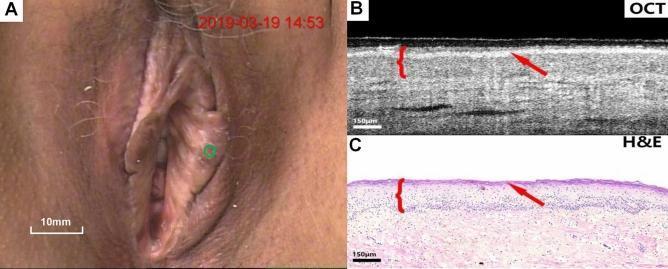
A three-panel figure demonstrating clinical and histological features of lichen sclerosus of the vulva. Panel A is a clinical colposcopy photograph showing the macroscopic appearance of the vulvar region, with a green circle indicating the specific site of interest. Panel B is a cross-sectional optical coherence tomography (OCT) image of the affected tissue. It shows well-defined stratification between the epithelium and stroma with uniform optical scattering. A key diagnostic feature highlighted is the loss of the serrated rete peg architecture at the epithelial-stromal junction. A red arrow indicates a hyper-reflective surface layer corresponding to hyperkeratosis, and a red bracket denotes the epithelial thickness. Panel C is a corresponding hematoxylin and eosin (H&E) stained histological section (150 μm scale) that correlates with the OCT findings. It confirms the presence of surface keratinization (arrow), a thinned or flattened epithelial layer (bracket), and the absence of rete pegs, consistent with lichen sclerosus pathology. This comparison serves as a diagnostic reference for utilizing non-invasive OCT imaging in identifying sub-surface morphological changes in gynecological dermatological conditions.
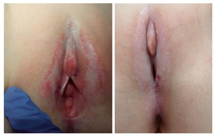
Clinical photograph presenting two cases of pediatric vulvar lichen sclerosus (VLS). The images demonstrate the characteristic 'figure-of-eight' distribution of the disease, involving the vulvar and perianal regions. Visible clinical signs include ivory-white, well-demarcated plaques with an atrophic, parchment-like or shiny skin texture. In the left image, there is prominent erythema, thinning of the labia minora, and early labial fusion. The right image shows more extensive hypopigmentation (porcelain-white appearance) affecting the clitoral hood and perianal area, with visible architectural changes including mild scarring and labial resorption. These cases highlight the typical presentation of VLS in young girls, emphasizing the hallmark features of cutaneous atrophy, hypopigmentation, and potential anatomical distortion of the external genitalia if left untreated.
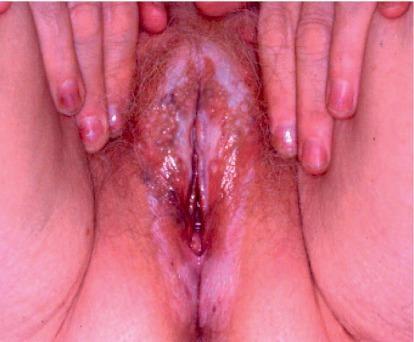
A clinical photograph showing the vulvar region of a patient with vulvar lichen sclerosus. The image demonstrates characteristic ivory-white, irregularly shaped patches (hypopigmentation) and plaques distributed across the labia majora, labia minora, and clitoral hood. The affected skin exhibits an atrophic, thin, and glistening texture, often described as having a 'parchment-like' or 'cigarette paper' appearance. Significant erythema and inflammation are visible alongside areas of subepithelial hemorrhage and dark, purpuric spots, which may indicate hemorrhagic bullae or excoriations from chronic pruritus. The anatomical landmarks show signs of architectural distortion, including possible early labial resorption and narrowing of the introitus. This visual content illustrates the progressive inflammatory nature of lichen sclerosus in the female genitalia, highlighting key diagnostic signs for dermatology and gynecology clinical education.
Vulvar Itching (Pruritus Vulvae) - Causes & Treatment
Common Causes
| Category | Examples |
|---|---|
| Infections | Candida vulvovaginitis, bacterial vaginosis, trichomoniasis, scabies, pubic lice, pinworms |
| Inflammatory dermatoses | Lichen sclerosus, lichen planus, lichen simplex chronicus, psoriasis, atopic/contact dermatitis |
| Hormonal | Atrophic vulvovaginitis (postmenopausal estrogen deficiency) |
| Neoplastic | Extramammary Paget disease, vulvar intraepithelial neoplasia (VIN), squamous cell carcinoma |
| Neuropathic | Lumbosacral radiculopathy (L4-S3 nerve compression), vulvodynia |
| Systemic | Diabetes mellitus, renal failure, liver disease, thyroid disorders |
Treatment by Cause
1. Candida Vulvovaginitis (most common infectious cause)
- First-line: Topical antifungals - miconazole or clotrimazole intravaginal cream/suppositories for 3-7 days
- Oral alternative: Fluconazole 150 mg single oral dose
- Severe cases: 2-3 doses of fluconazole 72 hours apart
- Recurrent cases (4+ episodes/year): Fluconazole 150 mg weekly for 6 months (suppressive therapy)
2. Lichen Sclerosus
- First-line (gold standard): Superpotent topical corticosteroids - clobetasol propionate 0.05% ointment once daily, tapering to 1-2x/week maintenance
- 95% of fully compliant patients achieve complete symptom control
- Maintenance therapy prevents scarring progression and reduces SCC risk
- Second-line: Topical calcineurin inhibitors (tacrolimus 0.1% or pimecrolimus 1%) - reserved for steroid non-responders or intolerance
- Neuropathic component (vulvodynia): Tricyclic antidepressants (amitriptyline), gabapentin, or duloxetine
- Surgical: Cryotherapy for refractory cases; photodynamic therapy
3. Contact/Irritant Dermatitis
- Identify and eliminate the offending agent (soaps, fragrances, synthetic fabrics, douches, panty liners, topical medications)
- Mild-potency topical corticosteroids (class 6-7, e.g., hydrocortisone 1%)
- Patch testing to identify allergens
- Barrier creams (zinc oxide paste)
4. Atrophic Vulvovaginitis (postmenopausal)
- Topical estrogen (cream, vaginal ring, or tablet) - restores epithelial integrity and relieves dryness/itching
- Non-hormonal: vaginal moisturizers and lubricants
5. Lichen Simplex Chronicus (chronic scratch-itch cycle)
- Mid-to-high potency topical corticosteroids
- Topical tacrolimus ointment
- Break the itch-scratch cycle: antihistamines (hydroxyzine) at night, cool compresses
6. Lichen Planus
- Potent topical corticosteroids
- Systemic immunosuppressants for erosive/severe forms
General (Non-Specific) Measures for All Causes
- Hygiene: Wash with plain water or mild, fragrance-free soap only; pat dry (do not rub)
- Avoid irritants: Scented products, tight synthetic clothing, panty liners, douches
- Emollients: Apply plain petrolatum (Vaseline) over moist skin as a barrier
- Topical anesthetics: Pramoxine (often combined with hydrocortisone) for symptomatic relief
- Sitz baths: Lukewarm water; cool compresses reduce acute pruritus
- Antihistamines: Helpful mainly for sleep disturbance from nocturnal itching
- When all topical medications seem to worsen symptoms, discontinue everything and use plain petrolatum only
When to Refer / Biopsy
- Diagnosis is uncertain
- There is a white, red, or pigmented lesion that does not respond to empirical treatment within 8-12 weeks
- Suspicion for VIN, Paget disease, or carcinoma
- Any ulcerated or persistent lesion in a postmenopausal woman
Key Points
- Always treat the underlying cause, not just the symptom
- Lichen sclerosus requires long-term maintenance therapy - stopping treatment leads to relapse and risk of SCC
- Candidal co-infection can develop during steroid therapy for lichen sclerosus - watch for it
- Recurrent or refractory pruritus warrants patch testing, biopsy, and specialist referral