
Explain about erythropoiesis in detail point wise
Reading File
Now let me search the medical library for erythropoiesis content.
Finding Sources
Searching the Web
erythropoiesis stages red blood cell development physiology
Good findings. Let me now read the most relevant textbook sections in parallel.
Reading File
Reading File
Reading File
Good content. Now let me search for more detail on erythropoiesis stages - the cell morphology stages and hemoglobin/iron aspects.
Finding Sources
Finding Sources
Reading File
Finding Sources
Now let me fetch the erythropoiesis diagram image and get a bit more detail from Guyton on regulation and stages.
Loading Image
Finding Sources
Now let me get the Guyton textbook content on erythropoiesis for the sites of production and regulation detail, plus read the histology plate image for cell stages.
Finding Sources
Loading Image
I now have comprehensive information from multiple authoritative textbooks to compile a detailed, point-wise explanation of erythropoiesis. Let me compile the answer.
Erythropoiesis - Detailed Point-wise Explanation
Erythropoiesis is the process by which red blood cells (erythrocytes) are produced from hematopoietic stem cells. Under normal conditions, approximately 2.5-3 million erythrocytes are produced every second, replacing ~1% of circulating erythrocytes daily.
(Source: Brenner and Rector's The Kidney; Basic Medical Biochemistry - A Clinical Approach; Histology: A Text and Atlas)
1. Definition and Overview
- Erythropoiesis is the regulated process by which erythrocyte concentration in peripheral blood is maintained in a steady state.
- It is a subset of hematopoiesis (general blood cell production).
- It occurs primarily in red bone marrow in adults.
- The entire process from stem cell to reticulocyte release takes approximately 7 days.
2. Sites of Erythropoiesis (Developmental Changes)
| Stage | Site | Period |
|---|---|---|
| Mesoblastic / Primitive | Yolk sac | First 2 months of gestation |
| Hepatic | Liver (mainly), spleen, lymph nodes | From 5th week of gestation |
| Medullary (Myeloid) | Bone marrow | From 5th month of gestation onwards; lifelong in adults |
- In children, erythropoiesis occurs in the bone marrow of most bones.
- In adults, it is restricted to axial skeleton bones (vertebrae, sternum, ribs, iliac crests) and proximal epiphyses of long bones.
- In states of extreme demand (e.g., severe hemolytic anemia), extramedullary erythropoiesis can resume in liver and spleen.
3. Stem Cell Hierarchy and Progenitor Cells
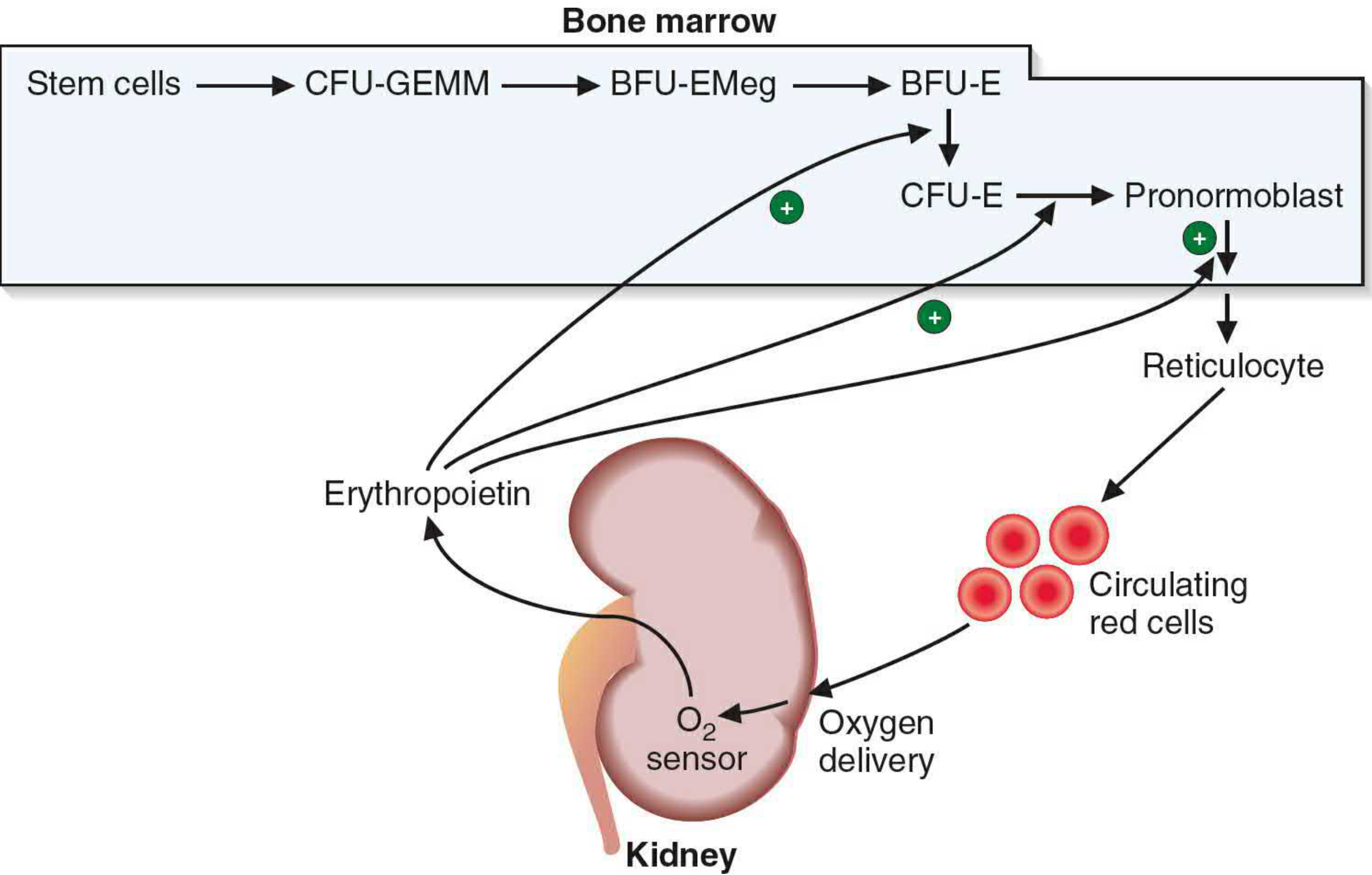
The progression from pluripotent stem cell to mature RBC follows this sequence:
Pluripotent Hematopoietic Stem Cell (HSC)
        ↓
Common Myeloid Progenitor (CMP)
        ↓
CFU-GEMM (Colony-Forming Unit - Granulocyte, Erythroid, Monocyte, Megakaryocyte)
        ↓
BFU-EMeg → BFU-E (Burst-Forming Unit - Erythroid)
        ↓
CFU-E (Colony-Forming Unit - Erythroid)   ← EPO acts here primarily
        ↓
Pronormoblast / Proerythroblast (first morphologically recognizable cell)
(Basic Medical Biochemistry, p.1536)
The diagram below from the textbook illustrates this pathway with the role of erythropoietin (EPO):
4. Morphological Stages of Erythroid Maturation
Each stage below occurs in the bone marrow. As cells mature, they decrease in size, their nucleus shrinks and condenses, and hemoglobin progressively accumulates.
Stage 1: Proerythroblast (Pronormoblast)
- Largest cell in the erythroid series
- Large nucleus with prominent nucleoli (2-4) and dense chromatin
- Cytoplasm is intensely basophilic (due to ribosomes)
- No hemoglobin detectable yet
- Golgi apparatus appears as a light-staining area
- Undergoes mitotic division → gives rise to basophilic erythroblasts
Stage 2: Basophilic Erythroblast (Early Normoblast)
- Smaller than proerythroblast (arises by mitotic division)
- Nucleus smaller, chromatin more condensed; nucleoli less prominent
- Cytoplasm shows strong basophilia due to large numbers of ribosomes involved in hemoglobin synthesis
- Hemoglobin synthesis begins here
- Still capable of mitosis
Stage 3: Polychromatophilic Erythroblast (Intermediate Normoblast)
- Hemoglobin accumulates progressively → cytoplasm begins staining with both blue (basophilic) and pink (eosinophilic) = "polychromasia" or gray-blue color
- Nucleus smaller than basophilic erythroblast; heterochromatin coarser
- Ribosome numbers decreasing, hemoglobin content increasing
- This is the last stage capable of mitosis (undergoes 1-2 more divisions)
Stage 4: Orthochromatic Erythroblast (Late Normoblast)
- Nucleus extremely condensed and pyknotic (ink-dot nucleus)
- Cytoplasm predominantly eosinophilic (pink) due to high hemoglobin content
- Slightly larger than a mature erythrocyte
- No longer capable of division
- The condensed nucleus is extruded from the cell → cell becomes a reticulocyte
- Nuclear extrusion is facilitated by macrophages in the "erythroblastic island"
Stage 5: Reticulocyte (Polychromatophilic Erythrocyte)
- Anucleate cell (nucleus has been expelled)
- Still contains residual ribosomes and mRNA → continues hemoglobin synthesis for 1-2 days
- Ribosomes give a slight blue tinge compared to mature RBCs (visible on supravital staining with brilliant cresyl blue as a "reticulum")
- Released from bone marrow into circulation
- Matures in the spleen over 1-2 days, where ribosomes and mRNA are removed
- Normal reticulocyte count: 0.5-1.5% of circulating RBCs
Stage 6: Mature Erythrocyte
- Biconcave disc, ~7-8 µm diameter
- No nucleus, no organelles
- Packed with hemoglobin (~280-340 million molecules per cell)
- Life span: ~120 days
- After ~120 days, senescent RBCs are phagocytosed by macrophages in the spleen, bone marrow, and liver (reticuloendothelial system)
5. Summary of Morphological Changes During Maturation
| Feature | Proerythroblast → Mature RBC |
|---|---|
| Cell size | Decreases (largest → smallest) |
| Nucleus | Present, large → progressively condensed → extruded |
| Nucleoli | Present → disappear |
| Cytoplasm color | Basophilic → polychromatophilic → eosinophilic |
| Ribosomes | Many → decreasing → absent in mature RBC |
| Hemoglobin | Absent → progressively increases → fully packed |
| Mitotic ability | Present in stages 1-3 → lost from stage 4 onwards |
6. Regulation of Erythropoiesis
A. Erythropoietin (EPO) - Primary Regulator
- EPO is a 30.4-kDa glycoprotein hormone produced predominantly by the peritubular interstitial cells of the kidney (90%), with a minor contribution from the liver (10%)
- Acts via a negative feedback loop:
- Reduced tissue oxygenation (anemia, hypoxia, high altitude) → kidney O2 sensor detects low O2 delivery → increased EPO production
- EPO acts on specific EPO receptors (EPO-Rs) on BFU-E and CFU-E progenitors in bone marrow
- Stimulates proliferation and differentiation of erythroid progenitors and accelerates maturation
- Under normal conditions: ~2.5-3 million RBCs/second are produced to replace 1% of daily turnover
- EPO production is mediated by HIF-1α (Hypoxia-Inducible Factor) - stabilized during hypoxia, drives EPO gene transcription
(Brenner and Rector's The Kidney, p.2465)
B. Other Hormones and Growth Factors
- Androgens - stimulate EPO production and directly stimulate erythroid progenitors (explains higher RBC count in males)
- Thyroid hormones - increase basal metabolic rate → increase O2 demand → stimulate erythropoiesis
- Growth hormone / IGF-1 - supportive role in erythroid progenitor proliferation
- IL-3, GM-CSF, SCF (Stem Cell Factor) - early-acting cytokines that support the BFU-E stage
- IL-3, IL-4, IL-9 - act at various stages alongside EPO
7. Nutritional Requirements for Erythropoiesis
| Nutrient | Role | Deficiency Effect |
|---|---|---|
| Iron | Core component of heme in hemoglobin | Iron deficiency anemia (microcytic, hypochromic) |
| Vitamin B12 (Cobalamin) | Required for DNA synthesis (thymidylate synthesis via folate cycle) | Megaloblastic anemia; impaired nuclear maturation |
| Folic Acid | Required for DNA synthesis (one-carbon transfer reactions) | Megaloblastic anemia |
| Vitamin B6 (Pyridoxine) | Cofactor in heme synthesis (ALA synthase reaction) | Sideroblastic anemia |
| Vitamin C | Aids iron absorption; antioxidant protection of RBCs | Mild anemia |
| Copper | Required for ferroxidase activity (ceruloplasmin) for iron mobilization | Anemia with iron accumulation |
| Protein/amino acids | Globin chain synthesis | Protein malnutrition anemia |
8. Kinetics of Erythropoiesis
- Mitoses occur in proerythroblasts, basophilic erythroblasts, and polychromatophilic erythroblasts
- Each normoblast undergoes 4 mitotic divisions from pronormoblast stage
- Takes approximately 7 days for a newly formed basophilic erythroblast's progeny to reach circulation
- Bone marrow is not a significant storage site for erythrocytes - RBCs are released almost immediately upon formation
- In high EPO states, transit time can be shortened (stress erythropoiesis), releasing reticulocytes earlier - called shift reticulocytes
(Histology: A Text and Atlas, p.746)
9. Fate of Senescent RBCs (End of Erythropoiesis Cycle)
- After ~120 days, RBCs become senescent (membrane changes, decreased ATP, loss of deformability)
- Phagocytosed by macrophages in spleen, bone marrow, and liver (reticuloendothelial system)
- Breakdown products:
- Globin → hydrolyzed to amino acids → recycled
- Iron → released from heme → stored as ferritin or hemosiderin in spleen → recycled for new hemoglobin synthesis
- Porphyrin ring of heme → converted to bilirubin → bound to albumin → transported to liver → conjugated → excreted as bilirubin glucuronide in bile
10. Erythroblastic Island - Microenvironmental Niche
- Erythroid precursors mature in specialized clusters in bone marrow called erythroblastic islands
- A central macrophage is surrounded by developing erythroblasts at various stages
- The macrophage:
- Provides iron directly to developing erythroblasts
- Phagocytoses extruded nuclei from late normoblasts
- Secretes cytokines supporting erythroid development
11. Clinical Correlations
| Condition | Mechanism | Effect |
|---|---|---|
| Chronic Kidney Disease | Reduced EPO production | Normocytic normochromic anemia |
| High altitude / Hypoxia | Increased EPO → increased erythropoiesis | Polycythemia |
| Iron deficiency | Insufficient heme synthesis | Microcytic hypochromic anemia |
| Vitamin B12 / Folate deficiency | Impaired DNA synthesis → large cells | Megaloblastic anemia |
| Polycythemia vera | Autonomous HSC proliferation (JAK2 mutation) | Overproduction of RBCs |
| Aplastic anemia | Bone marrow failure | Pancytopenia |
| EPO doping (athletes) | Exogenous EPO increases RBC mass | Increased O2-carrying capacity |
Sources:
- Basic Medical Biochemistry - A Clinical Approach, 6e, p.1536
- Histology: A Text and Atlas with Correlated Cell and Molecular Biology, pp.746, 776-777
- Brenner and Rector's The Kidney, p.2465
- Goodman & Gilman's Pharmacological Basis of Therapeutics
This is a shared conversation. Sign in to Orris to start your own chat.