
Vitamin deficiency deficiency
Which vitamin deficiency are you interested in?
"vitamin D deficiency"[MeSH Terms] AND treatment
vitamin D deficiency rickets osteomalacia bone
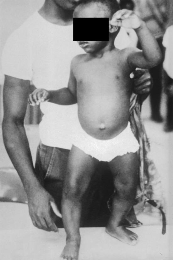
This clinical photograph displays the classic musculoskeletal manifestations of vitamin D deficiency rickets in a young child. The most prominent feature is significant genu varum (bowing of the legs), where the weight-bearing long bones show lateral curvature due to impaired mineralization of the growth plate and osteoid matrix. Additionally, the child exhibits a distended abdomen (potbelly), which is a common clinical sign associated with rickets due to hypotonia of the abdominal muscles. The upper extremities appear thin, suggesting poor muscle development or nutritional deficiency. This archival image serves as a pedagogical tool for identifying systemic skeletal deformities resulting from metabolic bone disease. Key educational concepts include the relationship between vitamin D deficiency, calcium homeostasis, and the softening of bones (osteomalacia) during childhood development. The clinical relevance extends to pediatrics, endocrinology, and public health nutrition.
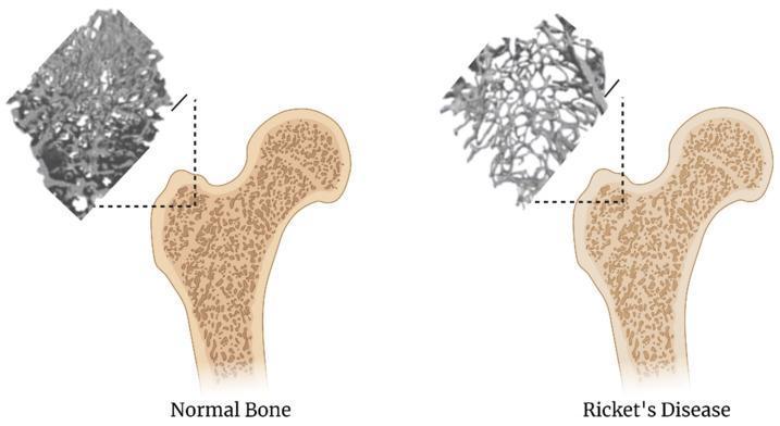
This comparison diagram illustrates the architectural differences between normal bone and bone affected by Rickets (osteomalacia) at the proximal femur. The image consists of two panels: 'Normal Bone' on the left and 'Ricket's Disease' on the right. Each panel features a cross-sectional anatomical diagram of the femoral head and neck, paired with a high-magnification micro-CT or radiographic inset demonstrating trabecular microarchitecture. In the normal bone, the trabeculae are thick, highly interconnected, and densely packed, indicating robust mineralization and structural integrity. Conversely, the Ricket's disease panel shows a significant reduction in bone mineral density. The trabeculae are visibly thinner, sparser, and exhibit increased porosity with larger marrow spaces, reflecting impaired mineralization and ossification typical of Vitamin D deficiency. A 1 mm scale bar is provided for reference. This educational visual is designed to highlight the pathophysiological impact of metabolic bone diseases on skeletal microstructure, useful for clinical training in orthopedics, endocrinology, and radiology.
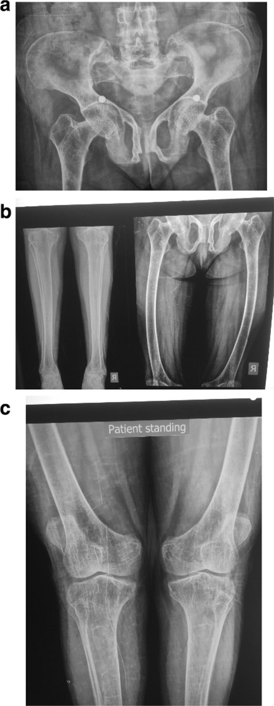
This composite diagnostic image comprises three sets of skeletal radiographs (a, b, and c) demonstrating manifestations of metabolic bone disease, consistent with Vitamin D resistant rickets. Image (a) displays an anteroposterior (AP) pelvic view showing diffuse osteopenia, particularly around the hip joints. The pelvic brim, pubic bones, and acetabular fossa exhibit abnormal, softened morphologies, though the hip joint spaces remain preserved. Image (b) consists of leg radiographs showing significant lateral bowing of the bilateral femoral shafts (bowing deformity) and mild osteopenia most prominent at the ankle joints. Image (c) provides a detailed AP view of the knees while standing, revealing bilateral genu valgus (knock-knee) deformity and prominent femoral epicondyles. There is notable periarticular osteopenia around the knee joints. The collection illustrates characteristic skeletal complications of chronic mineral deficiency, including bowing of weight-bearing long bones and structural pelvic remodeling, making it a key educational tool for identifying radiological signs of metabolic rickets and osteomalacia in a clinical context.
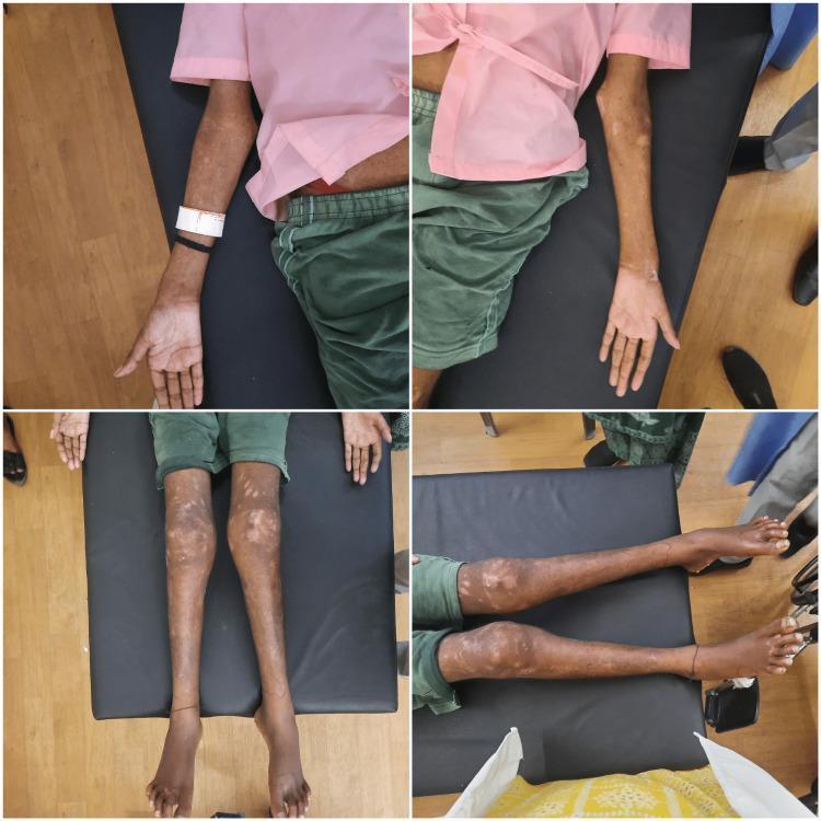
This four-panel clinical photograph illustrates multisystemic manifestations of a metabolic bone disease, likely rickets or osteomalacia, in a young patient. The top panels show the upper extremities, highlighting bilateral widening and enlargement of the elbow and wrist joints, which are hallmark signs of metaphyseal expansion. The bottom panels display the lower extremities in supine and lateral views, demonstrating significant genu valgum (knock-knee deformity) and flexion contractures at the knees. Accompanying these musculoskeletal findings are dermatological lesions characterized by diffuse, mottled hypopigmented and depigmented patches distributed symmetrically across the arms, legs, and periarticular regions. The patient exhibits an ectomorphic build with noticeable muscle wasting. These combined features serve as educational indicators for systemic conditions involving calcium/phosphorus dysregulation and chronic skin involvement, emphasizing the clinical presentation of vitamin D deficiency or similar metabolic disorders in an adolescent or young adult.
Vitamin D Deficiency
What is Vitamin D?
- Vitamin D3 (cholecalciferol) - synthesized in the skin from 7-dehydrocholesterol under UV-B radiation
- Vitamin D2 (ergocalciferol) - obtained from dietary plant sources
Causes of Vitamin D Deficiency
| Category | Specific Causes |
|---|---|
| Impaired skin production | Reduced sunlight exposure, dark skin, aging, sunscreen use |
| Dietary absence / malabsorption | Short gut syndrome, gastric bypass, fat malabsorption, terminal ileal disease |
| Accelerated loss/metabolism | Drugs: barbiturates, phenytoin, rifampin; CYP3A4 gain-of-function mutations |
| Impaired enterohepatic circulation | Terminal ileal disease, nephrotic syndrome |
| Impaired 25-hydroxylation | Severe liver disease, isoniazid, 25-hydroxylase mutations |
| Impaired 1α-hydroxylation | Hypoparathyroidism, CKD, FGF23 excess, ketoconazole, oncogenic osteomalacia |
| Target organ resistance | Vitamin D receptor (VDR) mutations, phenytoin |
| Other | Obesity (poorly understood mechanism) |
The elderly and nursing home residents are particularly at risk since both skin synthesis efficiency and intestinal absorption decline with age. - Harrison's Principles of Internal Medicine 22E
Clinical Features
In Children - Rickets
- Skeletal deformities: genu varum (bow legs) or genu valgum (knock knees), enlarged wrists/ankles (metaphyseal widening)
- Craniotabes - soft skull bones in infants
- Rachitic rosary - beading of costochondral junctions
- Harrison's sulcus - groove along the lower chest
- Pot belly (abdominal muscle hypotonia)
- Delayed tooth eruption, dental enamel defects
- Growth retardation
In Adults - Osteomalacia
- Bone pain - diffuse, often worse with weight bearing (back, hips, legs)
- Muscle weakness - proximal myopathy causing waddling gait
- Fragility fractures - especially pelvis, femoral neck, vertebrae
- Looser's zones (pseudofractures) - pathognomonic stress fractures seen on X-ray
- Hypocalcemia can cause tetany, paresthesias, Chvostek's and Trousseau's signs
Laboratory Findings
| Parameter | Finding in Vitamin D Deficiency |
|---|---|
| Serum 25(OH)D | Low (<20 ng/mL = deficient; 20-29 ng/mL = insufficient) |
| Serum calcium | Low or normal (compensated) |
| Serum phosphate | Low |
| PTH | Elevated (secondary hyperparathyroidism) |
| Alkaline phosphatase | Elevated (bone formation marker) |
| Urinary calcium | Low |
Radiological Findings
- Rickets: cupping, fraying, and widening of metaphyses; bowing of long bones; delayed bone age
- Osteomalacia: generalized osteopenia; Looser's zones (pseudofractures, especially at ribs, pubic rami, femoral neck, scapula)
Treatment
Vitamin D Supplementation
- Deficiency treatment: Vitamin D3 (cholecalciferol) 50,000 IU weekly for 8 weeks, then maintenance
- Maintenance: 1,500-2,000 IU/day (higher in obese, malabsorption, or elderly patients)
- Rickets: higher doses under pediatric/endocrine guidance
Special Situations
| Condition | Treatment |
|---|---|
| Chronic kidney disease | Calcitriol (active form, bypasses renal activation) |
| Hereditary 1α-hydroxylase deficiency | Calcitriol or alfacalcidol |
| VDR mutations (hereditary vitamin D-resistant rickets) | High-dose calcitriol + calcium infusions |
| Drug-induced (phenytoin, rifampin) | High-dose vitamin D supplementation |
- Calcium supplementation (1,000-1,500 mg/day) is given alongside vitamin D to fully correct osteomalacia
- Treat the underlying cause (e.g., correct malabsorption, adjust medications)
Broader Health Implications (Recent Evidence)
- Type 2 diabetes: A 2024 meta-analysis (PMID 39355942) found vitamin D supplementation may modestly improve glycaemic control in T2DM
- Preeclampsia: A 2024 meta-analysis (PMID 39716171) supports supplementation in pregnancy to reduce preeclampsia risk
- Depression: A 2025 systematic review (PMID 40322928) found a potential association between low vitamin D and depression in adults
Prevention
- Sunlight exposure: 10-30 minutes of midday sun several times per week on arms and legs
- Dietary sources: fatty fish (salmon, mackerel, tuna), egg yolks, fortified milk/cereals, cod liver oil
- Supplementation: recommended for infants (400 IU/day from birth), elderly (800-1000 IU/day), and high-risk groups