Why LAP score decrease in CML

Reading File
Finding Sources
Finding Sources
Finding Sources
Finding Sources
Reading File
Reading File
Finding Sources
Finding Sources
Finding Sources
Searching Images

LAP score leukocyte alkaline phosphatase CML neutrophil staining
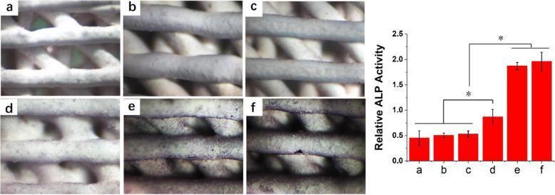
Educational comparison chart and graph detailing alkaline phosphatase (ALP) staining on 3D-printed hydroxyapatite (HA) scaffolds. The visual displays six panel images (a–f) showing MC3T3-E1 cells cultured for 7 days under various conditions: (a) HA only, (b) PSI10@HA, (c) PSI10/HABP@HA, (d) BMP2-MP@HA, (e) BMP2-MP/HABP@HA, and (f) a combination of PSI10/HABP and BMP2-MP/HABP@HA. Panels a–c show minimal staining, indicating baseline ALP activity. Panel d exhibits moderate purple staining, while panels e and f show significant, dense purple staining across the scaffold filaments, indicating robust osteogenic differentiation. Adjacent to the images is a red bar graph quantifying 'Relative ALP Activity' across the same groups (a–f), showing a progressive increase in activity that peaks in groups e and f. Asterisks indicate statistical significance (p < 0.05). This material illustrates early bone matrix mineralization and the efficacy of using HA-binding peptides (HABP) to deliver bone morphogenetic protein-2 (BMP2) for enhanced osteoinduction in regenerative medicine.
Educational comparison chart and graph detailing alkaline phosphatase (ALP) staining on 3D-printed hydroxyapatite (HA) scaffolds. The visual displays six panel images (a–f) showing MC3T3-E1 cells cultured for 7 days under various conditions: (a) HA only, (b) PSI10@HA, (c) PSI10/HABP@HA, (d) BMP2-MP@HA, (e) BMP2-MP/HABP@HA, and (f) a combination of PSI10/HABP and BMP2-MP/HABP@HA. Panels a–c show minimal staining, indicating baseline ALP activity. Panel d exhibits moderate purple staining, while panels e and f show significant, dense purple staining across the scaffold filaments, indicating robust osteogenic differentiation. Adjacent to the images is a red bar graph quantifying 'Relative ALP Activity' across the same groups (a–f), showing a progressive increase in activity that peaks in groups e and f. Asterisks indicate statistical significance (p < 0.05). This material illustrates early bone matrix mineralization and the efficacy of using HA-binding peptides (HABP) to deliver bone morphogenetic protein-2 (BMP2) for enhanced osteoinduction in regenerative medicine.

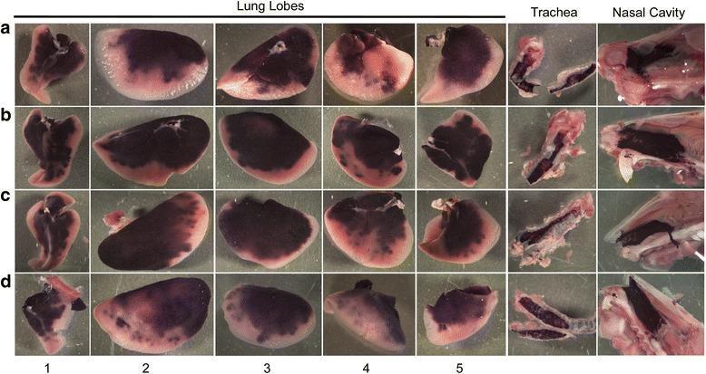
A comparative series of gross pathological images (rows a–d) showing human placental alkaline phosphatase (hPLAP) staining in the respiratory tract of mice following viral vector administration via a modified intranasal technique. The images display the nasal cavity, trachea, and five isolated lung lobes: (1) accessory, (2) left, (3) right cranial, (4) right caudal, and (5) right middle. Intense dark purple-black staining, representing successful hPLAP transgene expression, is consistently observed in the nasal epithelium and trachea across all mice. Within the lower respiratory tract, staining distribution is heterogeneous and multifocal. The lung lobes exhibit high-intensity staining primarily in the proximal and mid-distal regions, appearing as dense pigmented patches that contrast with the pale, unstained distal parenchymal edges. Variation in transduction efficiency is visible between individual mice (a–d) and across different lung lobes, ranging from nearly uniform staining in some mice to patchy, focal distribution in others. This visual data serves to evaluate the efficacy of modified intranasal delivery for targeting both upper and lower respiratory tract tissues in gene therapy models.
A comparative series of gross pathological images (rows a–d) showing human placental alkaline phosphatase (hPLAP) staining in the respiratory tract of mice following viral vector administration via a modified intranasal technique. The images display the nasal cavity, trachea, and five isolated lung lobes: (1) accessory, (2) left, (3) right cranial, (4) right caudal, and (5) right middle. Intense dark purple-black staining, representing successful hPLAP transgene expression, is consistently observed in the nasal epithelium and trachea across all mice. Within the lower respiratory tract, staining distribution is heterogeneous and multifocal. The lung lobes exhibit high-intensity staining primarily in the proximal and mid-distal regions, appearing as dense pigmented patches that contrast with the pale, unstained distal parenchymal edges. Variation in transduction efficiency is visible between individual mice (a–d) and across different lung lobes, ranging from nearly uniform staining in some mice to patchy, focal distribution in others. This visual data serves to evaluate the efficacy of modified intranasal delivery for targeting both upper and lower respiratory tract tissues in gene therapy models.

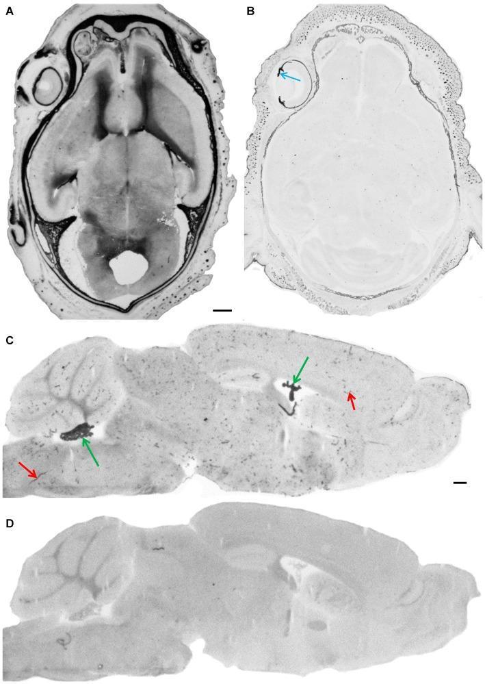
This set of diagnostic images demonstrates the presence and experimental blockade of endogenous alkaline phosphatase (AP) activity in mammalian brain tissue. Images A and B show horizontal sections of a mouse embryo head (E18). In A, standard fixation reveals intense AP staining throughout the meninges and brain parenchyma. In B, alkaline fixation with EDTA successfully inactivates endogenous AP, leaving only pigmented structures like the iris (blue arrow) visible. Images C and D display parasagittal sections of an adult brain. In C, positive AP staining is localized in the choroid plexus (green arrows) and cerebral blood vessels (red arrows), illustrating the enzyme's distribution in the blood-brain barrier and blood-cerebrospinal fluid barrier interfaces. Image D shows the corresponding section with inactivated AP activity, resulting in a loss of specific staining in these vascular and epithelial structures. Scale bars indicate 0.5 mm. The content illustrates histological techniques for blocking background enzymatic activity to improve the specificity of immunohistochemical staining for tight junction proteins.

This set of diagnostic images demonstrates the presence and experimental blockade of endogenous alkaline phosphatase (AP) activity in mammalian brain tissue. Images A and B show horizontal sections of a mouse embryo head (E18). In A, standard fixation reveals intense AP staining throughout the meninges and brain parenchyma. In B, alkaline fixation with EDTA successfully inactivates endogenous AP, leaving only pigmented structures like the iris (blue arrow) visible. Images C and D display parasagittal sections of an adult brain. In C, positive AP staining is localized in the choroid plexus (green arrows) and cerebral blood vessels (red arrows), illustrating the enzyme's distribution in the blood-brain barrier and blood-cerebrospinal fluid barrier interfaces. Image D shows the corresponding section with inactivated AP activity, resulting in a loss of specific staining in these vascular and epithelial structures. Scale bars indicate 0.5 mm. The content illustrates histological techniques for blocking background enzymatic activity to improve the specificity of immunohistochemical staining for tight junction proteins.

Loading Image
This is a shared conversation. Sign in to Orris to start your own chat.