
Head posture and ocular posture
Reading File
Head posture and ocular posture are topics in ophthalmology/strabismology. Let me search the medical library now.
Searching sources...
Finding Sources
Finding Sources
Finding Sources
Finding Sources
Finding Sources
Finding Sources
Good - the primary source is Kanski's Clinical Ophthalmology. Let me read the most relevant sections.
Reading File
Finding Sources
Reading File
Reading File
Reading File
Reading File
Now let me fetch the key image for head posture and also get the nystagmus treatment section:
Loading Image
Reading File
I now have comprehensive material. Let me compile the full answer.
Head Posture and Ocular Posture
Overview
In ophthalmology, head posture refers to the position a patient voluntarily or involuntarily adopts in relation to the visual axes, while ocular posture (also called the primary position) refers to the resting alignment of the eyes. The two are intimately linked: abnormal ocular posture (from muscle palsies, nystagmus, or strabismus) drives the adoption of an abnormal head posture to compensate.
Compensatory Head Posture (CHP)
The CHP is a motor adaptation to maintain binocular single vision (BSV) and eliminate diplopia by repositioning the head so the eyes deviate away from the field of action of the weakened muscle.
"Motor adaptation involves the adoption of a compensatory head posture (CHP) and occurs primarily in children with congenitally abnormal eye movements who use the CHP to maintain BSV. In these children loss of a CHP may indicate loss of binocular function and the need for surgical intervention." - Kanski's Clinical Ophthalmology, 10th ed.
The head is turned into the direction of the field of action of the weak muscle, so the eyes automatically deviate away from that field.
Three Types of CHP
1. Face Turn (Horizontal CHP)
- Adopted to control a purely horizontal deviation
- The face turns toward the side of the weak muscle, which rotates the eyes in the opposite direction
- Example: Left lateral rectus palsy → diplopia in left gaze → face turns left → eyes deviate right, away from the area of diplopia
- A face turn may also be adopted in vertical muscle paresis to avoid the side of greatest vertical deviation (e.g., right superior oblique weakness → face turns left)
2. Head Tilt (Torsional/Vertical CHP)
- Adopted to compensate for torsional and/or vertical diplopia
- Example: Right superior oblique weakness → right eye is relatively elevated and extorted → head tilts to the left (towards the hypotropic eye). This:
- Reduces vertical image separation, enabling fusion
- Reduces torsional component by invoking the righting reflex (placing the extorted eye in a position demanding extorsion)
3. Chin Elevation or Depression (Vertical CHP)
- Chin elevation compensates for weakness of an elevator muscle
- Chin depression compensates for weakness of a depressor muscle
- Also used to minimize horizontal deviation in A or V patterns (alphabet patterns)
CHP in Specific Conditions
Fourth Nerve (Trochlear) Palsy
The classic example of CHP. In a left superior oblique paresis:
- Left hypertropia in primary position, increasing on right gaze
- Limitation of left depression in adduction
- Left extorsion
- CHP: contralateral head tilt to the right, slight chin depression, slight face turn to the right
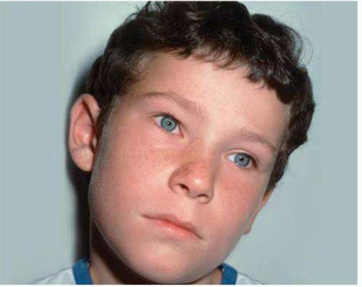
Fig. 18.17: Compensatory head posture in a right fourth nerve palsy - the head tilts to the left (away from the affected side). - Kanski's Clinical Ophthalmology, 10th ed.
Tip: Examination of old photographs for the presence of a CHP may help distinguish congenital from acquired fourth nerve palsy.
Brown Syndrome
- Brown syndrome causes limited elevation in adduction
- Mild/moderate: No hypotropia in primary position; down-shoot in adduction
- Severe: Hypotropia in primary position + down-shoot in adduction + chin-up head posture + face turn away from the affected eye
- Congenital cases do not usually require treatment as long as binocular function is maintained with an acceptable head posture
Nystagmus
- Most nystagmus has a null point - a direction of gaze where the oscillation is minimal
- Patients adopt a CHP to keep gaze at the null point in the primary position
- Surgery for nystagmus with a null point aims to move the null point so that the eyes and face are straight (Kestenbaum-Anderson procedure or similar)
Ocular Posture: Primary, Secondary, and Tertiary Positions
| Position | Definition |
|---|---|
| Primary position | Eyes looking straight ahead, head erect |
| Secondary positions | Pure elevation, depression, right gaze, left gaze |
| Tertiary positions | Oblique gaze directions (e.g. up-right, down-left) - require torsional adjustment |
Ocular posture is maintained by the coordinated action of the six extraocular muscles under neural control via CN III (oculomotor), CN IV (trochlear), and CN VI (abducens), integrated through the medial longitudinal fasciculus (MLF). The MLF connects vestibular nuclei to the nuclei governing conjugate eye movements, also influencing head posture via its superior vestibular nucleus contributions.
Sensory vs. Motor Adaptations to Abnormal Ocular Posture
| Adaptation | Type | Mechanism |
|---|---|---|
| Suppression | Sensory | Cortical suppression of the deviated eye's image to prevent diplopia; occurs mainly in children |
| Anomalous retinal correspondence (ARC) | Sensory | A new false correspondence develops between the fovea of the fixing eye and an extrafoveal point of the deviating eye |
| Compensatory head posture | Motor | Head repositioned so that eyes are in the position of maximum single vision |
Clinical Significance of CHP
- Loss of CHP in a child with congenital strabismus may signal decompensation of binocular function and warrant surgical review
- Adults may not be aware of their CHP, especially in long-standing congenital conditions
- Strabismus surgery can eliminate diplopia, expand binocular visual field, and correct a disfiguring CHP
- For nystagmus: recession of all horizontal recti has been successful in reducing amplitude in some patients without a significant null point
Source: Kanski's Clinical Ophthalmology: A Systematic Approach, 10th Edition - Chapters 18 (Strabismus) and 19 (Neuro-ophthalmology)
Demonstration
I'm not sure what you'd like me to demonstrate. Could you clarify what you have in mind?
Asking for Preferences
What would you like a demonstration of?
How to demonstrate head and occular posture
The clinical demonstration (examination) of head and ocular posture involves specific observation and testing techniques. Let me pull the detailed methodology from the textbook.
Searching sources...
Finding Sources
Finding Sources
Finding Sources
Finding Sources
Finding Sources
Finding Sources
Reading File
Finding Sources
Reading File
Reading File
Reading File
Reading File
Now let me fetch a couple of key clinical images:
Loading Image
Loading Image
Excellent - I now have everything needed for a complete clinical demonstration guide.
How to Demonstrate Head and Ocular Posture - Clinical Examination
Step 1: Observe the Head Posture First (Before Asking the Patient to Move)
Approach the patient from a distance and observe naturally. Note:
| Feature | What to Look For |
|---|---|
| Face turn | Is the face turned left or right? |
| Head tilt | Is the head tilted to one shoulder? |
| Chin position | Is the chin elevated (chin-up) or depressed (chin-down)? |
| Combination | Many palsies produce a combination (e.g. tilt + turn + chin depression) |
The patient turns the head into the field of action of the weak muscle so the eyes automatically deviate away from it - Kanski's Clinical Ophthalmology, 10th ed.
- Chin-up posture → suggests bilateral elevation weakness or Brown syndrome
- Head tilt to left → suggests right superior oblique palsy
- Face turn to left → suggests left lateral rectus weakness
Always examine old photographs to determine if the posture is longstanding (congenital) vs. new (acquired) - Wills Eye Manual, 7th ed.
Step 2: Assess Ocular Posture in Primary Position
With the patient's head held straight (neutralize the compensatory posture):
- Sit at eye level, 33 cm away with a penlight or fixation target
- Observe the corneal light reflex (Hirschberg test) - both reflexes should be centred symmetrically
- Look for any obvious deviation (esotropia, exotropia, hypertropia, hypotropia)
- Note any torsion (extorsion or intorsion) by observing the iris relative to the horizon
Step 3: Cover-Uncover Test
The most objective way to demonstrate manifest deviation (tropia):
- Ask patient to fixate a distant target (6 m)
- Cover one eye - watch the uncovered eye for movement:
- If the uncovered eye moves to take up fixation → tropia present
- No movement → no manifest deviation in that eye
- Uncover - watch the previously covered eye for movement:
- Movement indicates phoria (latent deviation) or tropia
- Repeat for the other eye
The higher eye comes down after being uncovered in hypertropia - Wills Eye Manual, 7th ed.
Step 4: The Parks-Bielschowsky Three-Step Test
Used specifically to identify a single paretic vertical muscle (especially CN IV palsy):
Step 1 - Primary position
- Identify which eye is hypertropic using the cover test
- This narrows the weak muscle to: depressors of the higher eye (SO or IR) OR elevators of the lower eye (SR or IO)
Step 2 - Right and left gaze
- Determine in which direction of horizontal gaze the hypertropia is greater
- WOOG rule: Worse on Opposite Gaze = superior oblique weakness
- Superior oblique weakness → deviation worse on contralateral gaze
- This isolates the culprit to 2 of the 4 muscles from Step 1
Step 3 - Bielschowsky Head Tilt Test (BHTT)
- With the patient fixating a distant target (3 metres), tilt the head to one shoulder, then the other
- Observe whether the hypertropia increases or decreases
- BOOT rule: Better on Opposite Tilt = fourth nerve palsy confirmed
- In left CN IV palsy: hypertropia worsens on left head tilt, improves on right head tilt
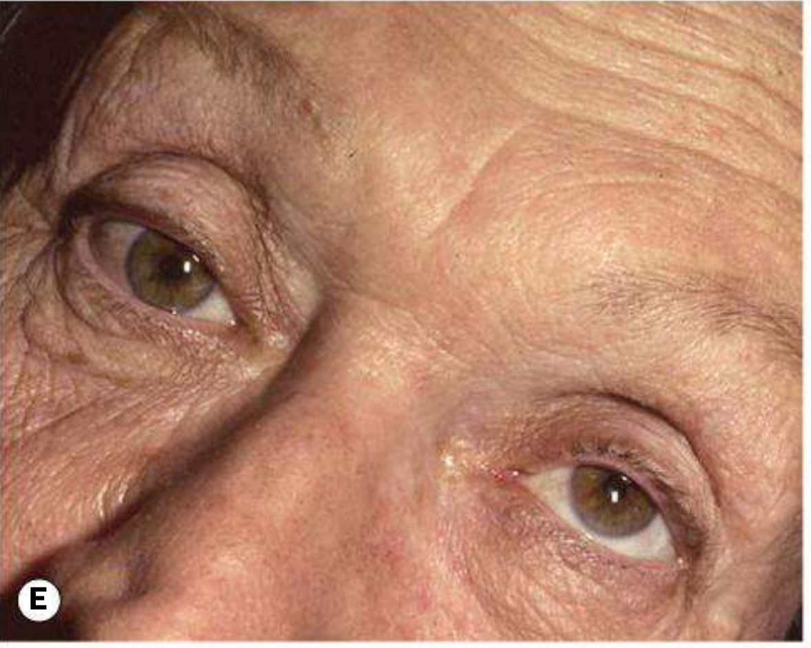
Fig. 19.70E: Positive Bielschowsky head tilt test - marked hypertropia on ipsilateral head tilt in left fourth nerve palsy. - Kanski's Clinical Ophthalmology, 10th ed.
Why does BHTT work? When the head tilts, the vestibulo-ocular reflex (righting reflex) invokes the intortor and extortor muscles to keep the eye level. If the intortor (superior oblique) is weak, tilting the head to its side demands intorsion it cannot provide - causing the eye to elevate, worsening the hypertropia.
Step 5: Assess Ocular Motility in Nine Positions of Gaze
Systematically move a target through 9 positions:
Up-Left Up Up-Right
Left Primary Right
Down-Left Down Down-Right
At each position:
- Look for limitation of movement (under-action of a muscle)
- Look for over-action (contralateral synergist over-shooting)
- Ask the patient if diplopia occurs and in which direction it is worst
Step 6: Additional Tests for Ocular Posture
| Test | When to Use | How |
|---|---|---|
| Double Maddox Rod test | Suspected bilateral CN IV palsy or torsion | White rod before one eye, red rod before the other; ask if the lines appear parallel; >10° total excyclotorsion = bilateral SO paresis |
| Vertical fusional amplitude | Distinguish congenital from acquired CN IV | >6 prism diopters = congenital; ≤6 = acquired |
| Red glass test | Subjective diplopia assessment | Red glass over one eye; patient reports position of red vs. white light |
| Maddox Wing/Rod | Measure phoria | Tests dissociated ocular deviation at near |
Step 7: Bilateral CN IV Palsy - What to Look For
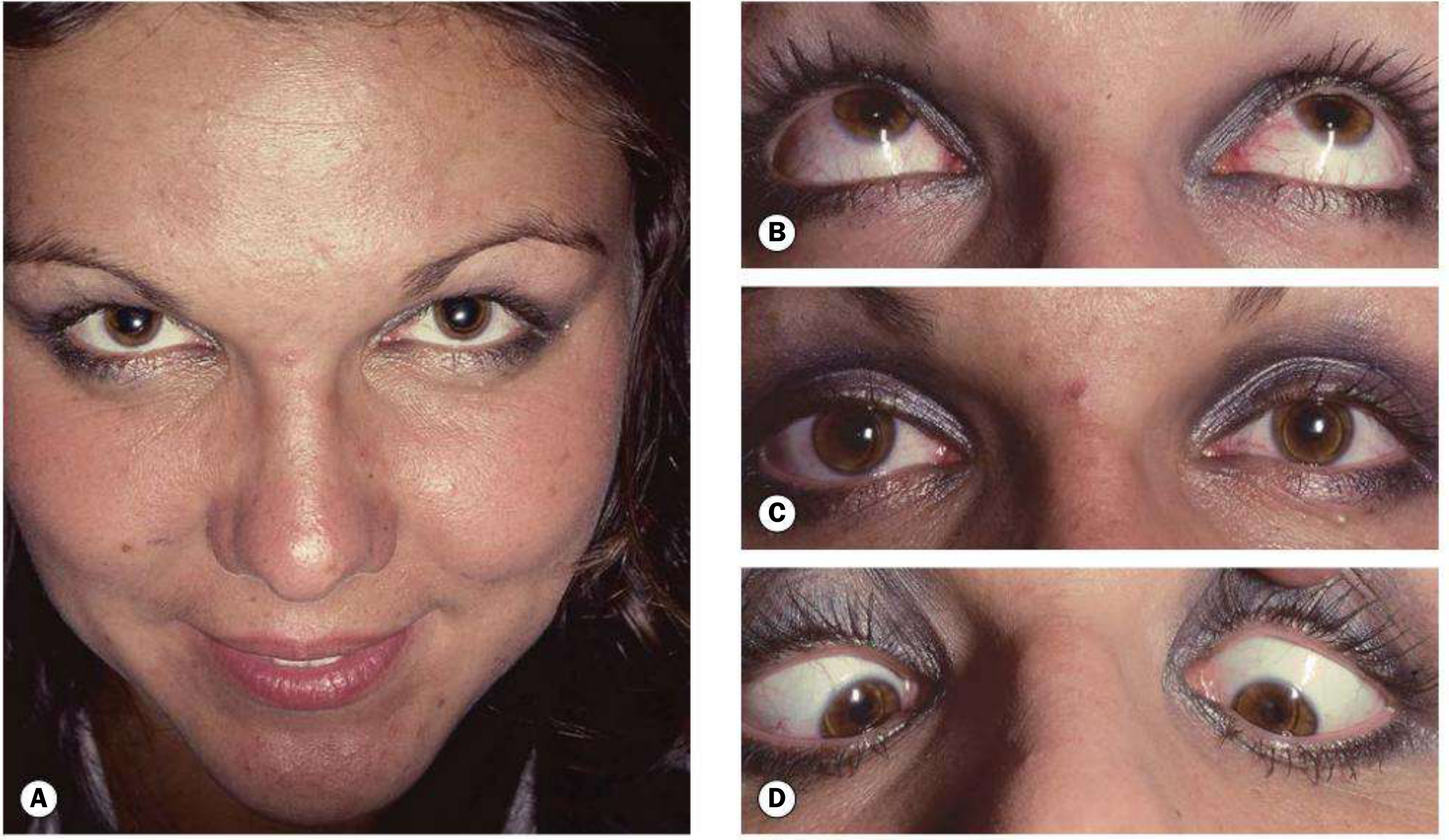
Fig. 19.71: Bilateral fourth nerve palsy showing chin-down compensatory head posture and V-pattern esotropia in downgaze. - Kanski's Clinical Ophthalmology, 10th ed.
- Right hypertropia in left gaze AND left hypertropia in right gaze
-
10° cyclodeviation on double Maddox rod
- "V" pattern esotropia (eyes cross more in downgaze)
- Bilaterally positive BHTT (hypertropia worsens on tilt to either shoulder)
Summary: CHP Direction vs. Likely Palsy
| Compensatory Head Posture | Likely Cause |
|---|---|
| Head tilt to left + face turn right + chin down | Right CN IV (superior oblique) palsy |
| Face turn to right | Left CN VI (lateral rectus) palsy |
| Chin-up | Bilateral elevator weakness; Brown syndrome (severe) |
| Chin-down | Bilateral CN IV palsy; depressor weakness |
| Head tilt to either side (bilateral BHTT +ve) | Bilateral CN IV palsy |
Sources:
- Kanski's Clinical Ophthalmology: A Systematic Approach, 10th ed., Chapters 18 & 19
- The Wills Eye Manual, 7th ed., Chapter 10.7 (Isolated Fourth Cranial Nerve Palsy)
- Bradley and Daroff's Neurology in Clinical Practice, Chapter 18
This is a shared conversation. Sign in to Orris to start your own chat.