
Solitary thyroid nodule
I'll start by reading the medical library skill, then search for comprehensive information on solitary thyroid nodules.
Reading File
Now let me search the medical library and PubMed in parallel for solitary thyroid nodule content.
Finding Sources
Finding Sources
Searching PubMed
"thyroid nodule" management evaluation
Excellent. Let me now read the primary textbook sections in parallel - Schwartz's Surgery on solitary thyroid nodule, K.J. Lee's Otolaryngology on evaluation and management, and Goldman-Cecil Medicine on clinical manifestations.
Reading File
Reading File
Reading File
Great content retrieved. Let me now read the Schwartz's continuation (which covers workup, FNAB, and management) and also fetch the differential diagnosis image.
Reading File
Reading File
Loading Image
Now let me read the management algorithm and FNAB/Bethesda section from Schwartz's and the Cummings rational approach continuation.
Reading File
Reading File
Excellent content. Let me fetch the management flowchart image and read the Bethesda classification continuation.
Loading Image
Reading File
I now have comprehensive content from multiple authoritative surgical textbooks. Let me compile the complete, well-structured answer.
Solitary Thyroid Nodule
A solitary thyroid nodule (STN) is a discrete lesion within the thyroid gland that is radiologically distinct from the surrounding thyroid parenchyma. STNs are present in approximately 4% of individuals in the United States, yet thyroid cancer has a much lower incidence (~40 new cases per 1 million). The core clinical challenge is distinguishing the rare malignant nodule from the common benign one.
Differential Diagnosis
The vast majority (~95%) of STNs are benign. The differential is broad:
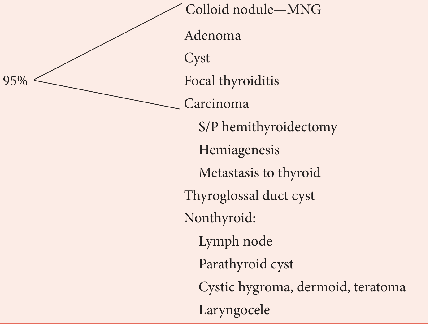
Thyroid origin (95%):
- Colloid nodule / multinodular goiter (MNG)
- Follicular adenoma
- Simple cyst
- Focal thyroiditis
- Carcinoma (papillary, follicular, medullary, anaplastic)
- Status post hemithyroidectomy
- Hemiagenesis
- Metastasis to thyroid
Non-thyroid origin:
- Thyroglossal duct cyst
- Lymph node
- Parathyroid cyst
- Cystic hygroma, dermoid, teratoma
- Laryngocele
(K.J. Lee's Essential Otolaryngology, Table 38-4)
History - Red Flags for Malignancy
| Less Concern | More Concern |
|---|---|
| Chronic stable examination | Age < 30 or > 60 years |
| Evidence of functional disorder (Hashimoto's, toxic nodule) | Male sex |
| Multinodular gland without dominant nodule | Rapid growth, pain |
| History of radiation exposure | |
| Family history of thyroid carcinoma | |
| Hard, fixed lesion | |
| Lymphadenopathy | |
| Vocal cord paralysis / hoarseness | |
| Size > 4 cm | |
| Aerodigestive tract compromise (stridor, dysphagia) |
(K.J. Lee's Essential Otolaryngology, Table 38-5)
Key history points:
- Radiation history: Low-dose therapeutic radiation (for tinea capitis, acne, tonsillar hypertrophy) confers ~40% risk of malignancy in any resulting nodule. Risk peaks 20-30 years after exposure. After Chernobyl, children showed a marked surge in papillary thyroid cancer within 4 years.
- Pain is usually hemorrhage into a benign nodule or thyroiditis; aching in the neck can also occur with medullary thyroid carcinoma (MTC).
- Hoarseness suggests possible recurrent laryngeal nerve (RLN) involvement by malignancy.
- Family history: MEN2 syndromes (medullary carcinoma + pheochromocytoma + hyperparathyroidism), Cowden disease (PTEN mutation - follicular thyroid carcinoma), Gardner syndrome (polyposis coli + papillary thyroid carcinoma).
(Schwartz's Principles of Surgery, 11th ed.; Cummings Otolaryngology)
Physical Examination
- Nodules are palpable when ≥1 cm; smaller ones are found incidentally on imaging.
- Firmness of the nodule increases the risk of malignancy 2-3 fold.
- Nodules >2 cm and solid lesions have higher incidence of carcinoma.
- Fixation to surrounding structures is strongly suspicious.
- Cervical lymphadenopathy adjacent to the nodule raises suspicion; it can be the only presenting sign of thyroid carcinoma.
- Check if the nodule moves with swallowing (thyroid origin) vs. non-thyroid.
- Assess for substernal extension: inferior aspect below the clavicle; Pemberton's maneuver (arms raised above head eliciting facial flushing + venous engorgement = thoracic inlet obstruction).
(Cummings Otolaryngology; Schwartz's Principles of Surgery)
Investigations
1. Serum TSH (First-Line)
This determines the diagnostic pathway:
- Suppressed TSH (hyperthyroid): Perform radionuclide scan - risk of malignancy is only ~1% in hot nodules.
- Elevated TSH (hypothyroid): Treat appropriately, then perform FNAC.
- Normal TSH (euthyroid): Most patients - proceed directly to ultrasound + FNAC.
2. Ultrasound (US) - Most Important Imaging
- Differentiates solid vs. cystic nodules.
- Identifies sonographic features raising malignancy risk: microcalcifications, hypoechoic solid nodule, irregular margins, taller-than-wide shape, absent halo, abnormal regional nodes.
- Guides FNAC for difficult-to-palpate nodules.
- Preferred for surveillance of benign nodules.
- Elastography: malignant nodules are stiffer (less deformable) than benign; newer technique, still being validated.
- Contrast-enhanced ultrasound (CEUS): A 2026 meta-analysis (Huo et al., PMID 42262145) shows emerging diagnostic utility for nodule characterization.
3. Fine Needle Aspiration Biopsy (FNAB) - Gold Standard
The cornerstone of evaluation. US guidance is recommended for:
- Difficult-to-palpate nodules
- Cystic/solid-cystic nodules recurring after prior aspiration
- Multinodular goiters
Adequate specimen = ≥6 follicular groups, each with ≥10-15 cells from ≥2 aspirates.
4. Radionuclide Scanning (¹²³I or ⁹⁹ᵐTc)
- NOT routine - use only if TSH is suppressed (to confirm hot/warm nodule) or after indeterminate FNAC.
- Cold nodules: 95% of all nodules; malignancy rate 10-15%.
- Hot nodules: malignancy rate ~4% (benign autonomous nodule usually).
- ⁹⁹ᵐTc: tests iodine transport only, faster (1 day), less radiation; cannot penetrate sternum.
- ¹²³I: tests transport AND organification; 2 days; preferred for substernal extension.
5. CT / MRI
- Not routine for thyroid nodule evaluation.
- Indicated for: substernal lesions, large/fixed tumors, suspected visceral compartment invasion, mediastinal adenopathy.
- Caution: iodinated CT contrast delays postoperative RAI therapy by 2-3 months.
- MRI superior to CT for distinguishing recurrent tumor vs. postoperative fibrosis.
6. Laboratory Tests
- Serum TSH - always
- Serum calcitonin - if MTC or MEN2 suspected (family history, FNAB suspicious for MTC)
- Serum thyroglobulin (Tg) - cannot differentiate benign from malignant preoperatively; used for post-thyroidectomy surveillance
- RET oncogene mutation testing - all patients with confirmed MTC; 24-hr urine VMA/metanephrines to rule out pheochromocytoma
Bethesda Classification of FNAB Results + Management
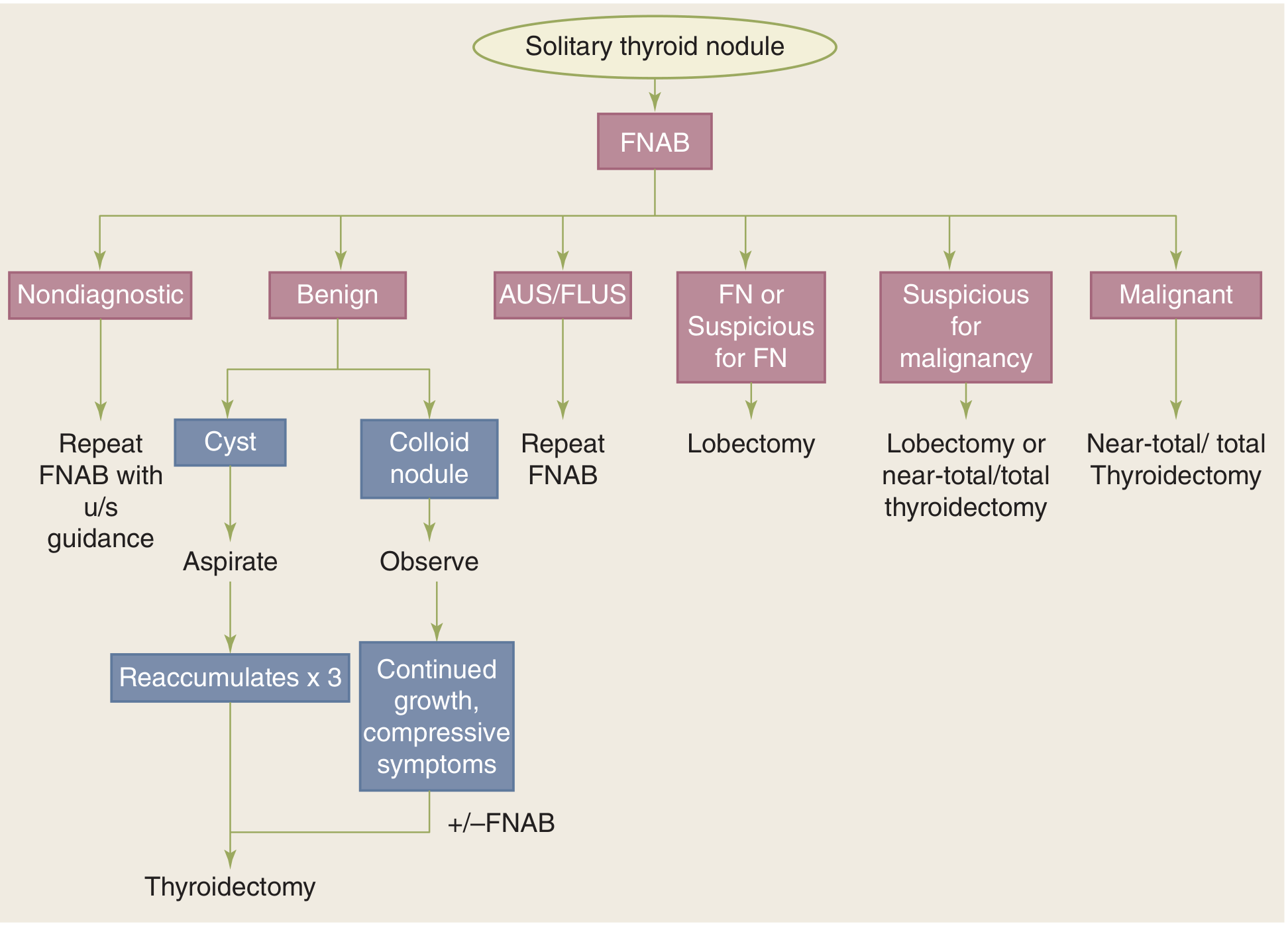
| Bethesda Category | Frequency | Malignancy Risk | Management |
|---|---|---|---|
| I - Nondiagnostic/Unsatisfactory | 2-20% | 1-4% | Repeat FNAB with US guidance |
| II - Benign | 60-70% | ~3% (false-negative) | Observe; surgery if growth/compressive symptoms |
| III - AUS/FLUS | 3-6% | 5-15% | Repeat FNAB; consider molecular testing |
| IV - Follicular Neoplasm (FN) | - | 15-35% | Diagnostic lobectomy |
| V - Suspicious for Malignancy | - | 60-75% | Lobectomy or near-total/total thyroidectomy |
| VI - Malignant | - | 97-99% | Near-total/total thyroidectomy |
(Schwartz's Principles of Surgery, 11th ed.; Cummings Otolaryngology)
Key details:
- Benign (II): Most common result. Includes colloid nodule, follicular adenoma, Hashimoto's, granulomatous thyroiditis. Cysts are aspirated; if they reaccumulate 3 times, thyroidectomy is recommended. Colloid nodules are observed; thyroidectomy if continued growth or compressive symptoms.
- FN/Suspicious for FN (IV): Up to 35% of Bethesda IV lesions are actually hyperplastic follicular proliferations, not true neoplasms. Lobectomy is preferred as it is both diagnostic and potentially therapeutic.
- Malignant (VI): Near-total or total thyroidectomy.
Molecular Marker Testing (for Indeterminate FNAB)
For Bethesda III/IV results, molecular testing helps avoid unnecessary surgery:
| Test | Role | Key Performance |
|---|---|---|
| Afirma Gene Expression Classifier | Rule-OUT malignancy | Sensitivity 92%, NPV 93% |
| ThyroSeq | Rule-OUT malignancy | NPV ~95% |
| ThyGenX/ThyraMIR | Rule-IN malignancy | Highest PPV ~66% |
(Cummings Otolaryngology)
Management Algorithm Summary
- History + Physical Exam - identify high-risk features
- Serum TSH
- Suppressed → Radionuclide scan → hot nodule = treat medically; cold nodule = FNAB
- Normal/elevated → Ultrasound + FNAB
- FNAB result guides surgical vs. conservative management (Bethesda criteria)
- Surgery for: malignant/suspicious cytology, follicular neoplasm, cyst reaccumulating ×3, symptomatic growth, history of radiation exposure, high-risk clinical features
Special Situations
- Radiation-exposed patients: Even a nodule showing benign FNAB cytology may harbor cancer in a separate nodule in 40% of cases - near-total thyroidectomy may be warranted.
- Children (<20 years): 20-50% malignancy rate with STN - aggressive workup mandatory.
- Elderly males: Higher incidence of nodular disease; malignancy if found has worse prognosis.
- MEN2 / Medullary thyroid carcinoma: Confirm RET mutation, rule out pheochromocytoma before surgery.
Recent evidence update: A 2025 systematic review (Lim et al., PMID 40658618) confirms efficacy and safety of thermal ablation (radiofrequency, microwave, laser) as a non-surgical option for indeterminate benign thyroid nodules, offering volume reduction while preserving thyroid function.
Sources: Schwartz's Principles of Surgery 11th ed. | K.J. Lee's Essential Otolaryngology | Cummings Otolaryngology Head and Neck Surgery
Answer to my last question?
Your last question was "Solitary thyroid nodule" — and I already answered it in full detail just above.
The response covered:
- Definition & epidemiology - present in ~4% of the US population
- Differential diagnosis - 95% benign (colloid nodule, adenoma, cyst, focal thyroiditis, carcinoma)
- History & red flags for malignancy (age <30/>60, male sex, radiation exposure, rapid growth, hoarseness, lymphadenopathy, fixation)
- Physical examination findings
- Investigations - TSH, ultrasound, FNAB (gold standard), radionuclide scan, CT/MRI, lab tests
- Bethesda classification (I-VI) with malignancy risk and management per category
- Molecular marker testing (Afirma, ThyroSeq, ThyGenX) for indeterminate FNAC
- Management algorithm flowchart
- Special situations (radiation exposure, children, MEN2)
Scroll up to read the full answer. Let me know if you'd like me to expand on any specific aspect.
Hodkin Lymphoma
Reading File
Now let me search the medical library and PubMed in parallel for Hodgkin Lymphoma.
Finding Sources
Searching PubMed
"Hodgkin lymphoma" treatment
Excellent. The best sources are Robbins Pathology (pathology/classification), Goldman-Cecil Medicine (clinical), and Cummings Otolaryngology. Let me read these in parallel.
Reading File
Reading File
Reading File
Good content. Let me now read the Robbins Pathology section on RS cell morphology/immunophenotype and the Goldman-Cecil clinical/treatment chapter, plus the Cummings staging/treatment continuation.
Reading File
Reading File
Reading File
Let me now read the Robbins Pathology subtypes section further and the Goldman-Cecil chapter on HL clinical features and treatment, and fetch key histology images.
Reading File
Reading File
Finding Sources
Excellent. Let me now fetch the RS cell histology image and read the Goldman-Cecil treatment section for the detailed treatment table.
Loading Image
Reading File
Let me read the Goldman-Cecil clinical features, diagnosis, and treatment sections.
Reading File
Reading File
Reading File
I now have comprehensive content from multiple authoritative textbooks. Let me compile the complete answer.
Hodgkin Lymphoma (HL)
Definition
Hodgkin lymphoma is a B-cell lymphoma consisting of two major types:
- Classic Hodgkin lymphoma (cHL) - characterized by the pathognomonic Hodgkin-Reed-Sternberg (HRS) cell
- Nodular lymphocyte-predominant Hodgkin lymphoma (NLPHL) - ~10% of cases; characterized by predominance of small lymphocytes and "popcorn cells"
Both types have an excellent response to treatment, with the large majority of patients being cured. (Goldman-Cecil Medicine)
Epidemiology
- Incidence: ~2.7 per 100,000; ~30,000 new cases annually in North America and Europe
- Bimodal age distribution: early peak at age 25-30 years (young adults), later peak at >50 years
- Slightly more common in males and in White populations
- Higher incidence in higher socioeconomic classes
- Lifetime cumulative risk: approximately 1 in 250-300 in North America
- In the Indian subcontinent, the age distribution is strongly shifted into childhood
(Goldman-Cecil Medicine)
Pathogenesis & Etiology
- RS cells arise from germinal center B cells (proven by single-cell microdissection showing clonal immunoglobulin gene rearrangements with somatic hypermutation in all RS cells)
- Epstein-Barr virus (EBV): Present in RS cells in up to 70% of mixed-cellularity subtype and smaller fractions of other classic forms. The EBV genome is clonally integrated - infection precedes transformation
- Immune evasion: RS cells express high levels of PD-L1 and PD-L2, which antagonize T-cell responses. Many tumors amplify the chromosome 9 region encoding these ligands - explaining why anti-PD-1 antibodies (checkpoint inhibitors) are so effective even in resistant disease
- RS cells also cause loss of β2-microglobulin function → failure to express Class I MHC molecules → hidden from cytotoxic T cells
- Cytokine-driven inflammation:
- IL-5 (from RS cells) → eosinophil chemoattraction
- TGF-β (from RS cells) → fibrosis (nodular sclerosis pattern)
- IL-13 (from RS cells) → autocrine RS cell growth stimulation
(Robbins & Kumar Basic Pathology; Goldman-Cecil Medicine)
WHO Classification of Subtypes
| Subtype | Frequency | Key Features |
|---|---|---|
| Nodular sclerosis (NS) | ~60% | Most common; M=F; mediastinal/cervical nodes; adolescents/young adults |
| Mixed cellularity (MC) | ~9% | Males >50y; EBV+ in 70%; most disseminated at presentation |
| Lymphocyte rich (LR) | ~3% | Rare; good prognosis |
| Lymphocyte depleted (LD) | ~1% | Rare; worst prognosis; EBV+; elderly/HIV |
| Nodular lymphocyte predominant (NLPHL) | ~8% | Different biology; CD20+, CD15-, CD30-; "popcorn cells" |
| HL, not otherwise classifiable | ~19% |
(Goldman-Cecil Medicine, Table 172-1)
Pathology: Reed-Sternberg Cell
The hallmark of Hodgkin lymphoma. The diagnosis rests on finding RS cells or their variants in an appropriate reactive background.
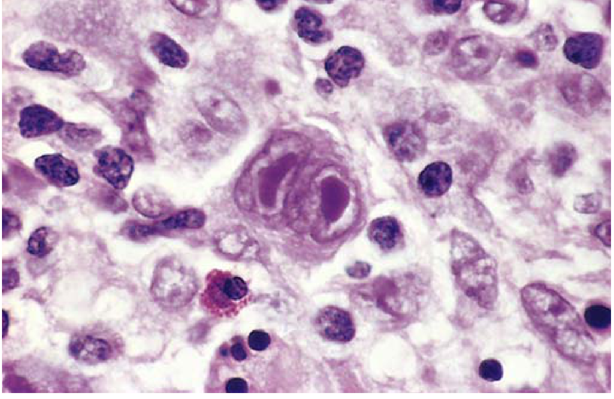
Reed-Sternberg cell: A binucleate giant cell with large, inclusion-like "owl-eye" nucleoli and abundant cytoplasm, surrounded by reactive lymphocytes, macrophages, and an eosinophil (Robbins Basic Pathology, Fig. 10.23)
RS cell characteristics:
- Very large cell: 15-45 μm in diameter
- Enormous multilobate nucleus with exceptionally prominent nucleoli
- Characteristic "owl-eye" appearance - two mirror-image nuclei/lobes, each with a large acidophilic nucleolus surrounded by a clear halo
- Abundant, slightly eosinophilic cytoplasm
- Tumor cells are typically a minor fraction of the total tumor mass; most cells are reactive host inflammatory cells
RS cell variants by subtype:
| Subtype | RS Cell Variant | Special Feature |
|---|---|---|
| Nodular sclerosis | Lacunar cells | Pale cytoplasm retracts in formalin (lacune); collagen bands divide tissue into nodules |
| Mixed cellularity | Classic RS cells (plentiful) | Surrounded by rich inflammatory infiltrate |
| NLPHL | "Popcorn" cells (L&H cells) | Delicate multilobed nucleus; CD20+, CD15-, CD30- |
Immunophenotype (Classic HL):
- CD30+ (90-100%) ← most reliable marker
- CD15+ (75-85%)
- CD45- (leukocyte common antigen negative)
- B and T cell markers usually negative
- CD20+ in ~40% (weak, minority of cells)
- PAX5/BSAP+ (>90%)
NLPHL immunophenotype:
- CD20+ (strongly, unlike cHL)
- CD45+, CD79a+
- CD30-, CD15-
(Robbins & Kumar Basic Pathology; Goldman-Cecil Medicine)
Clinical Features
Presentation
- Asymptomatic peripheral lymphadenopathy is the most common presentation - most commonly cervical and supraclavicular nodes
- Incidence peaks between 15-40 years
- Nodular sclerosis particularly involves lower cervical, supraclavicular, and mediastinal lymph nodes
B Symptoms (present in ~25% at diagnosis, uncommon in early-stage disease)
- Persistent fever >38°C
- Drenching night sweats
- Unexplained weight loss >10% body weight in 6 months
Other Symptoms
- Generalized pruritus - common in HL (rare in NHL); often precedes diagnosis by months; related to RS cell cytokine production
- Pel-Ebstein fever - characteristic cyclic fever pattern
- Alcohol-induced pain at lymph node sites (pathognomonic but rare)
- Mediastinal involvement → cough, dyspnea, SVC syndrome
- Bone pain if skeletal involvement
Spread Pattern
- HL spreads in a contiguous, stepwise fashion from one lymph node region to the anatomically adjacent one (unlike NHL, which spreads unpredictably) - this has important therapeutic implications for radiation field planning
(Cummings Otolaryngology; Robbins & Kumar Basic Pathology)
Staging: Ann Arbor System with Cotswolds Modification
| Stage | Definition |
|---|---|
| I | Involvement of a single lymph node region or lymphoid structure (spleen, thymus, Waldeyer's ring) |
| II | Involvement of 2+ lymph node regions on the same side of the diaphragm |
| III | Involvement of lymph node regions on both sides of the diaphragm (III₁ = splenic hilar/celiac/portal; III₂ = para-aortic/iliac/mesenteric) |
| IV | Multiple extranodal sites or lymph nodes + extranodal disease |
Suffixes:
- A = no B symptoms; B = B symptoms present
- E = limited, contiguous extranodal extension
- X = bulky disease (>1/3 mediastinal width or >10 cm nodal mass)
(Cummings Otolaryngology, Table 116.1)
Investigations / Staging Workup
- Complete history - B symptoms, bone pain, pruritus
- Physical examination - lymphadenopathy, organomegaly
- CBC - may show leukocytosis, lymphocytopenia, eosinophilia, thrombocytosis
- Serum biochemistry - albumin (<4 g/dL is a poor prognostic factor), LDH, alkaline phosphatase, bilirubin, creatinine, ESR
- Mediastinal Mass Ratio (MMR) - calculated on chest X-ray; MMR >0.33 (CXR) or >0.35 (CT) = bulky disease, worse prognosis
- CT neck/chest/abdomen/pelvis - standard staging; widely available
- ¹⁸F-FDG PET/CT - investigation of choice for staging; sensitivity 97%, specificity 100% for HL; changes management in 10-20% of HL cases vs CT alone; HL is FDG-avid
- Bone marrow biopsy - no longer routine if PET/CT performed
- Biopsy - excisional node biopsy preferred; RS cells + immunophenotyping required for diagnosis
(Cummings Otolaryngology; Goldman-Cecil Medicine, Table 172-2)
International Prognostic Score (IPS) - Advanced HL
Seven independent adverse prognostic factors (each scores 1 point):
| Factor | Threshold |
|---|---|
| Serum albumin | <4 g/dL |
| Hemoglobin | <10.5 g/dL |
| Sex | Male |
| Age | ≥45 years |
| Stage | IV disease |
| Leukocytosis | WBC >15,000/mm³ |
| Lymphocytopenia | Lymphocytes <600/mm³ OR <8% of WBC |
- IPS 0-2 (low risk): ~80% cure rate
- IPS ≥3 (high risk): ~60% cure rate (Goldman-Cecil Medicine; Cummings Otolaryngology)
Treatment
Hodgkin lymphoma is one of medicine's true success stories - it is usually curable.
Cure rates:
- Nonbulky Stage I-II: 90-95%
- Bulky Stage I-II: ~80%
- Stage III-IV: ~70%
- Overall 5-year survival (SEER): 86.4%
Treatment by Stage
| Group | Standard Treatment |
|---|---|
| Limited-stage favorable (Stage I-II, no B symptoms, no bulky disease) | 2 cycles ABVD + involved-field RT (20 Gy); OR 3 cycles ABVD alone if complete metabolic response on PET |
| Limited-stage unfavorable (Stage I-II with bulky disease, B symptoms, elevated ESR, ≥3 nodal sites) | 4 cycles ABVD + 30 Gy involved-field RT |
| Advanced-stage (Stage III-IV or bulky) | 6 cycles ABVD (standard); or escalated BEACOPP for high-risk (IPS ≥3) |
| Relapsed/Refractory | Salvage chemotherapy → high-dose chemotherapy + autologous HSCT (standard of care) |
ABVD Regimen (cornerstone of HL treatment)
- A - Adriamycin (doxorubicin)
- B - Bleomycin
- V - Vinblastine
- D - Dacarbazine
BEACOPP (escalated, for high-risk advanced HL)
- Bleomycin, Etoposide, Adriamycin, Cyclophosphamide, Oncovin (vincristine), Procarbazine, Prednisolone
Special Considerations
- Women aged 15-30 receiving mediastinal/axillary RT: high risk of secondary breast cancer - chemotherapy alone preferred
- Smokers: marked risk of lung cancer after mediastinal RT
- PET-adapted therapy: Interim PET scan after 2 cycles guides escalation or de-escalation of therapy
Relapsed/Refractory Disease
- Autologous HSCT: Standard of care for primary refractory or relapsed HL sensitive to salvage chemotherapy
- Brentuximab vedotin (anti-CD30 antibody-drug conjugate): Effective in relapsed/refractory cHL
- Anti-PD-1 checkpoint inhibitors (nivolumab, pembrolizumab): Highly effective even in multiply relapsed disease, leveraging the PD-L1 overexpression mechanism of RS cells
- Allogeneic HSCT: Option for patients relapsing after autologous HSCT
(Goldman-Cecil Medicine; Cummings Otolaryngology)
Long-Term Treatment Complications
A major focus of modern HL management is minimizing these while maintaining cure:
- Cardiopulmonary toxicity - anthracycline cardiomyopathy, bleomycin pulmonary toxicity, radiation pericarditis/coronary artery disease
- Secondary malignancies - breast cancer (mediastinal RT in young women), lung cancer (mediastinal RT + smoking), therapy-related AML/MDS (alkylating agents)
- Infertility / premature menopause (alkylating agents)
- Hypothyroidism (neck irradiation)
Differential Diagnosis
- Non-Hodgkin lymphoma
- Primary mediastinal B-cell lymphoma
- Mediastinal gray zone lymphoma
- Germ cell tumors
- Thymoma
- Sarcoidosis
- Tuberculosis
- Reactive lymphadenopathy (EBV infectious mononucleosis)
The specific diagnosis is established by excisional biopsy + expert hematopathology + immunohistochemistry.
Sources: Robbins & Kumar Basic Pathology | Goldman-Cecil Medicine 2-vol set (Chapter 172) | Cummings Otolaryngology Head & Neck Surgery
This is a shared conversation. Sign in to Orris to start your own chat.