
Topic: 3D Interactive Model of Kshara Sutra Therapy with Clinical Correlation Title "Interactive 3D Kshara Sutra Model: Anatomy of the Anal Canal and Clinical Correlation of Bhagandara (Fistula-in-Ano)" Aim To develop a book-style, layered 3D model demonstrating the anatomy of the anal canal, the fistulous tract, and the application of Kshara Sutra therapy for the management of Bhagandara (Fistula-in-Ano). Objectives To understand the anatomy of the anal canal and perianal region. To demonstrate the formation and course of a fistulous tract. To explain the preparation and application of Kshara Sutra. To illustrate the mechanism of cutting, curetting, and healing produced by Kshara Sutra. To correlate Ayurvedic principles with modern surgical management of fistula-in-ano. Concept of the Model The model will open like a book, with each page representing one anatomical layer. As each layer is lifted, the deeper structures become visible. The final layer will demonstrate the fistulous tract with Kshara Sutra placed through it. Layer 1 – Skin Perianal skin Gluteal folds External opening of fistula Layer 2 – Superficial Fascia Superficial fascia Ischioanal fat Superficial blood vessels Layer 3 – Muscular Layer External anal sphincter Internal anal sphincter Levator ani muscle Layer 4 – Anal Canal Anal canal Dentate line Anal columns Internal opening of fistula Layer 5 – Fistulous Tract Primary fistulous tract Secondary tract (if present) Abscess cavity Kshara Sutra placed through the tract Materials Required Foam sheets (different colours) EVA foam Cardboard/MDF base Transparent plastic sheet Flexible coloured pipes or IV tubing (blood vessels) Cotton thread coated as Kshara Sutra Clay or modelling putty Velcro or magnets Hinges (for book opening) Acrylic colours Labels Special Features 1. Layer-by-Layer Book Design Each anatomical layer can be lifted separately to visualize deeper structures. 2. Interactive Kshara Sutra Demonstration A removable Kshara Sutra thread will pass through the fistulous tract, allowing demonstration of its placement and weekly replacement. 3. Fistula Demonstration Use coloured silicone tubes or flexible pipes to represent the fistulous tract. Demonstrate: Low anal fistula High anal fistula Internal opening External opening Course of the fistulous tract 4. Healing Mechanism Demonstration Provide a removable flap to demonstrate: Placement of Kshara Sutra Cutting through unhealthy tissue Simultaneous healing by fibrosis Preservation of anal sphincters Clinical Correlation Bhagandara (Fistula-in-Ano) Perianal abscess Recurrent fistula Kshara Sutra therapy Fistulectomy and Fistulotomy (comparison) Importance of sphincter preservation Educational Advantages Easy visualization of anal canal anatomy. Interactive understanding of Kshara Sutra therapy. Better understanding of Ayurvedic parasurgical procedures. Correlates anatomy with clinical cases. Suitable for undergraduate Ayurveda (Shalya Tantra) demonstrations and exhibitions. Expected Outcome The completed project will function as a reusable educational model that enables students to understand the anatomy of the anal canal, appreciate the formation of fistulous tracts, and recognize the scientific principles and clinical application of Kshara Sutra therapy in the management of Bhagandara. Through an engaging, hands-on learning experience, the model will enhance understanding of both Ayurvedic and modern surgical concepts.
anal canal anatomy layers sphincter dentate line cross section diagram
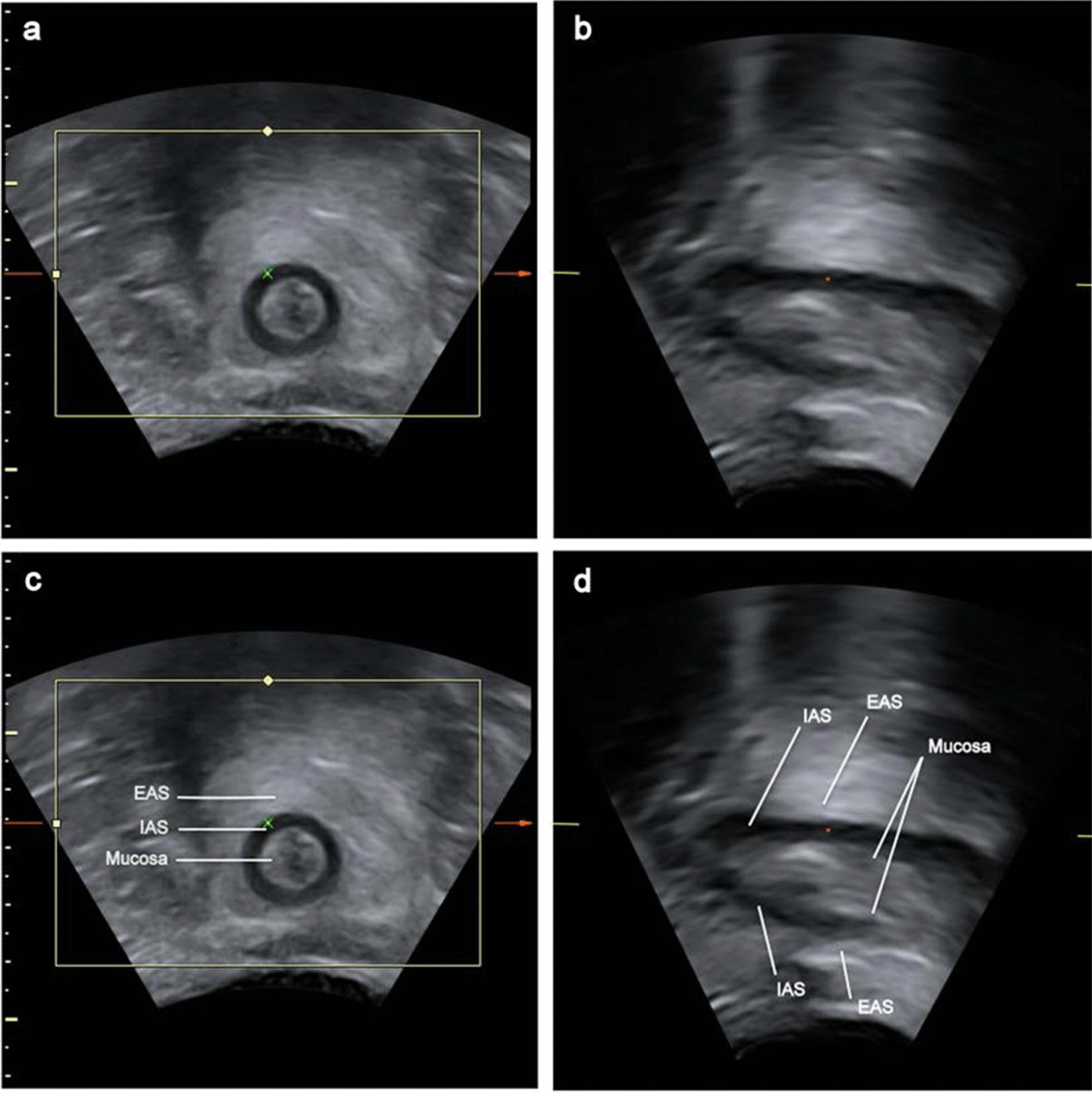
This set of diagnostic images shows perianal ultrasound (PAUS) views of the anal canal at rest. Images (a) and (c) display the coronal plane, while (b) and (d) display the midsagittal plane. In the coronal views, the anal canal is seen in cross-section with concentric layers: a central hypoechoic lumen representing the mucosa, surrounded by the dark, hypoechoic ring of the internal anal sphincter (IAS), which is further enclosed by the relatively hyperechoic external anal sphincter (EAS). In the midsagittal views, the longitudinal anatomy of the canal is visualized, showing the mucosa as a thin echogenic line, the IAS as a hypoechoic band running parallel to the canal, and the EAS as a thicker hyperechoic layer external to the IAS. Labels in (c) and (d) identify the IAS, EAS, and mucosa to highlight their relative spatial arrangement and echogenicity. These images are used in pelvic floor imaging to assess anorectal function, sphincter thickness, and anatomical integrity.
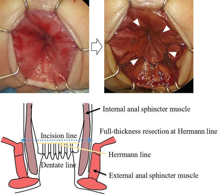
This composite educational resource comprises clinical photographs and a labeled anatomical diagram illustrating the use of Lugol's iodine staining during ileal pouch-anal canal anastomosis (IPACA). The top-left clinical photograph shows the anal canal pre-staining with an anal retractor in place. The top-right photograph demonstrates the 'after' effect following the application of 1% Lugol’s iodine; white arrowheads indicate Herrmann's line, where iodine-positive squamous epithelium stains a dark brown/red color, creating a distinct visual demarcation from the non-staining columnar mucosa. The bottom anatomical diagram provides a coronal cross-section of the anal canal, labeling the internal and external anal sphincter muscles. It identifies the dentate line and Herrmann's line (the upper margin of the surgical anal canal), indicating the specific incision line for full-thickness resection. This visual aid is designed for surgical education in pediatric surgery or gastroenterology, specifically highlighting the technique to accurately identify surgical margins to preserve anal sensation and ensure complete aganglionic tissue removal.
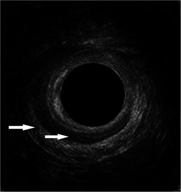
This diagnostic image is an axial cross-section from an endoanal ultrasonography (EAUS) focusing on the mid anal canal. The image demonstrates the characteristic concentric anatomy of the anal sphincter complex with two distinct muscular layers indicated by white arrows. The external anal sphincter (EAS) is visualized as the peripheral, hyperechoic (brighter) ring. A significant structural defect in the EAS is evident, extending from the 9 o'clock to the 3 o'clock position, characterized by a loss of echoic continuity. Internal to this, the internal anal sphincter (IAS) appears as a darker, hypoechoic ring. The IAS also exhibits a pathological defect, visible from the 11 o'clock to the 4 o'clock position. Such findings are clinically significant in the evaluation of obstetric anal sphincter injuries (OASI) and the assessment of fecal or anal incontinence. The image serves as an educational example of how ultrasound can identify localized muscular disruptions and quantify the extent of sphincter damage using clock-face orientation.
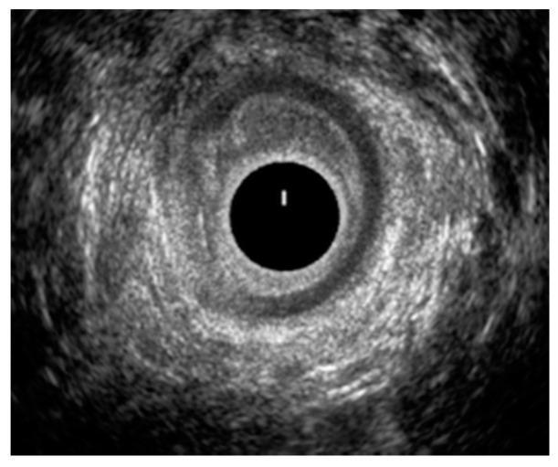
This diagnostic image is an endoanal ultrasound (EAUS) cross-section of the middle anal canal. The image shows the characteristic circular anatomy of the anal canal with concentric rings of varying echogenicity. At the center is a dark, anechoic circle representing the ultrasound probe within the lumen. Surrounding this are the distinct anatomical layers, primarily the hypoechoic (darker) internal anal sphincter (IAS) and the more hyperechoic (brighter) external anal sphincter. The primary clinical finding is a significant structural disruption and discontinuity of the internal anal sphincter, visible as a laceration between the 7 o'clock and 11 o'clock positions. This defect is characterized by a break in the normally continuous hypoechoic muscular ring, indicating tissue compromise. The image serves as a clinical illustration for diagnosing sphincter injuries, which are critical in the assessment of fecal incontinence and obstetric trauma. Target audience includes gastroenterology and colorectal surgery specialists focusing on pelvic floor integrity.
fistula in ano fistulous tract internal external opening perianal
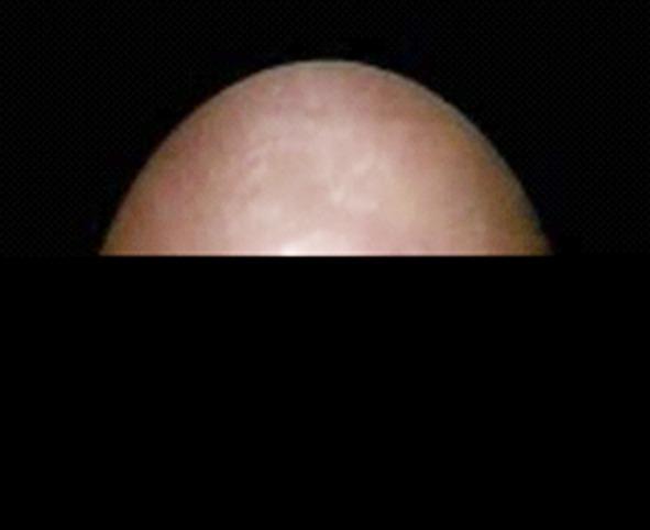
This clinical photograph shows a close-up view of an external fistula opening. The primary content type is a clinical photograph depicting a surgical or dermatological manifestation of perianal or abdominal disease. The tissue shown is pinkish-tan with a relatively smooth, dome-shaped contour against a dark, contrasting background. There are subtle variations in the surface texture and color, indicative of underlying soft tissue structures or the healing process of a fistulous tract. The image illustrates a stage of primary healing or the presence of a chronic external opening associated with a fistula-in-ano or similar tract. It is educationally relevant for medical students and clinicians studying colorectal surgery and wound healing, specifically focusing on the external morphology of fistulous communications between internal organs or tracts and the skin surface.
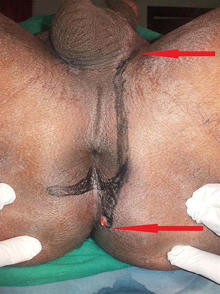
This clinical photograph displays the perianal and perineal region of a patient in the lithotomy position, used for surgical or diagnostic mapping of an anal fistula. The primary finding is an active external sinus tract opening located inferior to the anus, characterized by a visible nodule with slight erythema. Dark, linear markings have been applied to the skin to delineate the suspected subcutaneous course of the fistulous tracts. One marking extends superiorly from the anal region toward the base of the scrotum, while another extends laterally toward the right gluteal region. Red arrows highlight the distal and proximal extents of these tracts. This image is an educational example of preoperative fistula-in-ano assessment and the clinical manifestation of complex perianal disease, demonstrating how external markings correlate with underlying pathology for surgical planning or thread placement (e.g., Ksharasutra therapy).
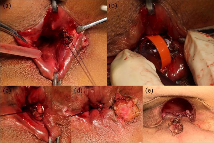
This composite of clinical photographs illustrates the intraoperative stages of treating a cryptoglandular fistula-in-ano using an Anal Fistula Clip (AFC) and an endorectal advancement flap (ERAF). (a) Preparation of the internal opening at the dentate line with the placement of two U-shaped traction sutures. (b) Advancement of the specialized AFC applicator into the anal canal, featuring a distinct orange band for visualization. (c) Deployment and release of the nitinol super-elastic clip at the internal opening to achieve mechanical closure. (d) Debridement and appropriate excision of the external fistula tract to facilitate adequate drainage and tissue healing. (e) Final view showing the sutured endorectal advancement flap (ERAF), providing a multi-layered closure of the anorectal defect. The series demonstrates a sphincter-preserving surgical technique aimed at managing complex perianal fistulous disease while minimizing the risk of incontinence.
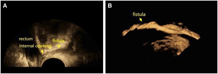
This diagnostic image provides a side-by-side comparison of 3D transperineal ultrasound (3D-TPUS) findings in the evaluation of a fistula-in-ano. Image A demonstrates a standard unenhanced ultrasound view where the rectum is identified, and the anal fistula appears as a subtle, linear hypoechoic tract extending towards an internal opening. Image B shows the same anatomical region after the administration of SonoVue (SVE) contrast. The contrast-enhanced ultrasound (CEUS) technique causes the fistulous tract to become markedly hyperechoic and lustrously reflective, clearly delineating the pathway and branching of the fistula against the dark background. The images illustrate the clinical utility of contrast-enhanced 3D imaging in improving the visibility and diagnostic accuracy of complex perianal fistulae and their internal openings. This educational material is relevant for proctology and radiology specialties, focusing on advanced imaging modalities for perianal disease assessment.
Kshara Sutra therapy Bhagandara fistula-in-ano mechanism preparation Ayurveda
Interactive 3D Kshara Sutra Model
Anatomy of the Anal Canal and Clinical Correlation of Bhagandara (Fistula-in-Ano)
Aim
Objectives
- To understand the anatomy of the anal canal and perianal region
- To demonstrate the formation and course of a fistulous tract
- To explain the preparation and application of Kshara Sutra
- To illustrate the mechanism of cutting, curetting, and healing produced by Kshara Sutra
- To correlate Ayurvedic principles with modern surgical management of fistula-in-ano
Part 1 - Anatomy of the Anal Canal
1.1 Surgical Anatomy (for the model)
- Posterior: Anococcygeal ligament separating it from the tip of the coccyx
- Anterior: Perineal body separating it from the membranous urethra and penile bulb (or lower vagina in females)
- Lateral: Ischiorectal (ischioanal) fossae
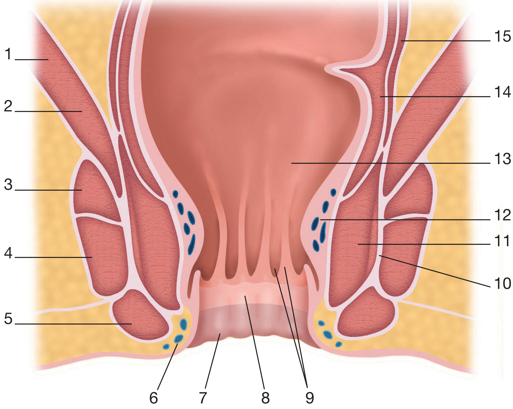
| No. | Structure |
|---|---|
| 1 | Levator ani - iliococcygeal part |
| 2 | Levator ani - puborectal part |
| 3-5 | External anal sphincter (deep, superficial, subcutaneous) |
| 6 | Inferior haemorrhoidal plexus |
| 7 | Perianal skin |
| 8 | Anoderm |
| 9 | Anal columns and crypts |
| 10 | Internal anal sphincter |
| 11 | Intersphincteric plane |
| 12 | Dentate line |
| 13 | Rectal mucosa (columnar epithelium) |
| 14 | Puborectalis sling |
| 15 | Rectal ampulla |
1.2 Layer-by-Layer Anatomy (Matching the 5 Model Layers)
Layer 1 - Perianal Skin
- Keratinized, hair-bearing skin surrounding the anal verge
- Contains perianal sebaceous glands and apocrine glands (cryptoglandular origin of fistulae)
- The external opening of the fistula appears here as a small pit with granulation tissue or indurated edges
- Gluteal folds mark the lateral limit of the perianal region
Layer 2 - Superficial Fascia and Ischioanal Fat
- Superficial fascia and ischioanal (ischiorectal) fossa fat occupy the lateral spaces
- The ischioanal fossa is a pyramidal fat-filled space bounded medially by the external anal sphincter and levator ani, laterally by the ischium and obturator fascia
- Contains the inferior rectal (haemorrhoidal) vessels and nerve - represented in the model by flexible coloured pipes/IV tubing
- The posterior anal space (deep postanal space) communicates with both ischioanal fossae - important for horseshoe fistulae
Layer 3 - Muscular Layer
- Forms the bulk of the sphincter complex
- Three components: deep, superficial, and subcutaneous
- Fused superiorly with puborectalis to form the anorectal ring - the palpable landmark most important in fistula surgery
- Nerve supply: inferior rectal branch of pudendal nerve (S2-S4)
- Cutting or dividing the EAS above its lower third risks incontinence
- A thickened continuation of the inner circular smooth muscle of the rectum
- Responsible for approximately 55% of resting anal canal pressure
- Separated from the EAS by the intersphincteric plane - the critical anatomical space in cryptoglandular abscess/fistula formation
- Puborectalis slings around the anorectal junction maintaining the anorectal angle (~90°)
- An important continence mechanism
- Division of puborectalis = major incontinence risk
Layer 4 - Anal Canal Interior
- Located approximately 2 cm above the anal verge
- Marks the junction between endodermal (upper) and ectodermal (lower) epithelium
- Above dentate line: columnar/cuboidal mucosa, autonomic innervation (painless), superior rectal lymphatics
- Below dentate line: squamous epithelium (anoderm), somatic innervation (painful), inguinal lymphatics
- Contains 8-14 anal crypts (recesses between anal columns)
- 4-8 anal glands open into the crypts at the dentate line
- They penetrate the IAS to lie in the intersphincteric plane
- Obstruction and infection of these glands = cryptoglandular abscess = origin of most fistulae
- The internal opening of the fistula is almost always at the dentate line level
- 8-14 longitudinal mucosal folds above the dentate line
- Contain the terminal ramifications of the superior rectal vessels (haemorrhoidal plexus)
1.3 Spaces Around the Anal Canal (Critical for Fistula Tracking)
| Space | Location | Clinical Importance |
|---|---|---|
| Perianal space | Immediately around anal canal below IAS | Low anal abscess, subanodermal fistula |
| Intersphincteric space | Between IAS and EAS | Primary site of cryptoglandular sepsis; intersphincteric fistula |
| Ischioanal (ischiorectal) fossa | Lateral to EAS, below levators | Transsphincteric fistula tracks through here |
| Supralevator space | Above levator ani | Suprasphincteric and extrasphincteric fistulae |
| Deep postanal space | Behind anal canal, below levators | Connects both ischioanal fossae - horseshoe abscess |
Part 2 - Bhagandara (Fistula-in-Ano)
2.1 Definition and Ayurvedic Correlation
- Shataponaka (Vataj)
- Ustragriva (Pittaj)
- Parisravi (Kaphaj)
- Sambukavarta (Tridoshaj)
- Unmargi (Agantuj)
2.2 Pathogenesis of Fistula Formation
Anal gland infection (cryptoglandular sepsis)
        ↓
Obstruction of anal duct → Abscess formation in intersphincteric plane
        ↓
Spontaneous/surgical drainage
        ↓
Persistent communication between internal opening (dentate line)
and external opening (perianal skin)
        ↓
Granulation tissue lines the tract → FISTULA-IN-ANO
2.3 Goodsall's Rule (Predicting Tract Course)
- External opening ANTERIOR to the transverse anal line: Tract runs radially in a straight line directly to the internal opening
- External opening POSTERIOR to the transverse anal line: Tract curves in a horseshoe pattern to a posterior midline internal opening
- Exception: Anterior external openings >3 cm from anal verge may still track posteriorly
2.4 Parks' Classification (for the Model - Layer 5)
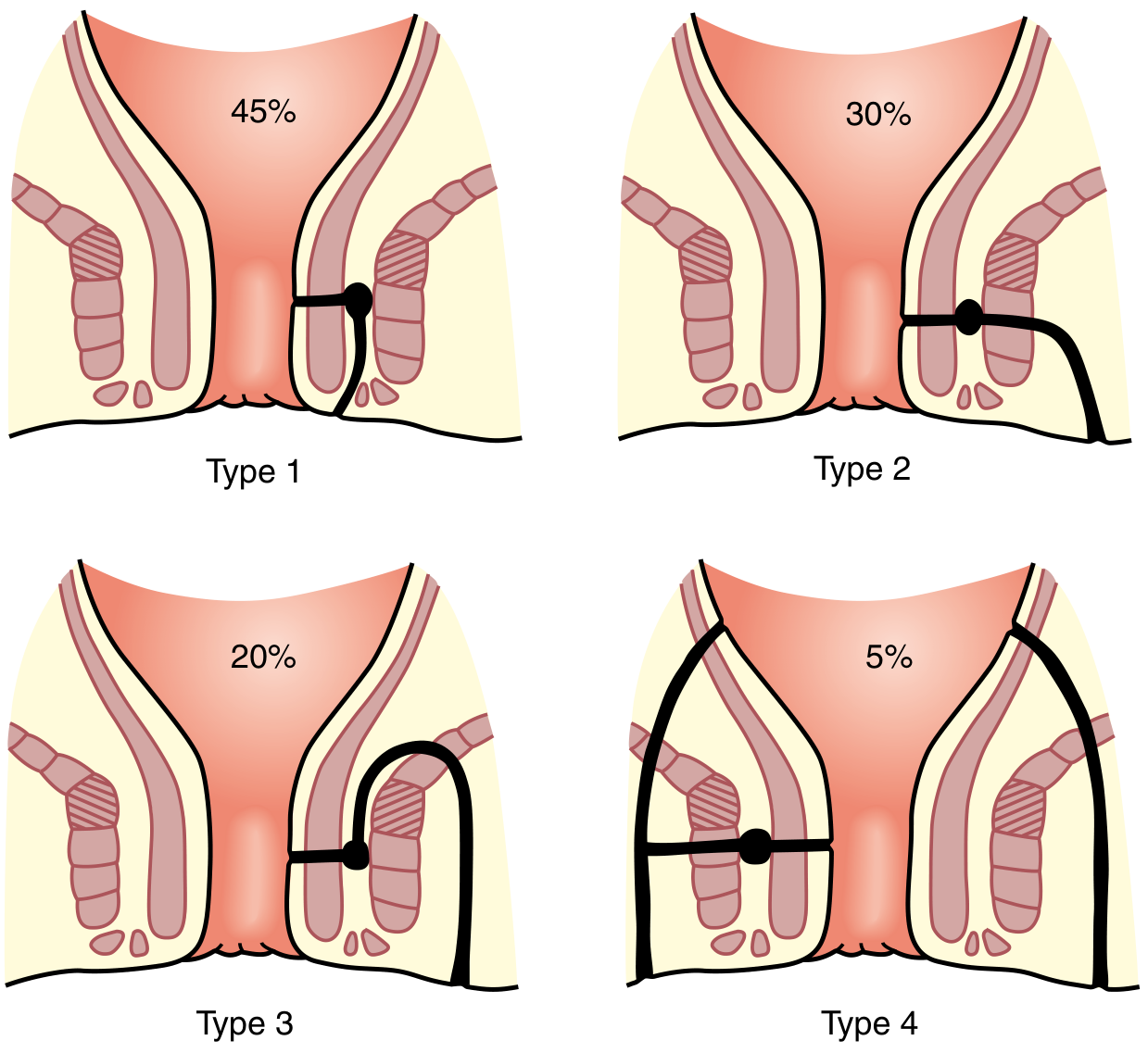
| Type | Course | Frequency | Incontinence Risk |
|---|---|---|---|
| Type 1 - Intersphincteric | Tracks between IAS and EAS; does NOT cross EAS | 45% | Low |
| Type 2 - Transsphincteric | Crosses both IAS and EAS; tracks through ischioanal fossa | 30% | Moderate |
| Type 3 - Suprasphincteric | Rises above puborectalis, curls down through levators and ischioanal fossa | 20% | High |
| Type 4 - Extrasphincteric | Passes from rectal wall through levator ani to perianal skin, bypassing sphincters entirely | 5% | Very High |
| Class | Features |
|---|---|
| Simple | Low (superficial/low intersphincteric/low transsphincteric); single external opening |
| Complex | High tract; extra/suprasphincteric; associated abscess; ano-vaginal fistula; anal stricture |
Part 3 - Kshara Sutra Therapy
3.1 Etymology and Historical Background
- First mentioned by Acharya Sushruta in Sushruta Samhita (Sutrasthana, Chapter 11) for the treatment of Nadi Vrana (sinus tracts), though the preparation was not detailed
- Chakrapani Datta later elaborated its preparation
- The ICMR (Indian Council of Medical Research) and CCRAS (Central Council for Research in Ayurvedic Sciences) validated and standardized Kshara Sutra in the 1980s-1990s
- The standard thread is now prepared at AIIMS New Delhi protocol
3.2 Preparation of Kshara Sutra
| Coating | Substance | Properties | Number of Coats |
|---|---|---|---|
| Primary (innermost) | Snuhi Ksheera (latex of Euphorbia neriifolia) | Vehicle/adhesive; mild Kshara | 11 coats |
| Secondary | Apamarga Kshara (Achyranthes aspera ash - alkaline) | Main caustic agent; pH 9-10; debrides, cuts tissue | 7 coats |
| Final (outermost) | Haridra (Curcuma longa, turmeric) powder | Anti-inflammatory, antimicrobial, promotes healing | 3 coats |
3.3 Mechanism of Action of Kshara Sutra
1. Chemical Cutting (Kshara Karma)
- Alkaline pH (from Apamarga Kshara) causes saponification of fats and protein denaturation in the fistulous tract wall
- Gradual, controlled chemical division of unhealthy granulation tissue
- Cutting rate: approximately 1 cm per week
2. Curettage of the Tract (Lekhana Karma)
- The coarse surface of the thread mechanically debrides the epithelialised/granulating fistula tract with each thread change (weekly)
- Removes unhealthy, infected tissue promoting fresh granulation
3. Simultaneous Healing (Ropana)
- While cutting anteriorly, fibrosis and healing occur behind the thread
- This is the key advantage over surgical fistulotomy - the sphincter is not suddenly divided, so fibrosis can bind and support muscle before the thread reaches it
- Haridra provides antimicrobial, anti-inflammatory, and wound-healing promotion
4. Drainage (Shodhana)
- The thread maintains the tract open, allowing continuous drainage of pus and infected material
- Prevents re-accumulation of the abscess
3.4 Clinical Application Procedure
- Patient in lithotomy position, spinal/local anaesthesia
- Probe the tract gently with Ksharasutra probe/malleable probe
- Identify internal and external openings
- Assess sphincter involvement
- Pass the Kshara Sutra through the fistulous tract from external to internal opening using a grooved director/probe
- The thread is tied in a loose loop (not tight) so it lies within the tract without strangulation
- Every 7 days, the old thread is removed and a new Kshara Sutra is passed
- Rate of cutting: ~0.5-1 cm per week
- Total duration: depends on tract length (typically 4-8 weeks for simple fistulae)
- Sitz bath with Panchavalkala Kwatha (bark decoction of 5 plants)
- Jatyadi oil local application
- Triphala Guggulu orally (anti-inflammatory, wound healing)
- High-fibre diet to avoid straining
Part 4 - Clinical Correlation: Kshara Sutra vs Modern Surgery
4.1 Comparison Table
| Parameter | Fistulotomy | Fistulectomy | Kshara Sutra |
|---|---|---|---|
| Principle | Lay-open tract surgically | Complete excision of tract | Gradual chemical cutting + healing |
| Sphincter risk | Low fistulae safe (<1/3 EAS); high fistulae risk incontinence | Same as fistulotomy | Minimal - simultaneous fibrosis preserves sphincters |
| Recurrence rate | 2-8% (low fistulae) | Similar | 3-6% (comparable) |
| Incontinence | <5% (low); significant for high | Similar | Very low (<2%) |
| Anaesthesia | General/spinal (OT) | General/spinal (OT) | Spinal/local (OPD/minor OT) |
| Hospital stay | 1-2 days | 2-3 days | Day care / nil |
| Healing time | 4-6 weeks | 6-8 weeks | 6-12 weeks (thread-dependent) |
| Suitable for | Low, simple fistulae | Low, simple fistulae | Low AND high fistulae; complex/recurrent |
| Cost | Moderate | High | Very low |
| Sphincter preservation | No guarantee for high | No | Yes - active healing prevents division |
4.2 Indications of Kshara Sutra (When to Choose It)
- High transsphincteric fistulae where fistulotomy would endanger continence
- Suprasphincteric fistulae
- Recurrent fistulae after failed surgery
- Patients with Crohn's disease-associated fistulae (with caution)
- High-risk patients (elderly, prior anorectal surgery, sphincter injury)
- When preservation of anal continence is the priority
4.3 Contraindications
- Fistula with active Crohn's disease flare
- Associated malignancy
- Radiation-induced fistula (relative)
- Extrasphincteric fistula originating from rectum (alternative procedures needed)
Part 5 - Model Construction Guide
5.1 Concept
5.2 Detailed Layer-by-Layer Construction
BASE (Book Spine)
- Material: MDF or thick cardboard (30 cm × 20 cm)
- Paint: Cream/off-white background
- Bind all layers with hinges on the left edge so they open right-to-left
- Label the spine: "Anatomy of Anal Canal and Kshara Sutra - 3D Interactive Model"
LAYER 1 - SKIN (Top Cover Page)
- Material: Light pink/peach EVA foam sheet (3 mm thick)
- Features:
- Cut an oval "anal verge" opening in the centre using scissors
- Paint gluteal folds with slightly darker peach acrylic on both sides
- Mark a small raised nodule (granulation bead) with clay = external fistula opening
- Place a small coloured dot or velcro button to mark the external opening location
- Attach a hinge at the left to allow this layer to lift off like a page
- Label: Perianal skin, Gluteal fold, External opening of fistula (arrow)
LAYER 2 - SUPERFICIAL FASCIA AND ISCHIOANAL FAT
- Material: Yellow EVA foam (to represent fat) on white/cream foam backing
- Features:
- Bulk out ischioanal fat bilaterally on each side of central canal with thick yellow foam pieces
- Thread thin red flexible silicone tubing (or IV tubing) in a branching pattern = inferior rectal vessels
- Thread yellow IV tubing = inferior rectal nerve
- The anococcygeal ligament can be shown as a thickened white strip posteriorly
- Label: Ischioanal fat, Inferior rectal vessels, Inferior rectal nerve, Anococcygeal ligament
LAYER 3 - MUSCULAR LAYER
- Material: Red/dark red EVA foam and putty/clay for muscle bellies
- Features:
- Mould levator ani as a broad flat sheet of red foam spanning the top of the layer (dome-shaped)
- Puborectalis: show as a U-shaped sling of clay/putty curling around the posterior anorectal junction
- External anal sphincter: show as three concentric ring layers (deep = innermost, subcutaneous = outermost) using rolled foam/putty of slightly varying shades of red
- Internal anal sphincter: pale pink/cream rolled foam ring inside EAS, separated by a thin white intersphincteric plane line
- Mark the anorectal ring at the top of the sphincter complex with a velcro marker or bold label
- Label: Levator ani, Puborectalis, EAS (deep/superficial/subcutaneous), IAS, Intersphincteric plane, Anorectal ring
LAYER 4 - ANAL CANAL
- Material: Transparent plastic sheet over a base layer; clay/putty for mucosal features
- Features:
- Cut a cylindrical "tunnel" (the anal canal lumen) vertically through the centre
- Line the upper half with pink/salmon clay = columnar mucosa
- Mark a horizontal ridge with gold/yellow clay = dentate line (clearly labelled with an arrow)
- Model 8-10 small vertical ridges above the dentate line = anal columns (of Morgagni)
- Between columns: small clay indentations = anal crypts
- Place a single red dot or tiny clay bead just above the dentate line at the 6 o'clock position = internal opening of fistula
- Line the lower anal canal (below dentate) with white/cream = squamous epithelium/anoderm
- Label: Dentate line, Anal columns, Anal crypts, Anal glands (marked below dentate), Internal opening of fistula
LAYER 5 - FISTULOUS TRACT WITH KSHARA SUTRA (Bottom Layer)
- Material: Transparent plastic base; silicone/flexible coloured tubing; cotton thread coated as Kshara Sutra
- Features:
- Primary tract: Use a flexible orange or red silicone tube (4-5 mm diameter) running from the internal opening (at dentate line) through the intersphincteric plane (or through the EAS for transsphincteric) and emerging at the external opening in the perianal skin
- Abscess cavity: At the intersphincteric plane junction, form a small pouch of yellow clay/putty = the source abscess
- Secondary (horseshoe) tract: Optional - add a second silicone tube arching around to the opposite side through the deep postanal space
- Kshara Sutra: Pass a cotton thread (stained yellow-brown with turmeric powder, coated with clay) through the lumen of the fistula tube. Tie it loosely at both ends. Make it removable (thread tip affixed with velcro at the external opening)
- Use velcro tabs so the Kshara Sutra thread can be pulled out and re-inserted to demonstrate weekly replacement
- Add a detachable foam flap to the tract area to show the healing zone (fibrosis behind the thread = white/cream zone; unhealthy tissue ahead = red/granulation)
- Label: Primary fistulous tract, Secondary tract, Intersphincteric plane abscess, Internal opening, External opening, Kshara Sutra thread (removable), Zone of healing (fibrosis), Zone of active cutting
5.3 Special Interactive Features
Feature 1 - Removable Kshara Sutra Demonstration
- Affix velcro at both ends of the fistula tube
- Pass a removable yellow-brown cotton Kshara Sutra thread through it
- Students can remove and reinsert the thread to simulate weekly thread replacement
- Include a small card: "Cutting rate: 1 cm/week. Replace every 7 days."
Feature 2 - Healing Zone Flap
- Attach a small hinged foam flap over the fistula tube in Layer 5
- One side (red = granulation tissue): "Active cutting zone - Kshara action"
- Other side (white/cream = fibrous tissue): "Healing zone - fibrosis sealing the divided tract"
- This demonstrates the simultaneous cut-and-heal mechanism
Feature 3 - Fistula Type Selector (Optional Add-on)
- Place 4 small removable tube segments on a card beside the model, each showing:
- Intersphincteric tract (stays between sphincters)
- Transsphincteric tract (crosses EAS)
- Suprasphincteric tract (arches above puborectalis)
- Extrasphincteric tract (arises from rectum)
- Students can compare and identify Parks' types
Feature 4 - Goodsall's Rule Plate
- Attach a clock-face diagram on the inside cover showing the anus from below
- Mark the transverse bisection line
- Use arrows: anterior external openings → straight radial tracts; posterior → curved to midline
5.4 Materials List (Detailed)
| Material | Purpose | Quantity |
|---|---|---|
| MDF board (30×20 cm) | Base/book spine | 1 piece |
| Cardboard sheets | Layer backing | 6 sheets |
| EVA foam (peach, yellow, red, white, pink, cream) | Anatomical layers | 10 sheets |
| Transparent plastic sheet | Anal canal lining | 2 sheets |
| Clay/modelling putty (multicolour) | Mucosal features, muscles, abscess | 4 packs |
| Silicone tubing (4-5 mm diameter, coloured) | Fistulous tract | 30 cm |
| IV tubing (red, yellow, blue) | Blood vessels, nerves | 50 cm |
| Cotton thread (No. 20 linen/thick cotton) | Kshara Sutra base | 1 reel |
| Turmeric powder + acrylic medium | Kshara Sutra coating | 1 tbsp |
| Velcro strips (both sides) | Layer attachment + thread fixation | 1 roll |
| Hinges (small) | Layer-by-layer opening | 6 sets |
| Acrylic paint set | Colours for structures | 1 set |
| White glue/fevicol | Bonding | 1 bottle |
| Labels and arrows (printed/handwritten) | Anatomical labelling | Per layer |
| Small magnets (optional) | Layer locking | 10 pairs |
5.5 Colour Coding Guide
| Colour | Structure |
|---|---|
| Peach/light pink | Perianal skin, anoderm |
| Yellow/cream | Ischioanal fat, subcutaneous tissue |
| Dark red | Skeletal muscle (EAS, levator ani) |
| Pale pink | Internal anal sphincter (smooth muscle) |
| Salmon/pale pink | Anal canal mucosa (columnar) |
| Gold/yellow | Dentate line |
| White | Fibrous tissue, intersphincteric plane, healing zone |
| Orange/red tube | Fistulous tract |
| Yellow-brown | Kshara Sutra thread |
| Red small bead | Internal opening |
| Pale nodule | External opening |
| Blue thin tubing | Veins |
| Red thin tubing | Arteries |
| Yellow thin tubing | Nerves |
Part 6 - Educational Content for Labels and Display Cards
Card 1 - Anal Canal (for display beside Layer 4)
"The anal canal is 3-4 cm long. It extends from the anorectal ring (palpable on digital rectal examination) to the anal verge. The dentate line divides the canal into upper (autonomic innervation, painless) and lower (somatic innervation, painful) halves. Anal glands open into the crypts at the dentate line - infection of these glands causes most anal abscesses and fistulae."
Card 2 - Bhagandara/Fistula-in-Ano (for display beside Layer 5)
"A fistula-in-ano is an abnormal hollow tract connecting the anal canal (internal opening at dentate line) to the perianal skin (external opening). It forms when a perianal abscess fails to heal. It is classified by its relation to the sphincters (Parks' classification). Kshara Sutra is indicated for high fistulae where conventional surgery risks incontinence."
Card 3 - Kshara Sutra (for interactive demonstration)
"Kshara Sutra = Alkaline medicated thread. Prepared with 11 coats of Snuhi latex + 7 coats of Apamarga Kshara + 3 coats of Haridra = 21 coats total. Mechanism: Chemical cutting (Kshara karma) + Curettage (Lekhana) + Simultaneous healing (Ropana) + Drainage (Shodhana). Thread is replaced every 7 days. Cutting rate: ~1 cm/week."
Card 4 - Goodsall's Rule
"Anterior external openings (anterior to transverse anal line) → Straight radial tract to internal opening. Posterior external openings → Curved horseshoe tract to posterior midline internal opening."
Card 5 - Why Kshara Sutra Preserves Continence
"Unlike surgical fistulotomy, where the sphincter is divided at once, Kshara Sutra cuts the sphincter muscle very slowly (1 cm/week). This allows progressive fibrosis to form behind the advancing thread. The scar tissue supports the muscle before it is divided, preventing incontinence. This 'cut-and-heal' simultaneity is the therapeutic genius of Kshara Sutra."
Part 7 - Expected Outcomes and Evaluation Criteria
7.1 Educational Objectives Assessment
- Name and identify all 5 anatomical layers of the perianal region
- Identify the dentate line and explain its significance in fistula pathogenesis
- Classify a given fistula using Parks' classification
- Describe the composition and preparation of Kshara Sutra
- Explain the mechanism of simultaneous cutting and healing
- Compare Kshara Sutra with fistulotomy/fistulectomy in terms of sphincter risk
7.2 Evidence Base
- A 2015 PMC comparative study (Dutta G et al.) showing Kshara Sutra is "simple, easy, and safe" with very low recurrence and anal incontinence rates
- ICMR multi-centre trials demonstrating equivalence to fistulotomy for simple fistulae and superiority for high complex fistulae in terms of continence preservation
- Meena RK et al. (PMC 2018) demonstrating Guggulu-based Kshara Sutra is more effective than standard formulation
7.3 Project Significance
- Provides hands-on anatomical learning impossible from 2D textbook diagrams
- Demonstrates a 2,500-year-old Ayurvedic surgical principle validated by modern RCT evidence
- Bridges Ayurvedic (Shalya Tantra) and modern surgical education
- Suitable for BAMS undergraduate exhibitions, faculty demonstrations, CME programmes, and inter-college competitions
References
- Williams NS, Bulstrode CJK, O'Connell PR. Bailey and Love's Short Practice of Surgery, 28th Ed. Chapter 80: Anal canal anatomy and physiology. CRC Press/Taylor & Francis; 2023.
- Townsend CM, Beauchamp RD, Evers BM, Mattox KL. Sabiston Textbook of Surgery: The Biological Basis of Modern Surgical Practice, 21st Ed. Chapter 97: Fistula-in-ano. Elsevier; 2022.
- Zinner MJ, Ashley SW. Maingot's Abdominal Operations, 13th Ed. Chapter 42: Anal canal anatomy and histology. McGraw-Hill; 2019.
- Sushruta. Sushruta Samhita - Sutrasthana, Chapter 11 (Kshara Sutra reference); Chikitsa Sthana, Chapter 17 (Bhagandara).
- Dutta G et al. Comparing Ksharasutra (Ayurvedic Seton) and open surgery in the management of fistula-in-ano. PMC4518420. J Ayurveda Integr Med. 2015.
- Meena RK et al. Comparative clinical study of Guggulu-based Ksharasutra. PMC6287408. AYU. 2018.
- CCRAS/ICMR. Standard Operating Procedure for Kshara Sutra Preparation and Application. New Delhi; 1990 (revised 2012).