attachment
attachment
attachment
attachment
attachment
attachment
attachment
attachment
attachment
attachment

Anlyse the face of this pt he has oily skin and acne white black heads what should be the line of treatment in detail

analyse_media
analyse_media
analyse_media
analyse_media
analyse_media
Reading File
Finding Sources
Finding Sources
Searching Images

acne vulgaris comedones oily skin forehead treatment
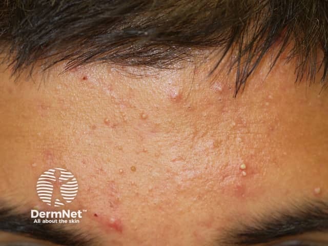
Clinical photography of the anterior forehead demonstrates an acneiform eruption with predominately follicular lesions. Imaging modality is digital clinical photography with a frontal, close-up view capturing the central facial T-zone. The skin surface shows multiple small erythematous papules and a mixture of open and closed comedones (blackheads and whiteheads), with mild superficial desquamation and subtle perifollicular erythema. The sebaceous-rich forehead displays uniform texture with hypertrophic pores and minimal scales, consistent with mild-to-moderate acne vulgaris. No scars or atrophic changes are evident in this field; overall complexion remains oily. The distribution is localized to the forehead, with extension toward the eyebrows and glabella in a typical seborrheic pattern; however, the lesions are most dense in the central forehead region. Visual features include pearly-to-red papules, comedonal plugs within pilosebaceous units, and occasional pinpoint pustules that suggest early inflammatory activity. Clinically, these findings correlate with increased sebum production, follicular hyperkeratinization, Propionibacterium acnes involvement, and inflammatory mediators. Diagnostic significance lies in recognizing common acne vulgaris to guide topical keratolytic and anti-inflammatory therapy, monitor response to treatment, and differentiate from rosacea, folliculitis, or keratosis pilaris. This image is suitable for educational demonstrations, dermatology training, primary care documentation, and therapeutic planning. Clinician education and patient counseling.
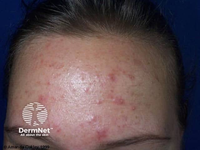
Clinical photography of the anterior forehead demonstrates an acneiform eruption with predominately follicular lesions. Imaging modality is digital clinical photography with a frontal, close-up view capturing the central facial T-zone. The skin surface shows multiple small erythematous papules and a mixture of open and closed comedones (blackheads and whiteheads), with mild superficial desquamation and subtle perifollicular erythema. The sebaceous-rich forehead displays uniform texture with hypertrophic pores and minimal scales, consistent with mild-to-moderate acne vulgaris. No scars or atrophic changes are evident in this field; overall complexion remains oily. The distribution is localized to the forehead, with extension toward the eyebrows and glabella in a typical seborrheic pattern; however, the lesions are most dense in the central forehead region. Visual features include pearly-to-red papules, comedonal plugs within pilosebaceous units, and occasional pinpoint pustules that suggest early inflammatory activity. Clinically, these findings correlate with increased sebum production, follicular hyperkeratinization, Propionibacterium acnes involvement, and inflammatory mediators. Diagnostic significance lies in recognizing common acne vulgaris to guide topical keratolytic and anti-inflammatory therapy, monitor response to treatment, and differentiate from rosacea, folliculitis, or keratosis pilaris. This image is suitable for educational demonstrations, dermatology training, primary care documentation, and therapeutic planning. Clinician education and patient counseling.

Clinical photography of the frontal face demonstrating mild to moderate inflammatory acne on the forehead during pregnancy. Imaging modality: Clinical photograph (dermatology), frontal/anterior view. The frontal region shows grouped erythematous papules with variable surface irregularity and a few discrete pustules, primarily in the central and upper forehead within the T-zone pattern. Comedones are partially visible as tiny non-inflammatory plugs. There is no overt nodular lesion or scarring in this image. The skin appears mildly oily with slight shine, consistent with hyperseborrhea commonly associated with pregnancy. The presentation is consistent with acne vulgaris in a hormonally driven context, i.e., gestational acne, characterized by increased sebum production from elevated androgens and pregnancy-related hormonal fluctuations. Differential considerations include rosacea, folliculitis, and irritant/contact dermatitis, but the predominant morphology and distribution favors inflammatory acne. Clinically, this image is relevant for dermatology and obstetric practice because it informs safe management during pregnancy: emphasize non-teratogenic therapies, gentle cleansing, noncomedogenic moisturizers, benzoyl peroxide use, possibly topical erythromycin or clindamycin, and avoidance of systemic retinoids (isotretinoin) and tetracyclines. The image supports educational discussion on pregnancy-safe dermatologic care, hormonal acne pathophysiology, and treatment tailoring for maternal-fetal safety. Clinician note: monitor acne progression; re-evaluate therapy if pregnancy course changes or complications.
Clinical photography of the frontal face demonstrating mild to moderate inflammatory acne on the forehead during pregnancy. Imaging modality: Clinical photograph (dermatology), frontal/anterior view. The frontal region shows grouped erythematous papules with variable surface irregularity and a few discrete pustules, primarily in the central and upper forehead within the T-zone pattern. Comedones are partially visible as tiny non-inflammatory plugs. There is no overt nodular lesion or scarring in this image. The skin appears mildly oily with slight shine, consistent with hyperseborrhea commonly associated with pregnancy. The presentation is consistent with acne vulgaris in a hormonally driven context, i.e., gestational acne, characterized by increased sebum production from elevated androgens and pregnancy-related hormonal fluctuations. Differential considerations include rosacea, folliculitis, and irritant/contact dermatitis, but the predominant morphology and distribution favors inflammatory acne. Clinically, this image is relevant for dermatology and obstetric practice because it informs safe management during pregnancy: emphasize non-teratogenic therapies, gentle cleansing, noncomedogenic moisturizers, benzoyl peroxide use, possibly topical erythromycin or clindamycin, and avoidance of systemic retinoids (isotretinoin) and tetracyclines. The image supports educational discussion on pregnancy-safe dermatologic care, hormonal acne pathophysiology, and treatment tailoring for maternal-fetal safety. Clinician note: monitor acne progression; re-evaluate therapy if pregnancy course changes or complications.

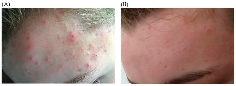
This clinical photograph illustrates a side-by-side comparison (A and B) of a 14-year-old patient with acne vulgaris on the forehead, demonstrating therapeutic progression. Image A shows baseline moderate inflammatory acne, characterized by a high density of erythematous papules, pustules with visible white centers, and underlying non-inflammatory comedones. The skin exhibits significant localized inflammation and an irregular texture. Image B depicts the same anatomical region after three months of treatment with oral minocycline and topical antimicrobial therapy. There is a marked reduction in lesion density and severity, with a transition from active inflammatory papulopustular lesions to sparse residual erythema. The overall skin texture is smoother and the inflammatory response is substantially resolved, showcasing the clinical efficacy of systemic and topical antibiotics in targeting Cutibacterium acnes (formerly Propionibacterium acnes). The content is relevant for dermatological education regarding the classification of acne lesions and the visual assessment of treatment outcomes.
This clinical photograph illustrates a side-by-side comparison (A and B) of a 14-year-old patient with acne vulgaris on the forehead, demonstrating therapeutic progression. Image A shows baseline moderate inflammatory acne, characterized by a high density of erythematous papules, pustules with visible white centers, and underlying non-inflammatory comedones. The skin exhibits significant localized inflammation and an irregular texture. Image B depicts the same anatomical region after three months of treatment with oral minocycline and topical antimicrobial therapy. There is a marked reduction in lesion density and severity, with a transition from active inflammatory papulopustular lesions to sparse residual erythema. The overall skin texture is smoother and the inflammatory response is substantially resolved, showcasing the clinical efficacy of systemic and topical antibiotics in targeting Cutibacterium acnes (formerly Propionibacterium acnes). The content is relevant for dermatological education regarding the classification of acne lesions and the visual assessment of treatment outcomes.

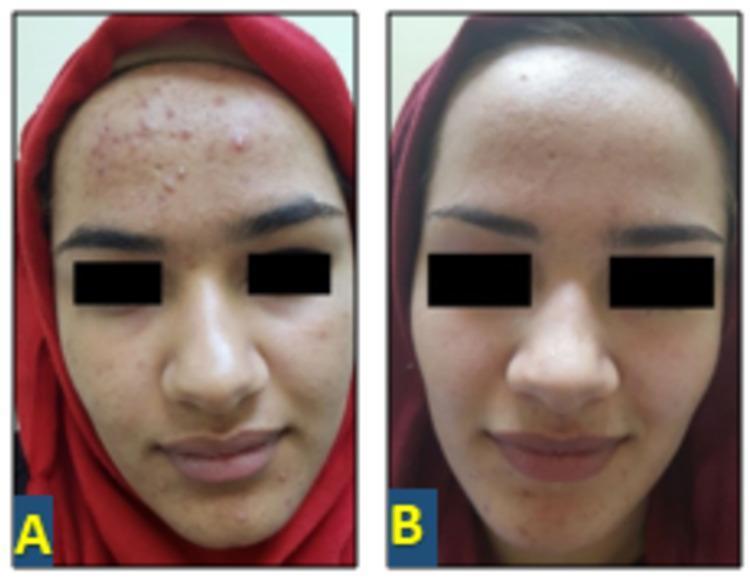
This clinical photograph exhibits a side-by-side comparison of a 17-year-old female with moderate acne vulgaris before and after treatment. Image A (baseline) demonstrates a high density of inflammatory and non-inflammatory lesions primarily concentrated on the forehead, with additional involvement of the cheeks and chin. Visible lesions include erythematous papules, pustules, and numerous closed and open comedones, set against a background of uneven skin texture and localized inflammation. Image B shows the same patient 12 weeks following the initiation of 2% topical spironolactone solution. This post-treatment image reveals a significant clinical improvement, characterized by a marked reduction in the total lesion count. The forehead appears notably smoother with a substantial clearance of papules and pustules. The overall skin tone is more uniform, and there is a visible decrease in inflammatory erythema, demonstrating the therapeutic efficacy of topical anti-androgen therapy in managing acne vulgaris and improving overall skin texture.

This clinical photograph exhibits a side-by-side comparison of a 17-year-old female with moderate acne vulgaris before and after treatment. Image A (baseline) demonstrates a high density of inflammatory and non-inflammatory lesions primarily concentrated on the forehead, with additional involvement of the cheeks and chin. Visible lesions include erythematous papules, pustules, and numerous closed and open comedones, set against a background of uneven skin texture and localized inflammation. Image B shows the same patient 12 weeks following the initiation of 2% topical spironolactone solution. This post-treatment image reveals a significant clinical improvement, characterized by a marked reduction in the total lesion count. The forehead appears notably smoother with a substantial clearance of papules and pustules. The overall skin tone is more uniform, and there is a visible decrease in inflammatory erythema, demonstrating the therapeutic efficacy of topical anti-androgen therapy in managing acne vulgaris and improving overall skin texture.

Your credits are exhausted. You can purchase a top-up or subscribe to a plan to continue using Orris. If you're already subscribed, your daily credits will reset tomorrow.
This is a shared conversation. Sign in to Orris to start your own chat.