
White discharge female
vaginal discharge leukorrhea diagnosis treatment
vaginal discharge types comparison normal candida bacterial vaginosis
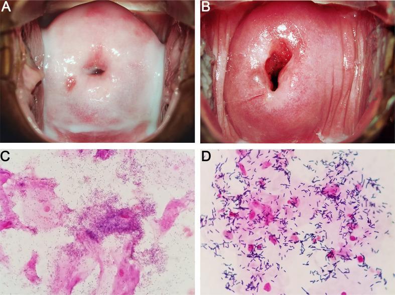
This four-panel image provides a side-by-side comparison of Bacterial Vaginosis (BV) and a normal vaginal microbiome through colposcopic and microscopic examinations. Panels A and B are colposcopic clinical photographs of the cervix and vaginal vault. Panel A (BV) displays characteristic thin, homogeneous, milk-white discharge coating the vaginal walls. In contrast, Panel B (Normal) shows a healthy cervix with clear, physiological secretions. Panels C and D show Gram-stained microscopic slides of vaginal smears. Panel C (BV) demonstrates the presence of 'clue cells'—vaginal epithelial cells heavily encrusted with coccobacilli (Gardnerella-like microbes) that obscure the cell margins. Panel D (Normal) shows a healthy microbiome dominated by large, purple-stained Gram-positive rods, consistent with Lactobacillus species, and clear epithelial cells without bacterial adherence. This comparison illustrates the clinical and laboratory findings used in Amsel criteria and Nugent scoring for the diagnosis of dysbiosis in the female reproductive tract.
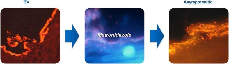
This comparison chart utilizes optical coherence tomography (OCT) or similar high-resolution diagnostic imaging to illustrate the progression of a polymicrobial bacterial vaginosis (BV) biofilm. The visual is divided into three sequential panels. The first panel, labeled 'BV', depicts a dense, continuous, and brightly fluorescent orange-red layer adhering to the vaginal epithelial surface, representing a mature pathogenic biofilm often composed of Gardnerella vaginalis and Atopobium vaginae. The middle panel shows the clinical intervention with 'Metronidazole', showing a blue-tinted transitional phase where the biofilm structure is being disrupted. The final panel, labeled 'Asymptomatic', demonstrates the state after treatment. In this state, the biofilm appears less dense, fragmented, and more diffuse, indicating successful thinning and disruption of the adhering microbial community. This educational visual emphasizes the persistence of subclinical biofilms even in asymptomatic states, which contributes to the high recurrence rates of BV. It is designed for medical professionals and students focusing on gynecology, microbiology, and infectious disease management.
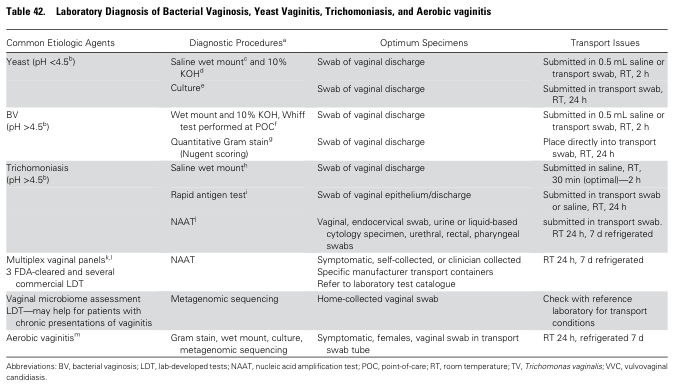
Table 42. Laboratory Diagnosis of Bacterial Vaginosis, Yeast Vaginitis, Trichomoniasis, and Aerobic vaginitis <table><thead><tr><th>Common Etiologic Agents</th><th>Diagnostic Procedures<sup>a</sup></th><th>Optimum Specimens</th><th>Transport Issues</th></tr></thead><tbody><tr><td>Yeast (pH <4.5<sup>b</sup>)</td><td>Saline wet mount<sup>c</sup> and 10% KOH<sup>d</sup></td><td>Swab of vaginal discharge</td><td>Submitted in 0.5 mL saline or transport swab, RT, 2 h</td></tr><tr><td></td><td>Culture<sup>e</sup></td><td>Swab of vaginal discharge</td><td>Submitted in transport swab, RT, 24 h</td></tr><tr><td>BV (pH >4.5<sup>b</sup>)</td><td>Wet mount and 10% KOH, Whiff test performed at POC<sup>f</sup></td><td>Swab of vaginal discharge</td><td>Submitted in 0.5 mL saline or transport swab, RT, 2 h</td></tr><tr><td></td><td>Quantitative Gram stain<sup>g</sup> (Nugent scoring)</td><td>Swab of vaginal discharge</td><td>Place directly into transport swab, RT, 24 h</td></tr><tr><td>Trichomoniasis (pH >4.5<sup>b</sup>)</td><td>Saline wet mount<sup>h</sup></td><td>Swab of vaginal discharge</td><td>Submitted in saline, RT, 30 min (optimal)—2 h</td></tr><tr><td></td><td>Rapid antigen test<sup>i</sup></td><td>Swab of vaginal epithelium/discharge</td><td>Submitted in transport swab or saline, RT, 24 h</td></tr><tr><td></td><td>NAAT<sup>j</sup></td><td>Vaginal, endocervical swab, urine or liquid-based cytology specimen, urethral, rectal, pharyngeal swabs</td><td>submitted in transport swab, RT 24 h, 7 d refrigerated</td></tr><tr><td>Multiplex vaginal panels<sup>k,l</sup> 3 FDA-cleared and several commercial LDT</td><td>NAAT</td><td>Symptomatic, self-collected, or clinician collected Specific manufacturer transport containers Refer to laboratory test catalogue</td><td>RT 24 h, 7 d refrigerated</td></tr><tr><td>Vaginal microbiome assessment LDT—may help for patients with chronic presentations of vaginitis</td><td>Metagenomic sequencing</td><td>Home-collected vaginal swab</td><td>Check with reference laboratory for transport conditions</td></tr><tr><td>Aerobic vaginitis<sup>m</sup></td><td>Gram stain, wet mount, culture, metagenomic sequencing</td><td>Symptomatic, females, vaginal swab in transport swab tube</td><td>RT 24 h, refrigerated 7 d</td></tr></tbody></table> Abbreviations: BV, bacterial vaginosis; LDT, lab-developed tests; NAAT, nucleic acid amplification test; POC, point-of-care; RT, room temperature; TV, Trichomonas vaginalis; VVC, vulvovaginal candidiasis.
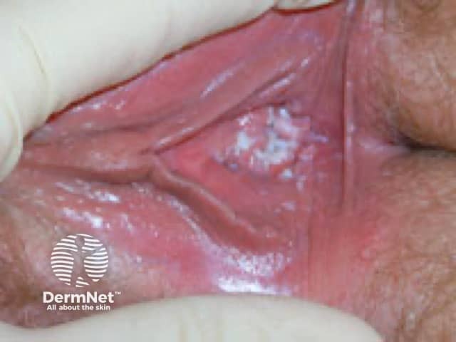
Clinical photography of the external female genitalia demonstrating vulvar mucosal inflammation with characteristic white curd-like plaques consistent with candidal vulvovaginitis. The view focuses on the labia majora and vestibular mucosa, highlighting diffuse erythema, edema, and superficial fissuring surrounding adherent, cottage-cheese-like plaques. The image captures the hallmark appearance of vulvovaginal candidiasis: intense itching, burning, and soreness often accompanying a thick, whitish discharge, though discharge quality may vary. From a diagnostic perspective, this photograph supports a clinical suspicion of Candida species infection, particularly Candida albicans, in an immunocompetent or mildly compromised host. Correlation with microscopy (KOH prep) and culture, or rapid antigen tests, can confirm budding yeast and pseudohyphae within mucosal epithelium. Normal vulvar variants are unlikely given erythema and plaque formation. Differential diagnoses include irritant contact dermatitis, bacterial vaginosis, and trichomoniasis, but the discrete white plaques favor fungal etiology. Timely antifungal therapy, such as azole antifungals, usually yields rapid improvement; recurrence prompts evaluation for diabetes, antibiotic exposure, or immunosuppression. This image is relevant for medical education, dermatology and gynecology training, clinical decision support, and research on vulvar infections, mucosal candidiasis pathophysiology, and patient counseling about symptom management and prevention. Educational value includes differential diagnosis, treatment guidelines, and patient education resources handouts.
White Vaginal Discharge (Leukorrhea)
Definition
1. Physiological Leukorrhea
- Clear to white, non-malodorous
- Thin, slippery consistency
- Scant to variable amount
- Microscopy shows normal epithelial cells with Lactobacillus predominating
- Vaginal pH < 4.5
- Neonates - first 2-3 weeks of life due to maternal estrogen influence
- Around ovulation - thin, egg-white type discharge
- Pregnancy - milky white, non-malodorous (due to increased estrogen-dependent blood flow to vagina)
- Sexual arousal - transient increase
2. Pathological Causes
| Feature | Physiologic | Candidiasis (VVC) | Bacterial Vaginosis (BV) | Trichomoniasis |
|---|---|---|---|---|
| Organism | - | Candida albicans (mostly) | Gardnerella vaginalis + anaerobes | Trichomonas vaginalis |
| Discharge color | Clear/white | White | White or gray | Yellow-green |
| Discharge type | Heterogeneous | Thick, curdy/clumped, adherent plaques | Thin, homogeneous, milky | Frothy, profuse |
| Odor | None | None | Fishy (amine odor) | May be present |
| Itching/inflammation | No | Yes (intense pruritus) | No | Yes (vulvar irritation) |
| Vaginal pH | <4.5 | <4.5 | >4.5 | >5.0 |
| Whiff test | Negative | Negative | Positive | May be positive |
| Microscopy | Normal flora, Lactobacilli | Yeast, pseudohyphae (KOH prep) | Clue cells, few WBCs | Motile trichomonads |
| Treatment | None | Azoles (fluconazole / clotrimazole) | Metronidazole / clindamycin | Metronidazole / tinidazole |
A. Vulvovaginal Candidiasis (VVC)
- Thick, white "cottage cheese" or curdy discharge
- Intense pruritus, vulvar soreness, dyspareunia, external dysuria
- Predisposed by: antibiotics, high estrogen states (pregnancy, OCP), poorly controlled diabetes, immunosuppression
- C. albicans is part of normal flora in ~20% of women; becomes pathogenic with predisposing factors
- Diagnosis: KOH wet mount shows budding yeast + pseudohyphae
- Uncomplicated: single dose oral fluconazole 150 mg OR topical azoles (clotrimazole, miconazole)
- Recurrent (≥4 episodes/year): fluconazole weekly for 6 months
B. Bacterial Vaginosis (BV)
- Most common vaginosis (accounts for ~40% of cases)
- Thin, homogeneous grayish-white discharge; low viscosity (milk-like)
- Fishy odor (worse after intercourse or menses)
- No inflammation of vulva/vagina
- Caused by overgrowth of anaerobes replacing Lactobacillus
- Amsel criteria (≥3 of 4 required for diagnosis):
- Homogeneous thin grayish-white discharge
- pH > 4.5
- Positive whiff-amine test (KOH)
- Clue cells on microscopy (epithelial cells encrusted with coccobacilli)
- Oral metronidazole 500 mg twice daily x 7 days
- Or intravaginal metronidazole gel or clindamycin cream
- Treat in pregnancy to reduce risk of preterm birth
C. Trichomoniasis
- Caused by the flagellated protozoan Trichomonas vaginalis
- Most common non-viral STI worldwide
- Profuse, frothy, yellow-green discharge (not truly white)
- Vulvar irritation; "strawberry cervix" on speculum exam (punctate hemorrhages)
- Often asymptomatic in men (can cause male urethritis)
- Diagnosis: wet mount (motile trichomonads) or NAAT (preferred - higher sensitivity)
- Tinidazole 2 g single dose (preferred) OR metronidazole 2 g single dose
- Treat sexual partner simultaneously
D. Other Causes of White/Abnormal Discharge
| Cause | Notes |
|---|---|
| Cervicitis (Chlamydia, Gonorrhea) | Mucopurulent cervical discharge; Chlamydia is most common STI pathogen (~40% of cervicitis); treat with azithromycin/doxycycline |
| Atrophic vaginitis | Postmenopausal; thin dry mucosa; treat with topical estrogen |
| Retained foreign body | Malodorous discharge; common in children |
| Cervical polyp/ectropion | Can cause increased mucoid discharge |
| Physiologic (pregnancy) | Milky white, non-malodorous - estrogen effect |
3. Diagnostic Approach
- Vaginal pH (sidewall swab)
- Saline wet mount - clue cells, trichomonads
- KOH wet mount - pseudohyphae (candida)
- Whiff test (KOH + discharge)
- NAAT for Chlamydia, Gonorrhea, T. vaginalis
- Nugent scoring (Gram stain) - gold standard for BV
Summary Table: Quick Differential
| White discharge + itching | Think Candidiasis |
|---|---|
| White/gray discharge + fishy smell | Think Bacterial Vaginosis |
| Frothy yellowish + frothy + STI risk | Think Trichomoniasis |
| White, no smell, no itch, pregnancy/newborn | Physiological |
| Mucopurulent + cervical tenderness | Think Cervicitis / Chlamydia / GC |