I now have comprehensive information from multiple authoritative sources. Let me compile the complete detailed answer.
Bile Metabolism - Detailed Study with Clinical Significance
Note: I've interpreted "Bike metabolism" as Bile metabolism (common autocorrect/typo). If you meant something else, please clarify.
1. Overview of Bile
Bile is a complex aqueous secretion produced by hepatocytes (~500-600 mL/day). Its major organic components are:
- Bile acids/salts (primary organic solute)
- Phospholipids (mainly phosphatidylcholine)
- Cholesterol (unesterified)
- Bilirubin (conjugated)
- Proteins and electrolytes
Bile acids are amphipathic molecules - they have both hydrophilic and hydrophobic faces - making them ideal detergents for fat digestion and absorption.
2. Bile Acid Synthesis
Primary Bile Acid Synthesis (in Liver)
Bile acids are synthesized from cholesterol in the pericentral hepatocytes of the hepatic acinus. Two primary (unconjugated) bile acids are produced:
| Bile Acid | Type | Hydroxyl Groups |
|---|
| Cholic acid (CA) | Trihydroxy | C-3, C-7, C-12 |
| Chenodeoxycholic acid (CDCA) | Dihydroxy | C-3, C-7 |
Normal hepatic bile acid synthesis = 0.2-0.6 g/day, but can rise to 4-6 g/day after small bowel resection.
Two Major Biosynthetic Pathways
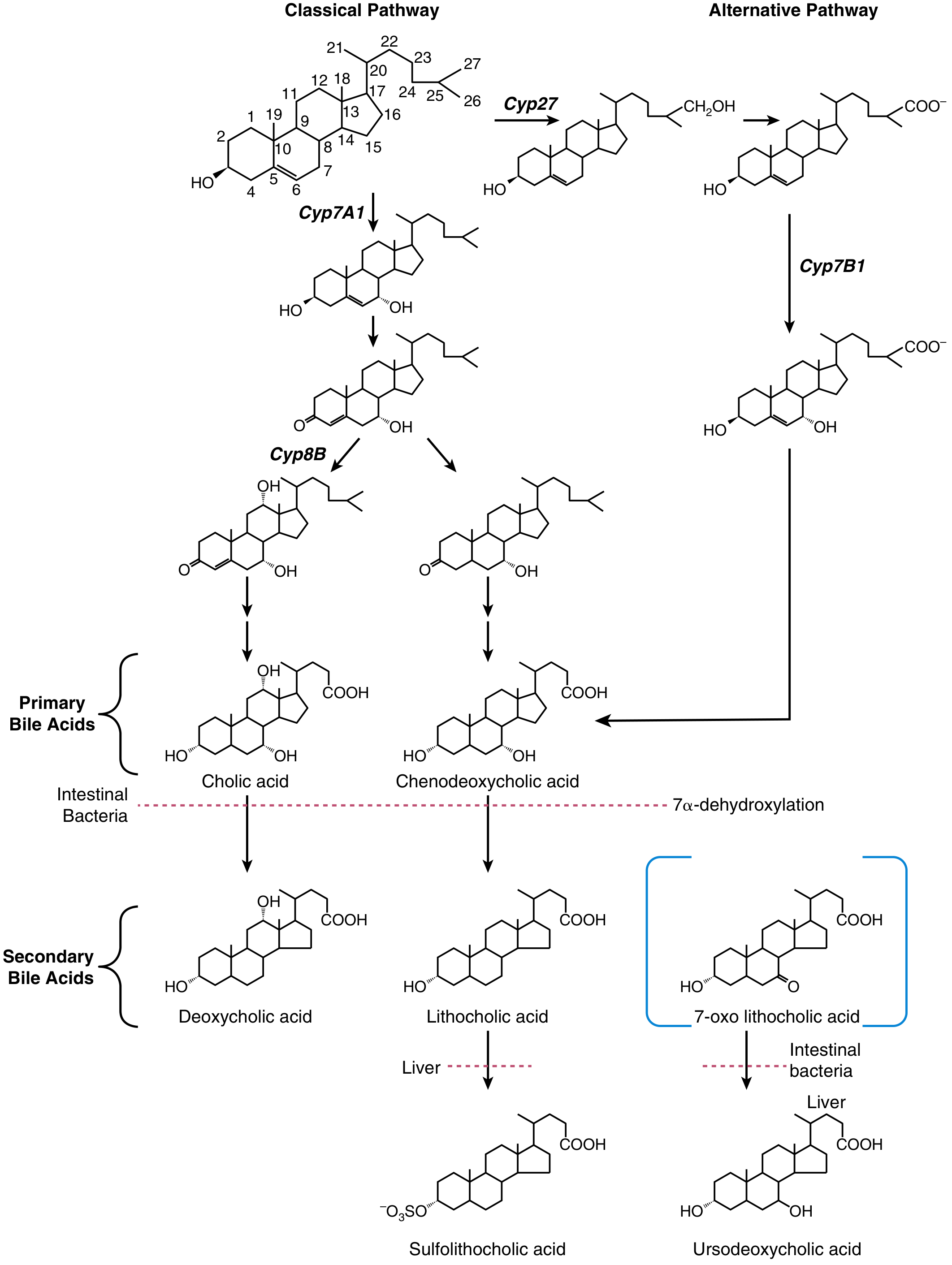
A. Classical (Neutral) Pathway - CYP7A1/CYP8B1
- Dominant pathway in humans
- Rate-limiting enzyme: Cholesterol 7α-hydroxylase (CYP7A1)
- Favors cholic acid synthesis (requires CYP8B1 for 12α-hydroxylation)
- Regulated by negative feedback from bile acids returning to the liver
B. Alternative (Acidic) Pathway - CYP27A1/CYP7B1
- Initiated by sterol 27-hydroxylase (CYP27A1)
- Favors chenodeoxycholic acid (CDCA) synthesis
- Produces oxysterol intermediates first
Fig. 64.2 - Bile acid synthesis pathways showing Classical (CYP7A1/CYP8B1) and Alternative (CYP27A1/CYP7B1) routes. Source: Sleisenger & Fordtran's GI and Liver Disease.
Conjugation (Amidation)
Before secretion, primary bile acids are conjugated (amidated) with:
- Glycine (75%) → glycocholic acid, glycochenodeoxycholic acid
- Taurine (25%) → taurocholic acid, taurochenodeoxycholic acid
Conjugation lowers the pKa, keeping bile acids ionized and water-soluble at intestinal pH, and prevents passive back-diffusion. Conjugated bile acids are the predominant form in bile.
3. Secondary Bile Acids (Bacterial Metabolism)
After reaching the colon, the intestinal flora transform primary bile acids into secondary bile acids:
| Reaction | Product | Enzyme |
|---|
| 7α-dehydroxylation of CA | Deoxycholic acid (DCA) | Intestinal bacteria |
| 7α-dehydroxylation of CDCA | Lithocholic acid (LCA) | Intestinal bacteria |
| Epimerization of CDCA | Ursodeoxycholic acid (UDCA) | Intestinal bacteria + liver |
| Sulfation of LCA | Sulfolithocholic acid | Liver/kidney |
- LCA is highly hepatotoxic and is largely sulfated at the C-3 position (detoxified) by liver/kidney
- UDCA (the 7β-epimer of CDCA) has become an important therapeutic agent
[Sleisenger & Fordtran's GI and Liver Disease, p.1201-1202]
4. Bile Acid Pool and Kinetics
| Bile Acid | Pool Size (mg) | Fractional Turnover (days⁻¹) | Hepatic Synthesis (mg/day) |
|---|
| Cholic acid | 500-1500 | 0.2-0.5 | 120-400 |
| Chenodeoxycholic acid | 500-1500 | 0.2-0.5 | 120-250 |
| Deoxycholic acid | 500-1000 | 0.3-0.6 | 0 (from CA) |
Total bile acid pool = ~2.5 g; fecal loss = ~600 mg/day (replaced by new synthesis).
5. Enterohepatic Circulation (EHC)
This is the cornerstone of bile acid conservation - a continuous recirculation loop between the intestine and liver.
Steps in EHC
- Hepatic secretion: Bile acids secreted into bile canaliculi via the Bile Salt Export Pump (BSEP/ABCB11) at the canalicular membrane
- Gallbladder storage: Bile concentrated 5-10x during fasting
- Meal-triggered release: Cholecystokinin (CCK) causes gallbladder contraction, releasing bile into duodenum
- Intestinal transit: Bile acids remain in lumen throughout the small intestine (high concentration = maximal fat digestion)
- Ileal reabsorption: Terminal ileum - Apical Sodium-dependent Bile Acid Transporter (ASBT/SLC10A2) actively recaptures bile acids
- Transcellular transport: IBABP (ileal bile acid binding protein) transports across enterocyte
- Basolateral efflux: OSTα-OSTβ heterodimer exports bile acids into portal blood
- Portal return: Bile acids travel to liver (portal vein concentration 20-50 μmol/L)
- Hepatic re-uptake: NTCP (Na⁺-taurocholate cotransporting polypeptide/SLC10A1) at the sinusoidal membrane - first-pass extraction 50-90%
- Re-secretion: Cycle repeats
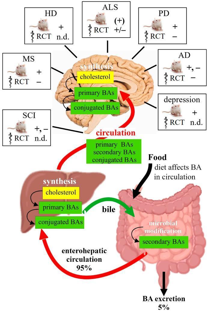
The EHC recycles bile acids 6-10 times/day. More than 95% of secreted bile acids are conserved per cycle; only 5% are excreted in feces.
[Costanzo Physiology, 7th Ed., p.379]
6. Regulation of Bile Acid Synthesis: FXR-FGF19 Axis
The Farnesoid X Receptor (FXR) - a nuclear receptor - is the master regulator of bile acid homeostasis. It mediates negative feedback through four key mechanisms:
-
Hepatic FXR → SHP pathway: FXR induces Small Heterodimer Partner (SHP), which inhibits LRH-1 (liver receptor homolog-1), suppressing CYP7A1 expression (rate-limiting enzyme)
-
Ileal FXR → FGF19 pathway: FXR in ileal enterocytes stimulates synthesis and secretion of Fibroblast Growth Factor 19 (FGF19) into portal blood → activates FGFR4 on hepatocytes → represses CYP7A1
-
Hepatic FXR on transporters: Upregulates BSEP (↑ biliary secretion) and downregulates NTCP (↓ uptake) → reduces intracellular bile acid accumulation
-
Ileal FXR on transporters: Downregulates ASBT (↓ intestinal absorption) and upregulates OSTα-OSTβ (↑ efflux) → reduces intracellular bile acids in enterocytes
Additionally, TGR5 (a G protein-coupled receptor on cholangiocytes and gallbladder epithelium) is activated by bile acids → ↑ cAMP → Cl⁻ secretion, and in brown adipose tissue → stimulates thyroid hormone activation and energy expenditure.
[Medical Physiology, p.473-488]
7. Hepatic Transport Proteins Summary
| Transporter | Location | Function |
|---|
| NTCP (SLC10A1) | Hepatocyte sinusoidal | Na⁺-dependent bile acid uptake |
| OATPs (SLCO family) | Hepatocyte sinusoidal | Na⁺-independent organic anion uptake |
| BSEP (ABCB11) | Canalicular | Primary bile acid secretion into bile |
| MRP2 (ABCC2) | Canalicular | Conjugated bilirubin, sulfated bile acids |
| MDR3 (ABCB4) | Canalicular | Phospholipid secretion |
| ASBT (SLC10A2) | Ileal brush border | Ileal bile acid reabsorption |
| OSTα-OSTβ | Basolateral of enterocyte | Bile acid export to portal blood |
8. Clinical Significance
8.1 Cholestasis
Cholestasis is defined as interruption of normal bile formation, subdivided into:
- Intrahepatic cholestasis: functional defect at hepatocyte level (transporter mutations)
- Extrahepatic cholestasis: mechanical obstruction in biliary tract
Disorders are classified into:
- Defects in bile acid synthesis and conjugation
- Defects in membrane transport (uptake/secretion)
- Disturbances involving bacterial transformation
- Disturbances in movement between organs
Key inherited cholestatic disorders:
- Progressive Familial Intrahepatic Cholestasis (PFIC):
- PFIC1: ATP8B1 mutations (FIC1 protein)
- PFIC2: ABCB11 mutations (BSEP)
- PFIC3: ABCB4 mutations (MDR3)
- PFIC5: NR1H4 mutations (FXR itself)
- Intrahepatic Cholestasis of Pregnancy (ICP): Elevated fasting serum bile acids are an early marker; UDCA is used for treatment
[Sleisenger & Fordtran's, p.1209]
8.2 Bile Acid Malabsorption (BAM)
- Caused by ASBT gene mutations (primary bile acid malabsorption) or ileal resection/disease (e.g., Crohn's disease)
- Results in steatorrhea (fat malabsorption), diarrhea, and vitamin deficiencies (fat-soluble vitamins A, D, E, K)
- When loss exceeds hepatic synthesis capacity → bile acid pool depleted → impaired fat digestion
- OSTβ gene (SLC51B) mutations cause congenital chronic diarrhea
8.3 Gallstone Disease (Cholelithiasis)
Two types:
- Cholesterol gallstones (~80%): result from imbalance between cholesterol, bile acids, and phospholipids in bile
- Supersaturation with cholesterol → nucleation of cholesterol crystals
- Risk factors: obesity, female sex, pregnancy, rapid weight loss, fibrates
- Mutations in ABCG5/G8 (cholesterol transporter) increase risk
- Pigment stones: excess bilirubin (hemolytic anemias, UGT1A1 polymorphisms)
Cholelithiasis pathophysiology: Insufficient bile acids relative to cholesterol → excess cholesterol-enriched vesicles → nucleation → crystal/stone formation. [Medical Physiology, p.493]
8.4 Primary Biliary Cholangitis (PBC)
- Autoimmune destruction of intrahepatic bile ducts
- Treatment: UDCA (first-line) - stimulates biliary bicarbonate secretion, delays progression, reduces liver transplant need
- Second-line: Obeticholic acid (OCA) - a semi-synthetic FXR agonist
- Combination with bezafibrate (PPAR agonist) also used in UDCA partial-responders
8.5 Primary Sclerosing Cholangitis (PSC)
- Fibro-inflammatory stricturing of intra/extrahepatic bile ducts
- norUDCA (C-23 analog of UDCA, resistant to conjugation) - undergoes cholehepatic shunting → showed benefit in a Phase 2 clinical trial
8.6 Bile Acid Synthesis Defects
- Rare inborn errors of bile acid biosynthesis
- Present with markedly reduced/absent CA and CDCA + elevated atypical bile acids in bile, serum, and urine
- Diagnosed by fast atom bombardment mass spectrometry or electrospray ionization-tandem MS of urine/bile
- Treatment: Unconjugated cholic acid administration - suppresses cytotoxic precursor synthesis and restores primary bile acids in the EHC
- Rare patients with conjugation defects require glycocholic acid (conjugated form)
8.7 Metabolic Syndrome - Bile Acids as Metabolic Regulators
Bile acids are now recognized as systemic metabolic hormones via FXR and TGR5:
| Receptor | Tissue | Metabolic Effect |
|---|
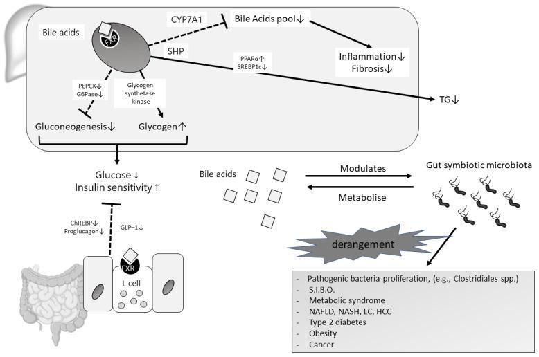
| FXR | Liver | ↓ Gluconeogenesis (inhibits PEPCK/G6Pase), ↑ glycogen synthesis → ↓ blood glucose, ↑ insulin sensitivity |
| FXR | Liver | ↓ Triglycerides (via PPAR-α, SREBP1c modulation) |
| FXR | Liver | ↓ Inflammation and fibrosis (via ↓ bile acid pool) |
| TGR5 | Intestinal L-cells | ↑ GLP-1 secretion → ↑ insulin release |
| TGR5 | Brown adipose/muscle | Activates DIO2 (type 2 deiodinase) → ↑ T3 → ↑ energy expenditure |
This explains why bariatric surgery (Roux-en-Y gastric bypass) - which markedly alters bile acid circulation - leads to metabolic improvements beyond mere caloric restriction.
8.8 Gut Microbiome - Bile Acid Axis
- The gut microbiome extensively modifies bile acids (7α-dehydroxylation, deconjugation, epimerization)
- Dysbiosis → altered secondary bile acid profile → disturbs FXR/TGR5 signaling → contributes to NAFLD, NASH, liver cirrhosis, HCC, T2DM, and obesity
- Emerging as a therapeutic target in inflammatory bowel disease, C. difficile infection, and colorectal cancer
8.9 Pruritus in Cholestasis
- Bile acid accumulation in skin was thought to cause itching, but the mechanism is more complex
- Current evidence implicates lysophosphatidic acid and endogenous opioids more than bile acids directly
- Bile acid sequestrants (cholestyramine, colestipol, colesevelam) provide modest relief
- ASBT inhibitors (blocking ileal reabsorption) - in clinical development for cholestatic pruritus
8.10 Hypercholesterolemia Treatment
- Bile acid sequestrants (cholestyramine, colesevelam) bind bile acids in gut → interrupt EHC → force liver to synthesize more bile acids from cholesterol → ↓ serum LDL cholesterol
- Colesevelam also approved for glycemic control in type 2 diabetes
9. Bile Acid-Based Therapeutics Summary
| Drug | Mechanism | Approved Indication |
|---|
| UDCA | Displaces hydrophobic bile acids, bicarbonate-rich choleresis | PBC, ICP, gallstone dissolution |
| Obeticholic acid (OCA) | Semi-synthetic FXR agonist | PBC (second-line) |
| norUDCA | Cholehepatic shunting, anti-fibrotic | PSC (investigational) |
| Cholestyramine/colestipol | Bile acid sequestrants | Hypercholesterolemia, pruritus, bile acid diarrhea |
| Colesevelam | Bile acid sequestrant | Hypercholesterolemia + T2DM |
| ASBT inhibitors | Block ileal bile acid reabsorption | Chronic constipation (Japan), cholestasis (investigational) |
| FGF19 mimetics | Inhibit CYP7A1 (mimic ileal FXR→FGF19 signal) | PSC, PBC (in development) |
10. Key Lab Values
| Parameter | Normal | Clinical Relevance |
|---|
| Serum bile acids (fasting) | 2-5 μmol/L | Elevated in cholestasis |
| Serum bile acids (fed) | 5-15 μmol/L | Elevated in ICP, liver disease |
| Fecal bile acid loss | ~600 mg/day | ↑ in BAM, ileal resection |
| Bile acid pool | ~2.5 g | ↓ in BAM, ileal resection, PBC |
Elevated fasting serum bile acids are an early sensitive marker of intrahepatic cholestasis of pregnancy (ICP), detectable before bilirubin rises.
Recent Evidence (2024-2026)
- A 2025 review (PMID 40602290) in Pharmacological Reviews highlights altered bile acid metabolism and signaling as a key driver in hepatocellular carcinoma (HCC) pathogenesis - both as a biomarker and therapeutic target
- A 2025 review (PMID 39419312) in Clinical Chimica Acta covers the emerging role of the bile acid profile as a clinical biomarker panel beyond total serum bile acids
- A 2026 Lancet Gastroenterology review (PMID 41547360) links small intestinal microbial dysbiosis to disturbed bile acid metabolism as a core pathophysiological mechanism
Sources:
- Sleisenger & Fordtran's GI and Liver Disease, Chapter 64 - bile acid synthesis, EHC, clinical disorders
- Costanzo Physiology, 7th Ed. - enterohepatic circulation mechanics
- Medical Physiology (Boron & Boulpaep) - FXR regulation, gallstones
- Harper's Illustrated Biochemistry, 32nd Ed. - CYP7A1 regulation