
Teratology of fallots
Reading File
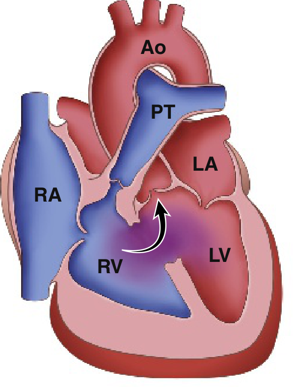
Tetralogy of Fallot (TOF) is the most common cyanotic congenital heart defect, accounting for about 10% of all congenital heart disease. It consists of four anatomical defects that result from a single developmental error - anterior and superior displacement of the infundibular (outlet) septum.
Searching sources...
Finding Sources
Searching PubMed
"tetralogy of Fallot"[MeSH Terms] AND management
Reading File
Reading File
Reading File
Reading File
Loading Image
Loading Image
Here is a comprehensive overview of Tetralogy of Fallot (TOF), drawing from Robbins Basic Pathology, Rosen's Emergency Medicine, and The Developing Human.
Tetralogy of Fallot (TOF)
TOF is the most common cause of cyanotic congenital heart disease beyond infancy, accounting for approximately 5-10% of all congenital cardiac malformations.
The Four Cardinal Features
All four defects stem from a single embryologic error: anterosuperior displacement of the infundibular (outlet/conus) septum, causing unequal division of the truncus arteriosus.
| Feature | Description |
|---|---|
| 1. Ventricular Septal Defect (VSD) | Large, unrestrictive, misaligned defect near the membranous septum |
| 2. Right Ventricular Outflow Tract (RVOT) Obstruction | Subpulmonic (infundibular) stenosis; sometimes pulmonary valve stenosis or complete atresia |
| 3. Overriding Aorta | Aortic valve straddles the VSD, receiving blood from both ventricles |
| 4. Right Ventricular Hypertrophy (RVH) | Secondary to the high pressure load from RVOT obstruction |
The degree of RVOT obstruction determines clinical severity. The first three features are structural; RVH is a secondary consequence.
Anatomical Diagram
Hemodynamics
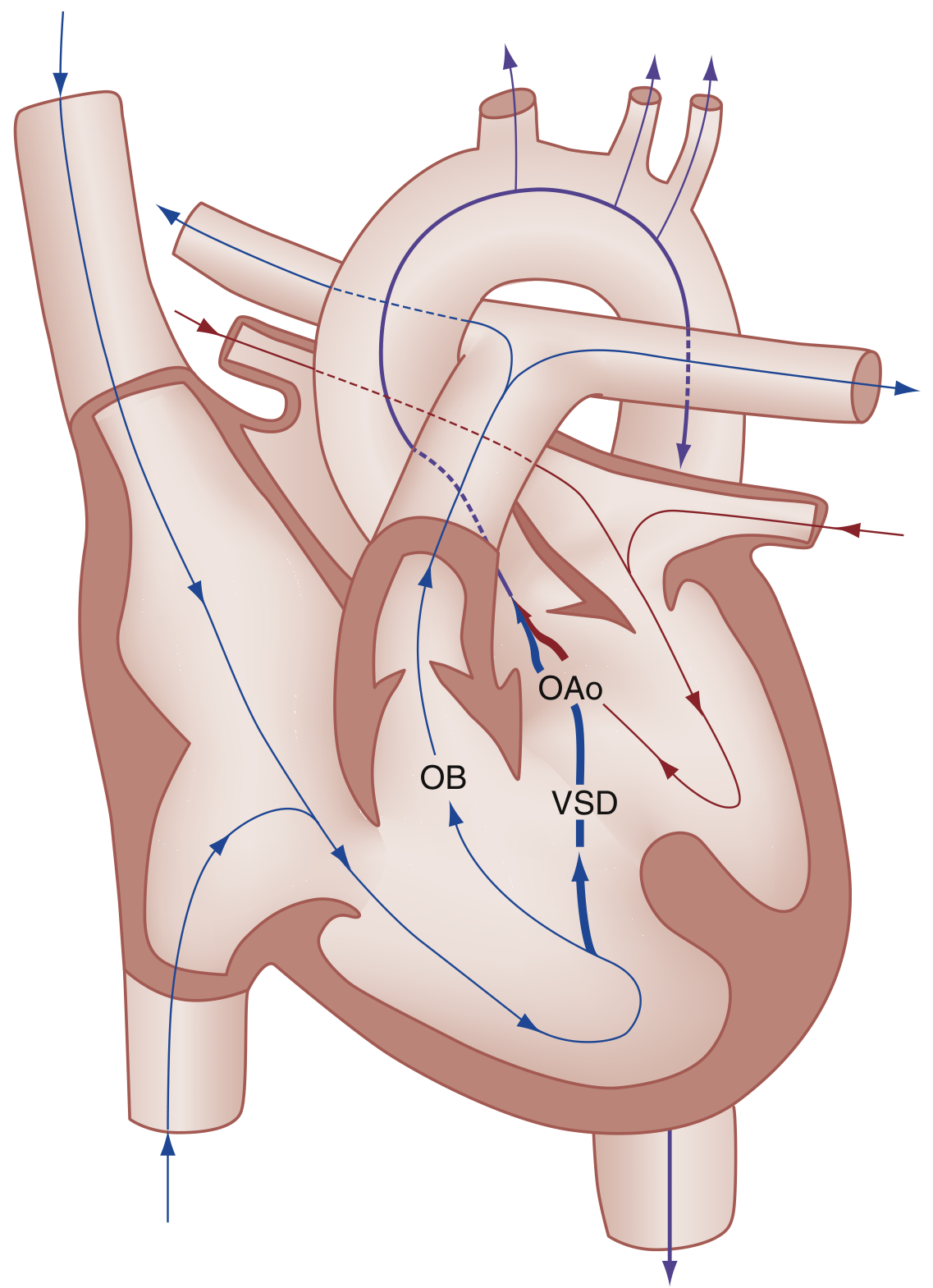
- Decreased pulmonary blood flow due to RVOT obstruction
- Right-to-left shunting across the VSD → deoxygenated blood enters systemic circulation
- Clinical severity directly proportional to the degree of RVOT obstruction
Clinical Features
At presentation:
- Cyanosis - typically not present at birth; worsens within weeks to months
- "Pink Tet" - mild RVOT obstruction causes only left-to-right shunt with no cyanosis initially
- Cyanosis worsens during crying and feeding (infants), or physical exertion (older children)
- Systolic ejection murmur along the left sternal border
- Chronic hypoxemia leads to compensatory polycythemia and finger/toe clubbing
Tet Spells (Hypercyanotic/Hypoxic Spells)
A life-threatening complication; peak incidence at 2-4 months of age.
Trigger: Any event that suddenly lowers systemic vascular resistance (SVR) - crying, defecation, hypovolemia, tachycardia.
Vicious cycle:
- SVR falls → increased R-to-L shunt across VSD
- ↓ PaO₂, ↑ PCO₂, ↓ arterial pH
- Stimulates respiratory center → hyperpnea
- Increased venous return to RV → more shunting → worsening hypoxia
Management of Tet Spells (Box 165.8 - Rosen's):
| Intervention | Details |
|---|---|
| Knee-to-chest position | Increases SVR, reduces R-to-L shunt |
| Supplemental oxygen | Limited value alone |
| Morphine | 0.1-0.2 mg/kg IV or IM - blunts hyperpnea drive |
| Fentanyl | 1 mcg/kg IV/IM (or 1.5-2 mcg/kg intranasal) as alternative |
| IV fluids | Correct hypovolemia |
| Phenylephrine | Increases SVR |
| Beta-blockers (propranolol) | Reduce heart rate and RVOT spasm |
Investigations
| Test | Finding |
|---|---|
| Chest X-ray | "Boot-shaped" heart (coeur en sabot) due to RVH; concave pulmonary artery segment; decreased pulmonary vascular markings |
| ECG | Right ventricular hypertrophy; right axis deviation |
| Echocardiography | Diagnostic - defines all four components |
| Right-sided aortic arch | Present in ~25% of cases |
Morphology Summary (Robbins)
- Enlarged, boot-shaped heart due to RVH
- Proximal aorta is dilated; pulmonary trunk is hypoplastic
- Left-sided chambers are normal size
- In pulmonary atresia (extreme form): blood reaches lungs only via patent ductus arteriosus or dilated bronchial arteries
Associated Defects
TOF is frequently associated with:
- Right-sided aortic arch (~25%)
- Atrial septal defect (ASD)
- Anomalous origin of the left coronary artery
Treatment
- Prostaglandin E1 (PGE1) infusion in severe neonatal cases - maintains PDA to preserve pulmonary blood flow
- Surgical repair is definitive: VSD closure + RVOT reconstruction
- Staged vs. primary repair in symptomatic neonates is an active area of research - recent meta-analysis (Bashir et al., 2026) of 4,628 neonates found outcomes data favoring primary repair in most centers
- In pulmonary atresia variant: initial shunt palliation (e.g., Blalock-Taussig shunt) may be needed before definitive correction
- Long-term: pulmonary regurgitation after repair often requires eventual pulmonary valve replacement (surgical or transcatheter)
Key memory hook: "PROVE" - Pulmonic stenosis, RVH, Overriding aorta, VSD, all from one embryologic defect (infundibular septal maldevelopment).
Sources: Robbins & Kumar Basic Pathology; ROSEN's Emergency Medicine; The Developing Human, Clinically Oriented Embryology
This is a shared conversation. Sign in to Orris to start your own chat.