
Tell me about peristalsis for physiology 1st year mbbs
Reading File
Finding Sources
Finding Sources
Searching PubMed
peristalsis AND physiology
Searching the Web
peristalsis physiology intestinal peristaltic reflex enteric nervous system
Reading File
Reading File
Peristalsis
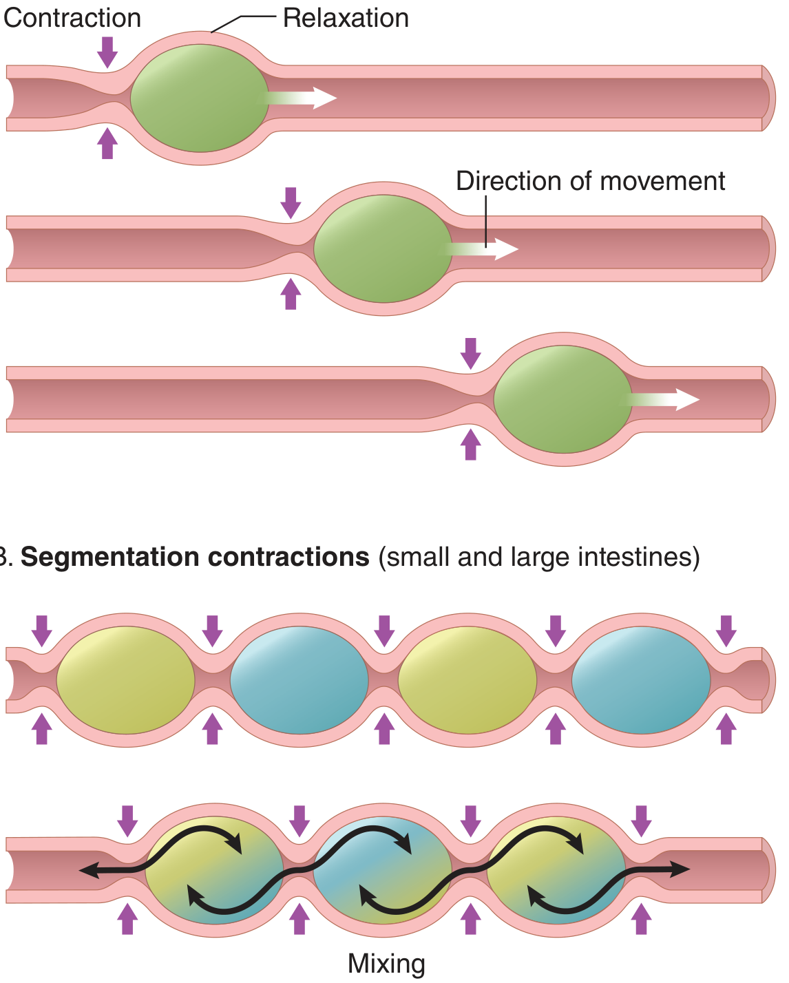
Peristalsis is the basic propulsive movement of the gastrointestinal tract. It moves food and intestinal contents forward, usually in the aboral direction (towards the anus).
Simple definition
It is a coordinated wave in which:
- The gut contracts behind a bolus of food.
- The gut relaxes in front of the bolus.
- This pushes the contents forward.
Think of squeezing toothpaste from behind the tube.
Mechanism: peristaltic reflex
The usual stimulus is distension of the gut wall by food or fluid.
- A bolus distends the intestinal wall.
- Sensory neurons in the enteric nervous system are stimulated.
- The circular muscle behind the bolus contracts.
- The circular muscle ahead of the bolus relaxes.
- The bolus is pushed forward.
Neural basis
The myenteric plexus (Auerbach plexus) is essential for effective peristalsis.
| Area relative to bolus | Motor response | Important mediators |
|---|---|---|
| Behind the bolus, oral side | Contraction of circular muscle | Acetylcholine, substance P |
| Ahead of the bolus, anal side | Relaxation of circular muscle | Nitric oxide, VIP |
The contraction behind and relaxation ahead together form the peristaltic reflex or myenteric reflex.
Law of the gut
The peristaltic wave normally proceeds towards the anus because the enteric nervous system is functionally polarized in the anal direction.
This is called the:
Law of the gut = peristaltic reflex + anal direction of movement.
In the intestine, the contractile ring begins about 2-3 cm proximal (oral) to the distended segment and propels contents distally. Downstream relaxation is called receptive relaxation. - Guyton and Hall Textbook of Medical Physiology, pp. 781-782.
Where does peristalsis occur?
Peristalsis occurs in smooth muscle tubes, including:
- Esophagus
- Stomach
- Small intestine
- Large intestine
- Bile ducts
- Ureters
Peristalsis versus segmentation
| Feature | Peristalsis | Segmentation |
|---|---|---|
| Main function | Propulsion | Mixing |
| Movement of contents | Forward, aboral | Little or no net forward movement |
| Pattern | A travelling wave | Local, intermittent ring contractions |
| Importance | Moves chyme along the gut | Mixes chyme with digestive juices and improves absorption |
Factors affecting peristalsis
Increases
- Gut distension
- Mucosal irritation
- Parasympathetic stimulation
- Local enteric reflexes
Decreases
- Sympathetic stimulation
- Anticholinergic drugs such as atropine
- Damage or absence of the myenteric plexus
Clinical correlation
- Hirschsprung disease: congenital absence of enteric ganglion cells, causing severely impaired peristalsis in the affected colon and functional obstruction.
- Paralytic ileus: intestinal motility, including peristalsis, becomes markedly reduced or absent.
- Atropine: blocks muscarinic cholinergic activity and can markedly depress intestinal peristalsis.
One-line exam answer
Peristalsis is a coordinated propulsive movement in which circular muscle contracts behind a bolus and relaxes ahead of it, moving gastrointestinal contents aborally through the peristaltic reflex mediated mainly by the myenteric plexus.
The NCBI overview of peristalsis similarly describes the importance of the myenteric plexus and cholinergic signaling in normal gut propulsion.
Now tell me about enteric nervous system
Reading File
Finding Sources
Finding Sources
Searching PubMed
"enteric nervous system" AND physiology
Searching the Web
enteric nervous system myenteric submucosal plexus physiology overview NCBI
Reading File
Reading File
Reading File
Reading File
Enteric nervous system (ENS)
The enteric nervous system is an intrinsic network of nerves located entirely within the wall of the gastrointestinal tract, extending from the esophagus to the anus. It is often called the "brain of the gut" because it has sensory neurons, interneurons, and motor neurons and can coordinate many gut functions independently of the CNS.
Its main functions are to regulate:
- Gastrointestinal motility and peristalsis
- Secretion of digestive juices
- Local blood flow
- Absorption and epithelial activity
- Local gut reflexes
Guyton and Hall Textbook of Medical Physiology, pp. 777-778.
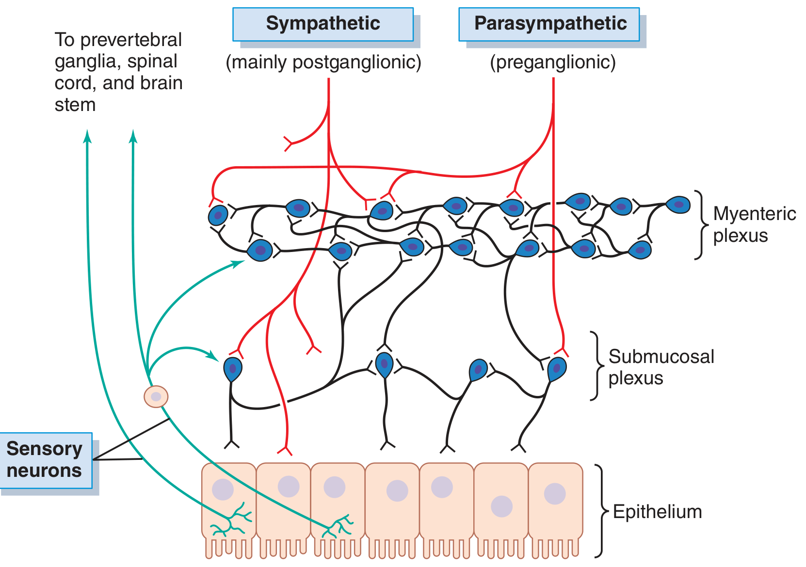
Two plexuses of ENS
| Feature | Myenteric plexus | Submucosal plexus |
|---|---|---|
| Other name | Auerbach plexus | Meissner plexus |
| Location | Between the longitudinal and circular muscle layers | In the submucosa |
| Main role | Motility of the gut | Secretion and local blood flow |
| Effect of stimulation | Increases tone, intensity and coordination of contractions | Regulates glandular secretion, absorption, and mucosal blood flow |
1. Myenteric plexus (Auerbach plexus)
It lies between the outer longitudinal and inner circular muscle layers.
It mainly controls GI motility by regulating:
- Muscle tone
- Strength of rhythmic contractions
- Frequency of contractions
- Peristalsis
- Relaxation of sphincters
It is therefore very important in the peristaltic reflex.
2. Submucosal plexus (Meissner plexus)
It lies in the submucosa, nearer the mucosa.
Its main functions are:
- Regulation of intestinal and glandular secretion
- Control of local blood flow
- Regulation of absorption
- Detection of chemical changes in the gut lumen
Components of ENS
The ENS has three functional classes of neurons:
-
Sensory neurons
Detect stretch, luminal chemicals, osmolarity, and irritation. -
Interneurons
Connect sensory neurons with motor neurons and coordinate local reflexes. -
Motor neurons
Act on smooth muscle, glands, blood vessels, and endocrine cells.
Important neurotransmitters
| Action | Important neurotransmitters |
|---|---|
| Excitatory, produces contraction | Acetylcholine, substance P |
| Inhibitory, produces relaxation | Nitric oxide, VIP |
For example, during intestinal peristalsis, excitatory neurons produce contraction behind the food bolus, while inhibitory neurons relax the segment ahead of it.
Relation with autonomic nervous system
Although the ENS can work independently, it is influenced by the autonomic nervous system:
- Parasympathetic stimulation via vagus and pelvic nerves generally increases gut motility and secretion.
- Sympathetic stimulation generally decreases motility and secretion and contracts sphincters.
Thus, the ENS is the local controller, while the autonomic nervous system modifies its activity.
Important local reflex
Distension of gut by food activates sensory neurons in the ENS:
- Ascending excitatory pathway causes contraction on the oral side.
- Descending inhibitory pathway causes relaxation on the anal side.
- Food is propelled towards the anus.
This is the basis of the peristaltic reflex and the law of the gut.
Clinical importance
- Hirschsprung disease: congenital absence of enteric ganglion cells in a segment of colon, causing failure of relaxation, constipation, and proximal dilatation.
- Achalasia: impaired inhibitory neural control in the lower esophageal sphincter, causing failure of sphincter relaxation.
- Diabetic autonomic neuropathy: may impair ENS regulation and contribute to gastroparesis or altered bowel motility.
- Anticholinergic drugs can reduce gut motility and produce constipation.
Exam-ready definition
The enteric nervous system is the intrinsic nervous system of the gastrointestinal tract, composed mainly of the myenteric and submucosal plexuses. It independently regulates gut motility, secretion, local blood flow, and gastrointestinal reflexes, while being modified by sympathetic and parasympathetic input.
An NCBI physiology review also summarizes the ENS role in controlling smooth muscle, secretion, and autonomic modulation.
Now tell me about pernicious anaemia
Reading File
Finding Sources
Finding Sources
Searching PubMed
"pernicious anemia"
Searching the Web
pernicious anemia diagnosis intrinsic factor antibodies treatment vitamin B12 guideline
Reading File
Reading File
Reading File
Reading Web Page
https://www.aafp.org/afp/2017/0915/p384
Pernicious anaemia
Pernicious anaemia is a megaloblastic anaemia caused by vitamin B12 deficiency due to autoimmune gastritis. The autoimmune process destroys gastric parietal cells and/or produces antibodies against intrinsic factor, so the body cannot absorb vitamin B12 properly.
It is not simply dietary B12 deficiency.
- Robbins, Cotran & Kumar Pathologic Basis of Disease, p. 609
- Henry's Clinical Diagnosis and Management by Laboratory Methods, pp. 1934-1935
Normal vitamin B12 absorption
- Vitamin B12 in food is released in the stomach.
- It binds to intrinsic factor (IF), made by gastric parietal cells.
- The B12-IF complex travels to the terminal ileum.
- It binds to receptors on ileal cells and is absorbed.
In pernicious anaemia, intrinsic factor is absent or ineffective, so B12 cannot be absorbed by the normal pathway.
Etiology and pathogenesis
Autoimmune destruction affects mainly the fundus and body of the stomach, producing chronic atrophic gastritis.
Autoantibodies include:
- Anti-parietal cell antibodies against H+/K+ ATPase
- Anti-intrinsic factor antibodies, which are more specific for pernicious anaemia
Consequences:
- Loss of parietal cells
- Reduced intrinsic factor
- Achlorhydria, or markedly decreased gastric acid
- Failure of B12 absorption
- Vitamin B12 depletion
- Impaired DNA synthesis leading to megaloblastic anaemia
Body stores of B12 are large, so symptoms may appear only years after the autoimmune process begins.
Clinical features
Due to anaemia
- Fatigue and weakness
- Pallor
- Dyspnoea on exertion
- Palpitations
- Light-headedness
Gastrointestinal features
- Loss of appetite
- Glossitis: smooth, painful, beefy-red tongue
- Mild diarrhoea or weight loss may occur
Neurological features of B12 deficiency
- Tingling or numbness in hands and feet
- Loss of vibration and position sense
- Unsteady gait or ataxia
- Cognitive or mood changes in severe deficiency
Neurological signs may occur even when anaemia is mild or absent.
Investigations
| Investigation | Typical finding |
|---|---|
| CBC | Macrocytic anaemia, increased MCV; sometimes pancytopenia |
| Peripheral smear | Macro-ovalocytes and hypersegmented neutrophils |
| Serum B12 | Low |
| Reticulocyte count | Low before treatment |
| LDH and indirect bilirubin | Increased due to ineffective erythropoiesis |
| Homocysteine | Increased |
| Methylmalonic acid | Increased in B12 deficiency |
| Anti-intrinsic factor antibody | Highly specific for pernicious anaemia |
| Anti-parietal cell antibody | Sensitive but less specific |
| Serum gastrin | May be raised because of achlorhydria |
An anti-intrinsic factor antibody test is useful when B12 deficiency has no clear dietary or other malabsorptive cause. A positive test strongly supports pernicious anaemia, as noted in the AAFP review.
Important associated conditions
Pernicious anaemia is associated with other autoimmune disorders, especially:
- Autoimmune thyroid disease
- Type 1 diabetes mellitus
- Vitiligo
Autoimmune gastritis can also reduce iron absorption. Therefore, iron deficiency may appear before B12 deficiency.
Treatment
The treatment is vitamin B12 replacement for life, because the intrinsic factor defect usually persists.
- Parenteral B12, such as intramuscular hydroxocobalamin or cyanocobalamin, is commonly used for initial replacement, particularly if there are neurological symptoms.
- High-dose oral B12 can also be effective in selected patients because a small amount is absorbed by passive diffusion.
- Response is monitored with symptoms, reticulocyte response, haemoglobin, MCV, and B12 status.
Important caution
Do not give folic acid alone if B12 deficiency is possible. Folate can correct the anaemia while neurological damage from B12 deficiency continues or worsens.
Pernicious anaemia versus nutritional B12 deficiency
| Feature | Pernicious anaemia | Nutritional B12 deficiency |
|---|---|---|
| Basic cause | Autoimmune loss of intrinsic factor | Low dietary B12 intake |
| Intrinsic factor | Reduced or absent | Usually normal |
| Anti-IF antibody | May be positive | Negative |
| Treatment duration | Usually lifelong B12 replacement | Treat deficiency and improve diet |
| Associated disorders | Other autoimmune diseases | Often strict vegan diet, malnutrition, etc. |
One-line exam answer
Pernicious anaemia is a megaloblastic anaemia caused by vitamin B12 malabsorption due to autoimmune atrophic gastritis with loss of intrinsic factor-producing parietal cells.
Flowchart for revision
Autoimmune gastritis
→ destruction of parietal cells
→ ↓ intrinsic factor
→ ↓ terminal ileal absorption of vitamin B12
→ impaired DNA synthesis
→ megaloblastic anaemia with possible neurological manifestations.
→ destruction of parietal cells
→ ↓ intrinsic factor
→ ↓ terminal ileal absorption of vitamin B12
→ impaired DNA synthesis
→ megaloblastic anaemia with possible neurological manifestations.
How about basal electrical rhythm
Reading File
Finding Sources
Finding Sources
Searching PubMed
gastrointestinal slow waves physiology
Searching the Web
gastrointestinal basal electrical rhythm slow waves interstitial cells Cajal physiology
Reading File
Reading File
Reading File
Basal electrical rhythm (BER)
Basal electrical rhythm, also called the basic electrical rhythm, slow-wave rhythm, or electrical control activity, is the spontaneous rhythmic fluctuation in membrane potential of gastrointestinal smooth muscle.
It sets the maximum possible frequency of phasic contractions in each part of the GI tract.
Slow waves themselves are not action potentials and usually do not cause contraction.
Origin
BER is generated by the interstitial cells of Cajal (ICC), which act as pacemaker cells of the gut.
- ICC generate rhythmic depolarization and repolarization.
- Slow waves spread to neighbouring smooth muscle cells through gap junctions.
- Therefore, smooth muscle in a region contracts in a coordinated rhythm.
Guyton and Hall Textbook of Medical Physiology, pp. 776-777.
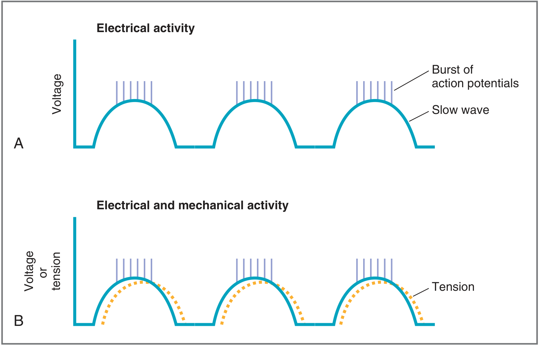
Nature of slow waves
- They are rhythmic oscillations of the resting membrane potential.
- Amplitude is approximately 5-15 mV.
- They move the membrane potential closer to, then away from, threshold.
- Their frequency varies from one GI segment to another.
When the crest of a slow wave reaches threshold, spike potentials occur on top of the slow wave. The spike potentials allow calcium entry into smooth muscle, causing contraction.
Sequence
Slow wave reaches threshold
→ Spike potentials occur
→ Ca2+ enters smooth muscle cell
→ Smooth muscle contracts
→ Spike potentials occur
→ Ca2+ enters smooth muscle cell
→ Smooth muscle contracts
Thus:
- BER determines frequency of possible contractions.
- Spike potentials determine whether contraction occurs and help determine its strength.
Costanzo Physiology, p. 353.
Frequency at different sites
| Site | BER frequency |
|---|---|
| Stomach | 3/min |
| Duodenum | 12/min, approximately 12-13/min in some texts |
| Jejunum | About 10-12/min |
| Terminal ileum | 8-9/min |
| Colon | About 2-6/min, variable |
The stomach has the slowest rhythm, whereas the duodenum has the fastest. Since action potentials can occur only at the peaks of slow waves, a segment cannot contract more frequently than its BER. - Guyton and Hall Textbook of Medical Physiology, p. 776.
Effect of nerves and hormones
The frequency of BER is intrinsic to each region and is usually not changed substantially by autonomic nerves or hormones.
However, nerves and hormones change the excitability of smooth muscle. Therefore, they determine whether slow waves reach threshold and produce spike potentials.
| Factor | Effect on membrane potential and contraction |
|---|---|
| Parasympathetic stimulation, acetylcholine, stretch | Depolarization, more spike potentials, increased contractions |
| Sympathetic stimulation, noradrenaline | Hyperpolarization, fewer spike potentials, decreased contractions |
So, parasympathetic activity does not create BER, but it increases the chance that slow waves will produce contractions.
Difference between slow waves and spike potentials
| Feature | Slow waves / BER | Spike potentials |
|---|---|---|
| Nature | Rhythmic oscillation in membrane potential | True action potentials |
| Main origin | Interstitial cells of Cajal | GI smooth muscle cells |
| Cause contraction? | Usually no | Yes |
| Main role | Sets maximum contraction frequency | Produces Ca2+-dependent contraction |
| Ion basis | Pacemaker ionic currents in ICC | Mainly Ca2+-Na+ channel opening |
Role in stomach
In the stomach, slow waves occur about 3 times per minute. They initiate mixing waves that travel towards the antrum. When action potentials are superimposed, these waves become strong peristaltic contractions that mix gastric contents and push them towards the pylorus. - Guyton and Hall Textbook of Medical Physiology, p. 783.
Clinical relevance
Loss or dysfunction of ICC can disturb slow-wave generation and GI motility. This contributes to motility disorders such as gastroparesis and intestinal dysmotility. The NCBI physiology overview also distinguishes slow waves from contractile spike potentials.
Exam-ready answer
Basal electrical rhythm is the spontaneous rhythmic slow-wave activity of gastrointestinal smooth muscle generated by interstitial cells of Cajal. It does not itself usually produce contraction but determines the maximum frequency of GI contractions. When a slow-wave crest reaches threshold, spike potentials occur, calcium enters smooth muscle, and contraction follows.
This is a shared conversation. Sign in to Orris to start your own chat.