
Renal autoregulation and relevance during preop fluid therapy 20 mark answer for anesthesia exam
Reading File
Now let me search the medical library in parallel for all relevant content.
Finding Sources
Finding Sources
Finding Sources
Excellent results. Now let me read the most relevant sections in parallel.
Reading File
Reading File
Reading File
Finding Sources
Great content from Miller's and Brenner & Rector. Let me now read more from Miller's (the anesthesia-specific content) and search for fluid therapy/perioperative renal protection.
Reading File
Finding Sources
Finding Sources
Finding Sources
Finding Sources
Finding Sources
Reading File
Finding Sources
Searching Images
renal autoregulation blood flow GFR mean arterial pressure curve
Searching PubMed
renal autoregulation perioperative acute kidney injury
I now have sufficient content from the authoritative textbooks to write a comprehensive 20-mark answer. Let me also fetch the autoregulation diagram that appeared in the textbooks.
Loading Image
I now have all the content needed to write a thorough 20-mark exam answer. Here it is:
Renal Autoregulation and Its Relevance During Preoperative Fluid Therapy
1. Introduction
The kidney receives approximately 20-25% of cardiac output (~1200 mL/min) despite constituting only 0.5% of body weight. This disproportionately high perfusion ensures adequate glomerular filtration, tubular reabsorption, and the maintenance of fluid and electrolyte homeostasis. Renal autoregulation is the intrinsic ability of the kidney to maintain relatively constant renal blood flow (RBF) and glomerular filtration rate (GFR) across a wide range of perfusion pressures - independent of systemic neurohormonal influences. Understanding this mechanism is fundamental to designing safe perioperative fluid strategies.
2. Definition and Autoregulatory Range
Renal autoregulation refers to the intrinsic capacity of the kidney to respond to changes in perfusion pressure by altering renal vascular resistance in a direction that maintains RBF and GFR. This is achieved primarily through changes in afferent arteriolar resistance.
The kidney autoregulates effectively over a mean arterial pressure (MAP) range of approximately 70 to 130 mmHg (some sources cite 80-180 mmHg), within which RBF and GFR remain relatively constant despite fluctuations in systemic pressure.
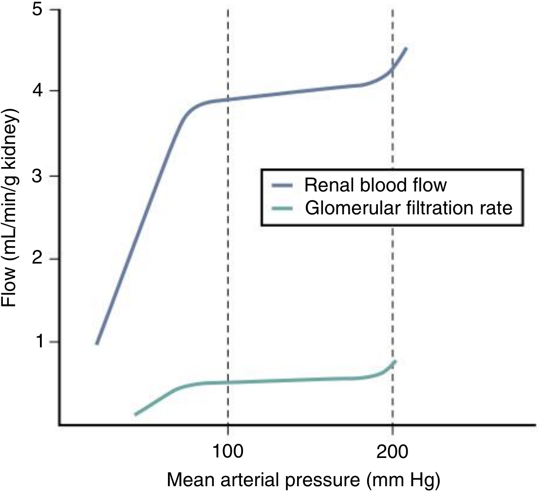
Fig: Autoregulation of renal blood flow and GFR across a range of mean arterial pressures. The flat portion of each curve represents the autoregulatory plateau. (Comprehensive Clinical Nephrology, 7th ed.)
Both RBF and GFR are autoregulated with high efficiency, indicating that the principal resistance change resides in the preglomerular vasculature, predominantly the afferent arterioles. - Brenner and Rector's The Kidney
3. Mechanisms of Renal Autoregulation
There are two primary intrinsic mechanisms:
3.1 Myogenic Mechanism
- Basis: Vascular smooth muscle cells of the afferent arteriole possess intrinsic mechanosensitive properties. When perfusion pressure rises, the vessel wall stretches, triggering depolarization via mechanosensitive cation channels and opening of L-type voltage-gated calcium channels. The resulting increase in intracellular Ca2+ causes arteriolar constriction, which raises resistance and buffers the transmission of elevated pressure to the glomerulus.
- Time course: Responds within 3 to 10 seconds. It is therefore the primary defense against rapid fluctuations in arterial pressure.
- Primary role: Protects glomerular capillaries from barotrauma during acute pressure surges.
- Contribution: Estimated to account for up to 50% of the total autoregulatory response.
- Calcium channel blockade (L-type) almost completely abolishes this mechanism. - Brenner and Rector's The Kidney
3.2 Tubuloglomerular Feedback (TGF)
- Basis: A negative feedback loop operating through the juxtaglomerular apparatus (JGA). The macula densa cells at the junction of the thick ascending limb and the early distal tubule sense luminal NaCl concentration. When perfusion pressure rises, GFR increases, delivering more NaCl to the macula densa.
- Mechanism: The macula densa takes up NaCl via a furosemide-sensitive Na-K-2Cl (NKCC2) cotransporter. This triggers ATP release into the periarteriolar space. ATP acts via P2X1 purinoceptors on the afferent arteriole causing vasoconstriction; ATP is also converted to adenosine, which acts on A1 receptors on the afferent arteriole to potentiate constriction.
- Time course: Responds over >20 seconds. More critical for sustained pressure changes.
- Modulation: TGF sensitivity is modulated by locally produced angiotensin II (Ang II, which augments TGF), nitric oxide (NO, which dampens TGF), and certain eicosanoids.
- The dynamic range of TGF is NaCl concentrations between 15 and 60 mmol/L, with maximal response above 60 mmol/L. - Miller's Anesthesia, 10th edition
3.3 Glomerulotubular Balance (GTB)
- Not to be confused with TGF. GTB ensures that a constant proportion (~67%) of filtered water and solutes is reabsorbed in the proximal convoluted tubule (PCT), regardless of the GFR level.
- This protects distal tubular segments from solute overload during GFR increases.
- The mechanism of GTB is incompletely understood. - Miller's Anesthesia, 10th edition
4. Cellular and Molecular Basis
| Feature | Myogenic | TGF |
|---|---|---|
| Stimulus | Wall stretch / pressure | Macula densa NaCl concentration |
| Mediator | Ca2+ via VGCC | ATP → Adenosine → A1 receptor |
| Effector | Afferent arteriole SM contraction | Afferent arteriole constriction |
| Time course | 3-10 sec | >20 sec |
| Blocked by | L-type Ca2+ channel blockers, furosemide (partially) | Furosemide (NKCC2 inhibition) |
- Both mechanisms share the same effector site (afferent arteriole), and are not mutually exclusive - most complete autoregulatory models incorporate both.
- Nitric oxide (NO) does not prevent autoregulation per se but reduces the plateau level of RBF at any given pressure. NO also modulates TGF sensitivity.
- Autoregulation persists even when TGF is blocked by furosemide, emphasizing the independent role of the myogenic mechanism. - Brenner and Rector's The Kidney
5. Limits and Failure of Autoregulation
Autoregulation is not absolute and can be overwhelmed or blunted by:
- Pressure outside the range: MAP <70 mmHg or MAP >130 mmHg breaches the plateau - RBF and GFR become pressure-dependent.
- Extrinsic neurohumoral override:
- High catecholamine states (surgical stress, hypovolemia): afferent arteriolar constriction; RBF falls even at normal MAP.
- Angiotensin II: preferentially constricts the efferent arteriole, temporarily preserving GFR at the cost of increased filtration fraction.
- Prostaglandins (PGE2, PGI2): vasodilatory; support RBF under hypovolemic/low-flow states. NSAIDs block these, dangerously reducing RBF in hypovolemic patients.
- Medullary circulation: Less efficiently autoregulated than the cortex. Under volume expansion, medullary blood flow autoregulation is diminished (contributing to pressure natriuresis), while cortical autoregulation is maintained. - Brenner and Rector's The Kidney
- Pre-existing disease: Diabetes, hypertension, chronic kidney disease - all shift or narrow the autoregulatory plateau.
6. Relevance in Preoperative Fluid Therapy
The perioperative period places multiple stresses on renal autoregulation. A structured approach to preoperative fluid therapy must account for the physiology described above.
6.1 Preoperative Hypovolemia and Its Renal Consequences
Patients present for surgery in various states of volume depletion:
- Prolonged nil-by-mouth (NBM) fasting
- Bowel preparation
- Diuretic therapy, uncontrolled diabetes, vomiting/diarrhea, burns
Consequences on renal autoregulation:
- Hypovolemia reduces MAP and renal perfusion pressure (RPP).
- If MAP falls below ~70-80 mmHg, the lower limit of autoregulation is breached. GFR and RBF become pressure-dependent - a linear fall occurs.
- Even before this threshold, RAAS activation and sympathetic surge cause afferent arteriolar constriction, which reduces RBF and GFR beyond what autoregulation can compensate.
- The combined effect is reduced oxygen delivery to the metabolically demanding tubular cells - setting the stage for ischemic acute kidney injury (AKI).
6.2 Goals of Preoperative Fluid Therapy in the Context of Renal Autoregulation
A. Restore intravascular volume to maintain perfusion pressure within the autoregulatory plateau (MAP 70-130 mmHg)
- The primary aim is to ensure MAP stays well within the autoregulatory range so that renal perfusion is autonomous and not dependent on systemic pressure fluctuations during induction and surgery.
- Fluid resuscitation before induction reduces the risk of hypotension on induction of general/regional anesthesia.
B. Optimize cardiac output (CO)
- Autoregulation maintains RBF at adequate cardiac output. If CO is reduced (e.g., heart failure, hypovolemia), renal perfusion pressure may be normal on the cuff but renal perfusion may still be inadequate due to increased renal venous pressure or low arterial flow.
- Goal-directed fluid therapy (GDFT) using arterial pulse contour analysis, esophageal Doppler, or transesophageal echocardiography is recommended for high-risk surgical patients to optimize CO and renal perfusion. - Morgan & Mikhail's Clinical Anesthesiology, 7th ed.
C. Avoid fluid overload
- Excess crystalloid causes tissue edema including renal interstitial edema, which raises renal capsular pressure and reduces the effective perfusion gradient.
- Large volumes of 0.9% normal saline cause hyperchloremic metabolic acidosis, which causes renal vasoconstriction and can reduce GFR.
- Volume expansion diminishes medullary autoregulatory efficiency, paradoxically impairing medullary perfusion.
D. Choice of fluid
| Fluid | Renal Consideration |
|---|---|
| Balanced crystalloids (Lactated Ringer's, PlasmaLyte) | Preferred - avoids hyperchloremia, maintains GFR |
| 0.9% Normal Saline | Hyperchloremic acidosis -> renal vasoconstriction |
| Colloids (albumin) | May be useful in hypoalbuminaemic patients to restore oncotic pressure; no established superiority over balanced crystalloids for renal outcomes |
| Hydroxyethyl starch (HES) | Contraindicated in at-risk renal patients - associated with AKI, osmotic nephrosis |
E. Renin-Angiotensin-Aldosterone System (RAAS) considerations
- Patients on ACE inhibitors or ARBs: efferent arteriolar tone is blunted. These patients lose the compensatory efferent constriction that maintains GFR during hypotension. This makes adequate preoperative volume loading especially important.
- Consider withholding ACE-I/ARB on the morning of surgery in elective cases where significant hemodynamic changes are anticipated.
F. Prostaglandin-dependent RBF states
- In hypovolemic patients, renal perfusion is prostaglandin-dependent. NSAIDs given pre- or intraoperatively reduce prostaglandin synthesis, causing unopposed afferent arteriolar constriction and precipitating AKI.
- Ensure adequate hydration before administering NSAIDs perioperatively.
6.3 Implications for Specific Patient Groups
- Chronic hypertension: Autoregulatory curve is shifted rightward. A "normal" MAP of 75 mmHg may be below their effective lower limit. These patients need higher perfusion pressure targets (MAP >80 mmHg, or approximately 20% above baseline) to preserve renal autoregulation.
- Diabetic nephropathy / CKD: Impaired myogenic and TGF responses. A narrower and shifted autoregulatory plateau. Greater susceptibility to ischemic AKI with perioperative hypotension.
- Elderly patients: Reduced afferent arteriolar responsiveness. Less efficient autoregulation. Careful titrated fluid administration is required.
- Sepsis: Altered autoregulation with vasodilation and loss of autoregulatory efficiency. Fluid resuscitation guided by dynamic markers is preferred.
7. Monitoring Renal Perfusion Adequacy
- Urine output: A minimum of 0.5 mL/kg/hr intraoperatively suggests adequate renal perfusion. Oliguria may indicate breach of autoregulatory limits.
- Serum creatinine and eGFR: Baseline assessment and postoperative trend.
- Urinary biomarkers (NGAL, KIM-1): Rising evidence for early AKI detection before creatinine rise (PMID: 38251619).
- Dynamic fluid responsiveness parameters: Stroke volume variation (SVV), pulse pressure variation (PPV) on controlled ventilation to guide fluid administration.
8. Summary Table
| Mechanism | Myogenic Reflex | Tubuloglomerular Feedback |
|---|---|---|
| Stimulus | Stretch of afferent arteriole wall | Macula densa NaCl sensing |
| Speed | Fast (3-10 sec) | Slow (>20 sec) |
| Role | Protects vs acute pressure surges | Maintains GFR with sustained hypotension |
| Pathway | Ca2+ influx via VGCC | ATP/Adenosine -> A1R |
| Anesthetic relevance | Volatile agents blunt myogenic response | RAAS inhibitors blunt TGF compensation |
Preoperative fluid therapy goals in context of autoregulation:
- Restore normovolaemia to keep MAP within the autoregulatory plateau (70-130 mmHg)
- Use balanced crystalloids; avoid large-volume normal saline and HES
- Goal-directed hemodynamic optimization for high-risk patients
- Account for shifted autoregulatory curves in hypertension, CKD, diabetes
- Time RAAS inhibitor and NSAID administration with adequate hydration
9. Conclusion
Renal autoregulation, operating through the myogenic reflex and tubuloglomerular feedback, provides robust protection against pressure-related fluctuations in RBF and GFR within a MAP range of 70-130 mmHg. This protection is not unlimited - it can be overridden by severe hypovolemia, systemic vasoconstrictors, nephrotoxins, and pre-existing renal disease. The anesthesiologist's primary tool to preserve autoregulation in the perioperative period is thoughtful preoperative fluid therapy: restoring euvolemia, maintaining adequate MAP, selecting appropriate fluids, and individualizing targets for patients with shifted autoregulatory curves. Failure to account for these principles underpins a significant proportion of perioperative AKI.
References (Textbooks):
- Miller's Anesthesia, 10th ed., Vol. 2 - Chapter 15, Renal Physiology
- Brenner and Rector's The Kidney, 2-Volume Set - Chapter 3, Intrinsic Mechanisms of Renal Autoregulation
- Comprehensive Clinical Nephrology, 7th ed. - Chapter 2, Autoregulation of RBF and GFR
- Morgan & Mikhail's Clinical Anesthesiology, 7th ed. - Chapter 5
- Costanzo Physiology, 7th ed. - Chapter 6, Renal Circulation
Recent Literature: Reviews on perioperative AKI and renal autoregulation: PMID 38251619, PMID 36872000
This is a shared conversation. Sign in to Orris to start your own chat.