
Non infectious granulomas of skin 10 marks md pathology exam
Reading File
Finding Sources
Finding Sources
Finding Sources
Finding Sources
Finding Sources
Finding Sources
Searching Images
cutaneous sarcoidosis naked granuloma histology skin
Non-Infectious Granulomas of Skin
Definition
A granuloma is a chronic inflammatory response characterized by focal collections of activated macrophages (epithelioid histiocytes), often with multinucleate giant cells, lymphocytes, and variable fibrosis, formed to contain a persistent but poorly degradable antigen or irritant. When no infectious organism can be demonstrated, the process is termed a non-infectious (sterile) granuloma.
Classification (by histological pattern)
| Pattern | Feature | Example diseases |
|---|---|---|
| Sarcoidal (epithelioid) type | Compact, well-formed epithelioid cell nodules with scanty peripheral lymphocytes ("naked tubercles") | Sarcoidosis |
| Necrobiotic (palisaded) type | Central zone of degenerated (necrobiotic) collagen surrounded by a palisade of histiocytes and giant cells | Granuloma annulare, Necrobiosis lipoidica, Rheumatoid nodule |
| Foreign body type | Histiocytes and giant cells surrounding recognizable foreign material | Foreign body granuloma, Tattoo granuloma, Silica/paraffin granuloma |
| Tuberculoid type (non-infectious mimics) | Well-formed granulomas with caseation-like necrosis but no organisms | Annular elastolytic giant cell granuloma, Crohn's (metastatic) skin lesions |
1. Granuloma Annulare (GA)
Etiology: Idiopathic; associated with diabetes mellitus, thyroid disease, and occasionally lymphoma/HIV in generalized forms.
Clinical features:
- Localized GA: skin-colored to erythematous, annular plaques with a beaded, firm border, central clearing; dorsa of hands/feet, extensor surfaces; common in children/young adults.
- Generalized GA: numerous papules/annular plaques, trunk and extremities, older adults.
- Subcutaneous ("pseudorheumatoid nodule") GA: deep nodules in children, scalp/pretibial area.
- Perforating and palmar variants also described.
Histopathology:
- Focal degeneration (necrobiosis) of dermal collagen and mucin deposition in the center.
- Palisade of histiocytes, lymphocytes, and occasional giant cells around the necrobiotic focus ("interstitial" pattern also seen, with histiocytes diffusely between collagen bundles without a distinct necrobiotic center).
- Increased dermal mucin (highlighted by colloidal iron/Alcian blue) helps distinguish GA from necrobiosis lipoidica (which shows less mucin, more layered necrobiosis).
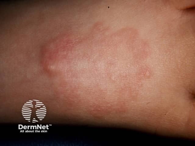
(Clinical annular plaque of granuloma annulare - Andrews' Diseases of the Skin, p. 978-989)
2. Necrobiosis Lipoidica (Diabeticorum)
Association: Strongly linked to diabetes mellitus (though not all patients are diabetic).
Clinical features: Yellow-brown, atrophic, telangiectatic plaques with violaceous, well-defined borders, classically on the pretibial region; can ulcerate.
Histopathology:
- Horizontal, "layered" (tiered) zones of necrobiotic collagen extending through the full thickness of the dermis (in contrast to the focal necrobiosis of GA).
- Granulomatous infiltrate of histiocytes, lymphocytes, plasma cells, and giant cells arranged in horizontal bands ("layer-cake" pattern).
- Thickened blood vessel walls, less mucin than GA.
3. Cutaneous Sarcoidosis
Etiology: Systemic granulomatous disease of unknown cause (immune-mediated, exaggerated cell-mediated response to poorly degradable antigen); skin involvement in ~25% of systemic sarcoidosis, may also be an isolated cutaneous disease.
Clinical features: Papules, plaques, subcutaneous nodules, lupus pernio (violaceous nodules on nose/cheeks/ears), scar sarcoidosis (granulomas within old scars).
Histopathology:
-
Well-formed, discrete, compact "naked" epithelioid granulomas in the dermis - i.e., nodules of epithelioid histiocytes with only a sparse rim of lymphocytes at the periphery (unlike the dense lymphocytic cuff of tuberculosis).
-
Multinucleated giant cells may contain asteroid bodies or Schaumann (calcified, laminated) bodies.
-
No caseation necrosis; special stains for AFB/fungi are negative - this negative workup is essential to exclude infectious granulomas before labeling as sarcoidal.
-
Fitzpatrick's Dermatology, Fig 35-14: "Histology of cutaneous sarcoidosis with naked granulomas."
-
Dermatology (Bolognia), Fig 93.6: "Nodular aggregates of epithelioid histiocytes forming tubercles filling the dermis... a sparse admixture of lymphocytes (naked tubercle)."
4. Foreign Body Granuloma
Etiology: Reaction to exogenous material implanted in the dermis - splinters, sutures, silica, talc, paraffin, tattoo pigment, ruptured cyst/follicle contents, injected filler material, or endogenous material (keratin, hair).
Histopathology:
- Histiocytes and multinucleated (foreign-body type) giant cells surround and often engulf the foreign material.
- Material may be visible directly (refractile under polarized light for silica/suture) or in giant cell cytoplasm.
- Mixed suppurative and granulomatous inflammation is common when a follicle/cyst has ruptured.
Clinical relevance: Diagnosis usually made on history + biopsy; removal of the foreign material is curative - Andrews' Diseases of the Skin, p. 1784 ff.
5. Rheumatoid Nodule
Association: Seropositive rheumatoid arthritis.
Histopathology: Similar palisading granuloma to subcutaneous GA - central fibrinoid necrosis surrounded by a palisade of histiocytes, with a peripheral zone of granulation tissue and lymphocytes/plasma cells; located in deep dermis/subcutis over pressure points (elbows).
6. Annular Elastolytic Giant Cell Granuloma (Actinic Granuloma)
Annular plaques on sun-exposed skin with elastophagocytosis: giant cells engulf and destroy degenerated (solar) elastic fibers, leaving an elastin-free center.
7. Metastatic (Cutaneous) Crohn's Disease
Non-caseating sarcoidal granulomas in skin distant from the gut, in a patient with known Crohn's disease - histologically identical to sarcoid granulomas, diagnosis rests on clinical correlation.
Key Differentiating Points (exam-focused table)
| Feature | Granuloma Annulare | Necrobiosis Lipoidica | Sarcoidosis | Foreign Body Granuloma |
|---|---|---|---|---|
| Pattern | Focal palisading necrobiosis | Diffuse/layered necrobiosis | Sarcoidal ("naked" tubercles) | Foreign body giant cells around material |
| Mucin | Increased (key clue) | Minimal | Absent | Absent |
| Necrosis | Focal | Extensive, tiered | None (no caseation) | Variable |
| Typical site | Dorsa of hands/feet | Pretibial | Face, trunk, scars | Site of implantation/injury |
| Association | Diabetes (some), idiopathic | Diabetes mellitus | Systemic sarcoid | Trauma, injected material, ruptured cyst |
Approach to Diagnosis
- Clinical morphology and distribution.
- Skin biopsy - essential to define granuloma pattern (sarcoidal vs necrobiotic vs foreign body) and to exclude infection.
- Special stains (AFB, PAS, GMS) and cultures to rule out mycobacterial/fungal etiology before calling a granuloma "non-infectious."
- Polarization of tissue to detect foreign material.
- Systemic work-up as indicated (fasting glucose/HbA1c for necrobiosis lipoidica and GA; chest imaging, ACE level for sarcoidosis).
Sources: Andrews' Diseases of the Skin (Clinical Dermatology), p. 978-1052; Dermatology 2-Volume Set (Bolognia) 5e, p. 1295-1310, 1401, 1784-1788; Fitzpatrick's Dermatology, Ch. 34-35.
This is a shared conversation. Sign in to Orris to start your own chat.