
https://chatgpt.com/share/6a9a2383-f88c-83ec-b417-dabcef2baeb3?ogimg=plain Use this link make a ppt with the content in it and with appropriate pictures
https://chatgpt.com/share/6a9a2383-f88c-83ec-b417-dabcef2baeb3
https://chatgpt.com/share/6a9a2383-f88c-83ec-b417-dabcef2bae…
~/
"primary ciliary dyskinesia"
Primary ciliary dyskinesia Kartagener syndrome chest x ray dextrocardia bronchiectasis
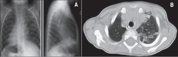
This composite clinical imaging set features a posteroanterior (PA) chest X-ray (A) and an axial CT chest scan (B) of a patient with Kartagener syndrome. Image A demonstrates dextrocardia, with the cardiac apex and right-sided aortic arch oriented toward the right hemithorax. The lung fields show diffuse airspace opacities and signs of chronic lung disease, including interspersed areas of hyperinflation. Image B, the axial CT scan, confirms dextrocardia and reveals findings consistent with situs inversus, including visceral transposition. Significant pulmonary pathology is visible, characterized by extensive chronic lung disease with a predominance in the left lung. Specific findings include diffuse bronchial dilatation (bronchiectasis), volume loss in the left upper lobe, and patchy areas of atelectasis. This case illustrates the classic triad of Kartagener syndrome: situs inversus, bronchiectasis, and chronic sinusitis (implied by clinical context), highlighting the associated pulmonary complications of primary ciliary dyskinesia.
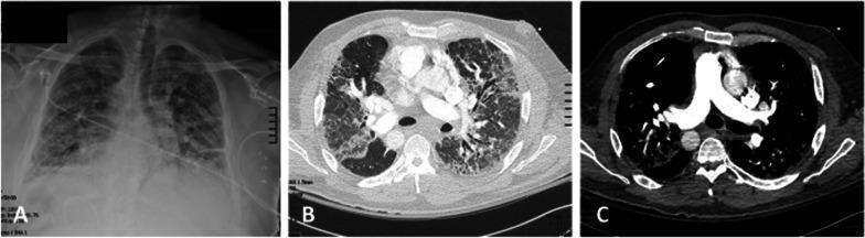
This set of diagnostic images includes a chest X-ray (A) and axial computed tomography (CT) scans (B and C) demonstrating the findings of Kartagener syndrome. In image A, a posteroanterior chest radiograph reveals dextrocardia (heart apex pointing to the right) and diffuse, bilateral reticulonodular opacities consistent with chronic interstitial lung disease. Image B is a CT lung window showing extensive bilateral bronchiectasis, thickened bronchial walls, and cystic changes, characteristic of advanced suppurative lung disease. Image C is a contrast-enhanced CT mediastinal window clearly illustrating situs inversus totalis, with the heart and great vessels positioned as a mirror image of normal anatomy (dextrocardia with the aortic arch on the right). These images highlight the cardinal features of primary ciliary dyskinesia: situs inversus and severe bronchiectasis, providing critical pre-operative context for patients requiring advanced interventions such as lung transplantation.
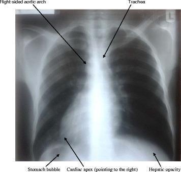
This diagnostic image is a posterior-anterior (PA) view chest X-ray demonstrating situs inversus totalis. The radiograph reveals a complete mirror-image reversal of the thoracic and upper abdominal viscera. Key findings include dextrocardia, where the cardiac apex is oriented toward the right hemithorax, and a right-sided aortic arch. The trachea shows a slight deviation to the left. In the subdiaphragmatic region, the stomach gas bubble (gastric bubble) is abnormally positioned below the right hemidiaphragm, while the hepatic opacity (liver shadow) is located under the left hemidiaphragm. The lung fields appear clear without remarkable pathology. This visual serves as a primary example of situs inversus, an autosomal recessive condition that may be associated with primary ciliary dyskinesia (Kartagener syndrome) when clinical symptoms like chronic sinusitis or bronchiectasis are present.
bronchiectasis high resolution CT scan diagram
ciliary axoneme 9 plus 2 microtubule dynein arm diagram
~/
mkdir -p "/tmp/workspace/kartagener_presentation/src" "/tmp/workspace/kartagener_presentation/assets" "/tmp/workspace/kartagener_presentation/output" && python "/tmp/skills/shared/scripts/fetch_images.py" "https://cdn.orris.care/cdss_images/pmc_clinical_VQA_5caac03a26c3a7939e8da00d4e7eb994d9e7ed51712ce7d8f160522bc49ac3ae.jpg" > "/tmp/workspace/kartagener_presentation/assets/image_fetch.json" && python -c "import json,base64; d=json.load(open('/tmp/workspace/kartagener_presentation/assets/image_fetch.json')); open('/tmp/workspace/kartagener_presentation/assets/kartagener_imaging.jpg','wb').write(base64.b64decode(d[0]['base64'].split(',',1)[1])); print(d[0]['error'])"
~/kartagener_presentation/src/create_presentation.py
from pptx import Presentation
from pptx.util import Inches, Pt
from pptx.dml.color import RGBColor
from pptx.enum.text import PP_ALIGN, MSO_ANCHOR
from pptx.enum.shapes import MSO_SHAPE
from pptx.enum.dml import MSO_THEME_COLOR
from pptx.enum.text import MSO_AUTO_SIZE
from PIL import Image
import os

OUT = '/tmp/workspace/kartagener_presentation/output/Kartagener_Syndrome_Presentation.pptx'
IMG = '/tmp/workspace/kartagener_presentation/assets/kartagener_imaging.jpg'

prs = Presentation()
prs.slide_width = Inches(13.333)
prs.slide_height = Inches(7.5)
blank = prs.slide_layouts[6]

NAVY = RGBColor(12, 38, 64)
TEAL = RGBColor(27, 153, 139)
SKY = RGBColor(64, 159, 206)
INK = RGBColor(32, 47, 62)
MUTED = RGBColor(88, 108, 124)
PALE = RGBColor(239, 247, 248)
PALE_BLUE = RGBColor(234, 244, 251)
WHITE = RGBColor(255,255,255)
CORAL = RGBColor(224, 100, 77)
GOLD = RGBColor(241, 180, 55)

# ---------- helpers ----------
def rect(slide, x,y,w,h, color, radius=False, line=None):
    shp=slide.shapes.add_shape(MSO_SHAPE.ROUNDED_RECTANGLE if radius else MSO_SHAPE.RECTANGLE, Inches(x), Inches(y), Inches(w), Inches(h))
    shp.fill.solid(); shp.fill.fore_color.rgb=color
    shp.line.color.rgb = line if line else color
    if radius: shp.adjustments[0] = 0.08
    return shp

def text(slide, s,x,y,w,h,size=20,color=INK,bold=False,align=PP_ALIGN.LEFT,font='Aptos',valign=MSO_ANCHOR.TOP):
    tb=slide.shapes.add_textbox(Inches(x), Inches(y), Inches(w), Inches(h))
    tf=tb.text_frame; tf.clear(); tf.word_wrap=True; tf.margin_left=Inches(.03); tf.margin_right=Inches(.03); tf.margin_top=Inches(.02); tf.margin_bottom=Inches(.02); tf.vertical_anchor=valign
    p=tf.paragraphs[0]; p.alignment=align
    r=p.add_run(); r.text=s; r.font.name=font; r.font.size=Pt(size); r.font.bold=bold; r.font.color.rgb=color
    return tb

def rich_lines(slide, items,x,y,w,h,size=18,lead=1.25, color=INK):
    tb=slide.shapes.add_textbox(Inches(x), Inches(y), Inches(w), Inches(h)); tf=tb.text_frame; tf.clear(); tf.word_wrap=True
    tf.margin_left=Inches(.08); tf.margin_right=Inches(.03); tf.margin_top=Inches(.03)
    for i, (head, body) in enumerate(items):
        p=tf.paragraphs[0] if i==0 else tf.add_paragraph(); p.space_after=Pt(10); p.level=0
        p.text='• '
        r=p.add_run(); r.text=head; r.font.name='Aptos'; r.font.size=Pt(size); r.font.bold=True; r.font.color.rgb=TEAL
        r=p.add_run(); r.text=body; r.font.name='Aptos'; r.font.size=Pt(size); r.font.color.rgb=color
    return tb

def title(slide, kicker, heading, sub=''):
    text(slide,kicker.upper(),.65,.38,8,.3,10,TEAL,True)
    text(slide,heading,.65,.72,12,0.62,28,NAVY,True)
    if sub: text(slide,sub,.67,1.37,11.8,.35,12,MUTED)
    rect(slide,.65,1.84,1.1,.04,TEAL)

def footer(slide, num):
    text(slide,'KARTAGENER SYNDROME  |  PRIMARY CILIARY DYSKINESIA',.65,7.13,7.4,.2,8,MUTED)
    text(slide,f'{num:02d}',12.12,7.08,.55,.25,10,TEAL,True,PP_ALIGN.RIGHT)

def add_img_crop(slide,path,x,y,w,h):
    # crop to target aspect ratio using python-pptx crop attributes
    pic=slide.shapes.add_picture(path, Inches(x), Inches(y), width=Inches(w), height=Inches(h))
    return pic

def icon_circle(slide,label,x,y,fill=TEAL):
    shp=slide.shapes.add_shape(MSO_SHAPE.OVAL,Inches(x),Inches(y),Inches(.62),Inches(.62)); shp.fill.solid(); shp.fill.fore_color.rgb=fill; shp.line.color.rgb=fill
    text(slide,label,x,y+.11,.62,.3,19,WHITE,True,PP_ALIGN.CENTER)

def arrow(slide,x,y,w=.5):
    text(slide,'→',x,y,w,.35,21,TEAL,True,PP_ALIGN.CENTER)

# 1 COVER
s=prs.slides.add_slide(blank); rect(s,0,0,13.333,7.5,NAVY)
# side abstract cilia
for i in range(8):
    xx=9.45+i*.34
    rect(s,xx,1.25+(i%2)*.17,.1,4.65,TEAL if i%2==0 else SKY,True)
    rect(s,xx-.11,1.0+(i%2)*.17,.32,.12,WHITE,True)
text(s,'CLINICAL PATHOLOGY PRESENTATION',.72,.72,4.8,.3,11,RGBColor(170,221,219),True)
text(s,'Kartagener\nSyndrome',.7,1.28,7.4,1.65,38,WHITE,True)
text(s,'A clinical variant of primary ciliary dyskinesia',.75,3.18,6.8,.35,18,RGBColor(209,226,236))
rect(s,.75,4.0,2.35,.42,TEAL,True)
text(s,'Cilia cannot move',.85,4.1,2.15,.18,12,WHITE,True,PP_ALIGN.CENTER)
text(s,'Mucus cannot move  •  Organs may reverse',.75,4.72,6.7,.3,18,WHITE)
text(s,'Bronchiectasis  •  Chronic sinusitis  •  Situs inversus',.75,5.3,7.8,.3,14,RGBColor(170,221,219))
text(s,'Prepared from the shared-chat content',.75,6.72,4.3,.2,9,RGBColor(170,221,219))

# 2 WHAT
s=prs.slides.add_slide(blank); title(s,'01 | Definition','What is Kartagener syndrome?','The memorable phenotype of an inherited mucociliary-clearance disorder.')
rect(s,.65,2.22,5.8,3.9,PALE,True)
text(s,'Primary ciliary dyskinesia (PCD)',.96,2.58,4.9,.35,23,NAVY,True)
text(s,'An autosomal-recessive disorder in which motile cilia are absent, immotile, or beat abnormally.',.97,3.15,4.9,.8,18,INK)
text(s,'Result:',.97,4.38,1,.25,14,TEAL,True)
text(s,'Impaired mucociliary clearance → mucus retention → recurrent respiratory infection.',.97,4.7,4.95,.7,17,INK)
rect(s,6.8,2.22,5.85,3.9,PALE_BLUE,True)
text(s,'Kartagener syndrome',7.12,2.58,4.8,.35,23,NAVY,True)
text(s,'The classic clinical triad:',7.13,3.18,3.6,.25,15,MUTED)
for i,(a,b,c) in enumerate([('🫁','Bronchiectasis',TEAL),('👃','Chronic sinusitis',SKY),('♥','Situs inversus',CORAL)]):
    yy=3.72+i*.68; icon_circle(s,a,7.14,yy,c); text(s,b,7.95,yy+.16,3.7,.26,18,INK,True)
text(s,'About half of people with PCD have situs inversus.',.67,6.45,7.2,.3,13,MUTED,False)
footer(s,2)

# 3 case
s=prs.slides.add_slide(blank); title(s,'02 | Clinical recognition','The pattern that should trigger suspicion')
rect(s,.65,2.25,3.55,3.9,NAVY,True); text(s,'PATIENT CLUES',.94,2.58,2.3,.25,12,RGBColor(154,220,215),True)
text(s,'Recurrent chest\ninfections',.94,3.05,2.4,.65,27,WHITE,True)
text(s,'Persistent wet cough, chronic nasal symptoms, or repeated bacterial infections from childhood.',.95,4.25,2.55,1.1,15,RGBColor(220,235,241))
for i,(h,b) in enumerate([('Upper airway','Chronic rhinosinusitis, nasal congestion, otitis media.'),('Lower airway','Productive cough and recurrent infections leading to bronchiectasis.'),('Reproductive','Reduced sperm motility may cause male infertility.')]):
    y=2.25+i*1.25; rect(s,4.55,y,7.85,1.0,WHITE,True,line=RGBColor(210,224,230)); text(s,h,4.88,y+.18,1.65,.25,16,TEAL,True); text(s,b,6.46,y+.16,5.5,.5,15,INK)
text(s,'Think PCD when the respiratory history is chronic and multisite, especially with dextrocardia or situs inversus.',.75,6.55,11.4,.28,14,NAVY,True)
footer(s,3)

# 4 cilia
s=prs.slides.add_slide(blank); title(s,'03 | Microscopy','Normal cilia versus primary ciliary dyskinesia')
rect(s,.65,2.25,5.85,3.95,PALE,True); text(s,'NORMAL MOTILE CILIUM',.95,2.55,3.2,.25,13,TEAL,True)
# 9+2 diagram
cx,cy=3.55,4.35
for angle in range(0,360,40):
    import math
    x=cx+1.18*math.cos(math.radians(angle)); y=cy+1.18*math.sin(math.radians(angle))
    sh=s.shapes.add_shape(MSO_SHAPE.OVAL,Inches(x-.16),Inches(y-.16),Inches(.32),Inches(.32)); sh.fill.solid(); sh.fill.fore_color.rgb=SKY; sh.line.color.rgb=WHITE
for x in [cx-.15,cx+.12]:
    sh=s.shapes.add_shape(MSO_SHAPE.OVAL,Inches(x),Inches(cy-.12),Inches(.25),Inches(.25)); sh.fill.solid(); sh.fill.fore_color.rgb=GOLD; sh.line.color.rgb=WHITE
text(s,'9 peripheral microtubule doublets\n+ 2 central microtubules',.94,5.38,4.95,.58,15,INK,True,PP_ALIGN.CENTER)
rect(s,6.83,2.25,5.85,3.95,PALE_BLUE,True); text(s,'PCD: AXONEMAL DEFECTS',7.13,2.55,3.4,.25,13,CORAL,True)
text(s,'Common sites of abnormality',7.14,3.05,3.4,.25,18,NAVY,True)
rich_lines(s,[('Dynein arms',' → defective ciliary movement'),('Radial spokes',' or central apparatus'),('Functional outcome',' → immotile or poorly motile cilia')],7.1,3.5,5.1,1.75,16)
text(s,'Transmission electron microscopy can demonstrate classic ultrastructural defects.',7.14,5.48,5.0,.4,13,MUTED)
footer(s,4)

# 5 pathogenesis
s=prs.slides.add_slide(blank); title(s,'04 | Pathogenesis','How a ciliary defect becomes bronchiectasis')
steps=[('1','AR mutation','Dynein / axonemal protein'),('2','Cilia do not beat','Immotile or dyskinetic cilia'),('3','Clearance fails','Mucus is retained'),('4','Infections recur','Neutrophilic inflammation'),('5','Airway wall injury','Bronchiectasis')]
for i,(n,h,b) in enumerate(steps):
    x=.67+i*2.52
    rect(s,x,2.58,2.13,2.35,WHITE,True,line=RGBColor(204,222,230))
    icon_circle(s,n,x+.76,2.82,TEAL if i<3 else CORAL)
    text(s,h,x+.18,3.64,1.78,.42,16,NAVY,True,PP_ALIGN.CENTER)
    text(s,b,x+.17,4.22,1.8,.45,12,MUTED,False,PP_ALIGN.CENTER)
    if i<4: arrow(s,x+2.16,3.55,.3)
rect(s,.67,5.47,11.98,.52,PALE,True); text(s,'In parallel during embryogenesis: defective nodal cilia → abnormal left-right orientation → situs inversus.',.9,5.62,11.45,.2,15,INK,True,PP_ALIGN.CENTER)
footer(s,5)

# 6 imaging
s=prs.slides.add_slide(blank); title(s,'05 | Imaging','The radiologic signature: dextrocardia plus bronchiectasis','Representative imaging from a published clinical-image collection.')
add_img_crop(s,IMG,.68,2.2,7.1,4.32)
rect(s,8.15,2.2,4.48,4.32,PALE_BLUE,True)
text(s,'Look for:',8.47,2.55,2.5,.3,21,NAVY,True)
rich_lines(s,[('Dextrocardia',' heart apex on the right'),('Situs inversus',' mirror-image viscera'),('Bronchiectasis',' dilated thick-walled bronchi'),('Chronic lung change',' atelectasis / hyperinflation may coexist')],8.37,3.12,3.8,2.5,15)
text(s,'Imaging caption: PA chest X-ray and axial CT, showing dextrocardia, situs inversus and bronchiectasis in Kartagener syndrome.',.69,6.65,11.85,.27,9,MUTED)
footer(s,6)

# 7 pathology
s=prs.slides.add_slide(blank); title(s,'06 | Bronchiectasis','What repeated infection does to the bronchial wall')
rect(s,.65,2.25,5.2,3.9,NAVY,True)
text(s,'MICROSCOPY',.95,2.55,2,.23,12,RGBColor(154,220,215),True)
rich_lines(s,[('Destruction',' of bronchial wall'),('Loss',' of ciliated epithelium'),('Chronic infiltrate',' with neutrophils during acute infection'),('Ulceration & fibrosis',' of the bronchial wall'),('Loss',' of smooth muscle and elastic tissue')],.88,3.05,4.5,2.65,15, color=WHITE)
# override rich heading may be teal but okay
rect(s,6.2,2.25,6.45,3.9,PALE,True)
text(s,'The vicious cycle',6.55,2.6,3,.3,21,NAVY,True)
flow=['Mucus retention','Bacterial infection','Chronic inflammation','Wall destruction','Dilated bronchi']
for i,v in enumerate(flow):
    y=3.12+i*.52; rect(s,6.58,y,3.25,.34,TEAL if i in [0,4] else SKY,True); text(s,v,6.73,y+.07,2.95,.16,12,WHITE,True,PP_ALIGN.CENTER)
    if i<4: text(s,'↓',7.95,y+.33,.5,.23,15,NAVY,True,PP_ALIGN.CENTER)
text(s,'The structural damage further worsens secretion clearance, perpetuating the cycle.',10.1,3.52,2.05,1.2,15,INK,True)
footer(s,7)

# 8 diagnosis
s=prs.slides.add_slide(blank); title(s,'07 | Investigation','Confirming the diagnosis')
cols=[('Clinical phenotype',['Chronic wet cough','Sinus disease / otitis','Situs inversus or dextrocardia','Family history']),('Functional assessment',['Nasal nitric oxide may be low','High-speed video microscopy','Assess ciliary beat pattern']),('Structural / genetic',['TEM for axonemal defects','Genetic testing','CT chest to stage bronchiectasis'])]
for i,(h,items) in enumerate(cols):
    x=.7+i*4.17; rect(s,x,2.38,3.77,3.88,WHITE,True,line=RGBColor(205,223,231)); icon_circle(s,str(i+1),x+.28,2.68,[TEAL,SKY,CORAL][i]); text(s,h,x+1.05,2.83,2.25,.27,18,NAVY,True)
    for j,it in enumerate(items):
        text(s,'• '+it,x+.35,3.48+j*.56,3.08,.38,15,INK)
text(s,'Diagnosis is based on a compatible phenotype plus objective evidence of ciliary dysfunction and/or a pathogenic genetic variant.',.75,6.55,11.5,.28,14,NAVY,True)
footer(s,8)

# 9 management
s=prs.slides.add_slide(blank); title(s,'08 | Management','Treat the complications. Support the clearance.')
text(s,'There is no therapy that restores the underlying defective cilia. Care is directed at airway health, infection control and complications.',.7,2.2,11.7,.45,17,INK)
items=[('Airway clearance','Chest physiotherapy, postural drainage, breathing exercises and clearance devices.',TEAL),('Respiratory infection','Culture-guided antibiotics where possible. Common bronchiectasis pathogens include H. influenzae, P. aeruginosa and S. aureus.',SKY),('Sinus disease','Saline nasal irrigation, intranasal therapy when indicated, antibiotics for bacterial sinusitis and ENT input.',CORAL),('Prevention & escalation','Influenza and pneumococcal vaccination. In advanced disease, lung transplantation may be considered.',GOLD)]
for i,(h,b,c) in enumerate(items):
    x=.7+(i%2)*6.15; y=3.0+(i//2)*1.55; rect(s,x,y,5.78,1.18,PALE if i%2==0 else PALE_BLUE,True); icon_circle(s,str(i+1),x+.28,y+.27,c); text(s,h,x+1.1,y+.2,4.2,.23,17,NAVY,True); text(s,b,x+1.1,y+.53,4.25,.43,12,INK)
footer(s,9)

# 10 role play summary
s=prs.slides.add_slide(blank); title(s,'09 | Role-play wrap-up','A concise clinical diagnosis')
rect(s,.7,2.25,12,2.35,NAVY,True)
text(s,'“Based on recurrent respiratory infections and chronic sinusitis, bronchiectasis with dextrocardia, and evidence of impaired ciliary function, this patient most likely has Kartagener syndrome, a form of primary ciliary dyskinesia.”',1.15,2.78,11.05,1.18,23,WHITE,True,PP_ALIGN.CENTER,valign=MSO_ANCHOR.MIDDLE)
text(s,'Suggested roles',.78,5.1,2.5,.28,18,NAVY,True)
roles=['Doctor','Patient','Examiner','Pathologist','Radiologist']
for i,r in enumerate(roles):
    x=.8+i*2.45; rect(s,x,5.6,2.1,.55,[TEAL,SKY,CORAL,GOLD,NAVY][i],True); text(s,r,x,5.77,2.1,.17,14,WHITE,True,PP_ALIGN.CENTER)
text(s,'ONE-LINE MEMORY: Cilia cannot move → mucus cannot move → organs may reverse.',.78,6.55,11.5,.3,17,TEAL,True,PP_ALIGN.CENTER)
footer(s,10)

# 11 sources
s=prs.slides.add_slide(blank); title(s,'References','Source notes')
text(s,'Core content',.75,2.25,2,.3,18,NAVY,True)
rich_lines(s,[('Shared chat',' User-provided ChatGPT shared conversation on primary ciliary dyskinesia / Kartagener syndrome.'),('Scott-Brown’s Otorhinolaryngology',' Primary ciliary dyskinesia: abnormal ciliary beating, impaired clearance and the clinical triad. p. 1088.'),('Robbins, Cotran & Kumar',' Pathogenesis of bronchiectasis and primary ciliary dyskinesia. p. 477.'),('Clinical image',' Representative Kartagener imaging: chest radiograph and CT demonstrating dextrocardia, situs inversus and bronchiectasis.')],.78,2.76,11.6,2.5,16)
rect(s,.78,5.72,11.78,.6,PALE,True); text(s,'Educational presentation only. Diagnosis and management should be individualized by qualified clinicians.',.97,5.92,11.4,.2,12,MUTED,False,PP_ALIGN.CENTER)
footer(s,11)

prs.save(OUT)
print(OUT)
python "/tmp/workspace/kartagener_presentation/src/create_presentation.py" && ls -lh "/tmp/workspace/kartagener_presentation/output/Kartagener_Syndrome_Presentation.pptx" && python - <<'PY' from pptx import Presentation p='/tmp/workspace/kartagener_presentation/output/Kartagener_Syndrome_Presentation.pptx' prs=Presentation(p) print('slides:',len(prs.slides)) for n,s in enumerate(prs.slides,1): tx=' '.join(sh.text for sh in s.shapes if getattr(sh,'has_text_frame',False)).replace('\n',' ') print(n, len(tx), tx[:80]) PY
Kartagener Syndrome Presentation
Presentation · PPTX