
Teach me according to DMSF and exam must know skill to crack DMSF EXAMS
Reading File
Now let me extract the full content of the PDF transcript.
Running Command
cd /home/daytona/workspace/attachments/d4204966-f1ee-4b45-823d-1afd327a0a50/ && python3 -c " import pdfplumber with pdfplumber.open('[TRANS] CP-PREFI LEC#4 - SYNOVIAL FLUID.pdf') as pdf: for i, page in enumerate(pdf.pages): text = page.extract_text() print(f'=== PAGE {i+1} ===') print(text) print() "
I have the full content. Now let me craft a comprehensive, exam-focused teaching guide based entirely on the DMSF transcript.
🧪 SYNOVIAL FLUID ANALYSIS - DMSF EXAM MASTER GUIDE
CP-PREFI Lec #4 | Clinical Pathology NMD2, 2nd Semester
1. WHAT IS SYNOVIAL FLUID?
- "Joint fluid" - transparent, viscous liquid found in the cavities of movable joints
- It is an ultrafiltrate of plasma (chemical constituents are similar to plasma)
- Chemical constituent is similar to that of plasma concentration
Functions (3 key points):
- Reduce friction / Lubricates the joint
- Provides nutrients for cells lining the synovial membrane
- Lessens shock - acts as a buffer zone
Synovial Membrane:
- Composed primarily of synoviocytes which secrete hyaluronic acid + small amount of protein (chiefly Lubricin)
- Hyaluronan complex protein maintains viscosity and helps moisten/lubricate the joint
2. NORMAL SYNOVIAL FLUID VALUES ⭐ (HIGH-YIELD TABLE)
| Parameter | Normal Value |
|---|---|
| Volume | < 3.5 mL |
| Color | Colorless to pale yellow |
| Clarity | Clear |
| Viscosity | String 4-6 cm long |
| Leukocyte Count | < 200 cells/uL |
| Neutrophils | < 25% of differential |
| Crystals | None present |
| Glucose : Plasma Difference | < 10 mg/dL lower than blood glucose |
| Total Protein | < 3 g/dL |
3. CLASSIFICATION OF JOINT DISORDERS ⭐⭐ (MOST EXAM-TESTED TABLE)
| Group | Disorders | Color/Clarity | Viscosity | WBC Count | Neutrophils | Glucose |
|---|---|---|---|---|---|---|
| Non-Inflammatory | Osteoarthritis, Degenerative joint disorders | Clear, Yellow | Good | < 1,000/uL | < 30% | Normal (similar to blood) |
| Inflammatory | RA, Lupus, Scleroderma, Polymyositis, Ankylosing spondylitis, Rheumatic fever, Lyme arthritis | Cloudy, Yellow | Poor | 2,000 - 75,000/uL | > 50% | Decreased; Possible autoantibodies |
| Crystal-induced | Gout, Pseudogout | Cloudy, Milky | Low | Up to 100,000/uL | > 70% | Decreased; Crystals present |
| Septic | Microbial infection | Cloudy, Yellow-green | Variable | 50,000 - 100,000/uL | > 75% | Decreased; Positive culture + Gram stain |
| Hemorrhagic | Trauma, Tumors, Hemophilia, Coag disorders, Anticoagulant overdose | Cloudy, Red | Low | WBCs = blood levels | Neutrophils = blood levels | Normal glucose |
📝 Exam Trick: Crystal-induced and Septic have the HIGHEST WBC counts. Septic is the only group with Positive culture. Hemorrhagic is the ONLY group with Normal glucose.
4. SPECIMEN COLLECTION & HANDLING ⭐
Procedure:
- Arthrocentesis = collection of synovial fluid from a joint cavity
- Done by fine needle aspiration (can be punctured at any site)
- Aseptic technique is critical - synovial fluid is a good culture medium for organisms (especially gram positives)
Tube Distribution (in order):
| Tube | Test |
|---|---|
| Sterile heparinized tube | Gram stain and Culture |
| Heparinized/EDTA tube | Cell count |
| Plain tube | Other tests |
| Sodium fluoride tube | Glucose Analysis |
Key Rules:
- Synovial fluid normally does NOT clot
- May contain fibrinogen in diseased joints (increased plasma ultrafiltrate permeability)
- Fluid collected in a heparinized syringe
- Powdered anticoagulant = NOT used (interferes with crystal examination)
- All tests should be done ASAP
5. MACROSCOPIC EXAMINATION
A. Volume
- Normal: up to 3.5 mL
- Results recorded at bedside/patient's chart
B. Color and Clarity ⭐
| Color/Clarity | Significance |
|---|---|
| Colorless and clear | Normal |
| Dark yellow and clear | Non-inflammatory effusion |
| Dark yellow and turbid | Inflammatory effusion |
| Greenish tinge | Bacterial infection |
| Red, brown or xanthochromic | Hemorrhage into the joint |
| White and cloudy (milky) | Crystals |
C. Inclusion Bodies ⭐
- Rice bodies - associated with Rheumatoid arthritis; degenerated synovium enriched with fibrin
- Ochronotic shards - debris from joint prosthesis
D. Viscosity ⭐⭐
Source: Comes from polymerization of hyaluronic acid - essential for lubrication
String Test:
- Normal: String measures 4-6 cm before it breaks
- Thin fluid = water-like when poured (poor viscosity)
- Thick fluid = honey-like; unbroken string (good viscosity)
Ropes/Mucin Test (Clot test):
- Add 2-5% acetic acid
- Good/Normal: Solid clot with clear fluid - seen in Normal, Osteoarthritis, Trauma, Bleed
- Fair: Soft clot - seen in Lupus, RA, Reiter's
- Poor: Clots cannot be seen - seen in Gout, Gonorrhea
For very viscous fluids (to reduce viscosity for counting):
- Add a pinch of hyaluronidase to 0.5 mL of fluid, OR
- One drop of 0.05% hyaluronidase in phosphate buffer per mL of fluid
- Incubate @ 37°C for 5 minutes
6. MICROSCOPIC EXAMINATION
A. Cell Count
- Normal: < 200 cells/uL
- Counting chamber: Neubauer counting chamber
- Clear fluid = charged directly and counted undiluted
Diluents used:
- Normal saline
- Hypotonic saline
- Saline + Saponin
- Saline + Methylene blue (makes cells more visible - stained)
B. Differential Count
- Performed on cytocentrifuged preparations or thinly smeared slides
- Fluid is incubated with hyaluronidase
Primary (Normal) cells:
| Cell | Normal % |
|---|---|
| Monocytes | Mostly seen |
| Macrophages | Present |
| Synovial tissue cells | Present |
| Neutrophils | < 25% |
| Lymphocytes | < 15% |
Abnormal/Pathologic Cells ⭐⭐:
| Cell | Description | Significance |
|---|---|---|
| LE cells | Neutrophils engulfing rounded matter (degenerating nucleus of lymphocytes) | SLE (Lupus) |
| RA cells / Ragocytes | Neutrophils containing bluish inclusions composed of precipitated Rheumatoid factor | Rheumatoid Arthritis |
| Reiter cells | Macrophage engulfing degenerating neutrophil | Reiter's syndrome |
| Lipid droplets | Lipid droplets in macrophages | Inflammatory conditions |
| Hemosiderin granules | Hemosiderin-laden macrophages | Pigmented villonodular synovitis |
| Cartilage cells | Present | Joint destruction |
| Multinucleated giant cells | Most likely osteoclasts |
📝 Exam Trick: RA cells = Ragocytes = Neutrophils with RF inclusions. Reiter cells = Macrophage eating a neutrophil.
C. Crystal Identification ⭐⭐⭐
Gout = crystal deposition in articular tissue
| Crystal | Shape | Birefringence | Disease |
|---|---|---|---|
| Monosodium urate (MSU) | Needle-like | Positive birefringence (greenish with polarized light) | Gout (Urate gout) |
| Calcium pyrophosphate dihydrate (CPPD) | 4-sided (square, rhomboid, rectangle) | Birefringent | Pseudogout / Chondrocalcinosis |
| Cholesterol crystals | Staircase/ladder pattern | - | (same pattern as in urine) |
| Calcium oxalate dihydrate | Cross/envelope within squares | - | Oxalate gout |
| Basic Calcium phosphates (BCP) | - | - | Apatite gout |
| Lipid crystals | - | - | Lipid gout |
7. CHEMICAL EXAMINATION ⭐
Glucose
- Compare with serum level (requires 8 hours fasting)
- Normal serum-synovial glucose difference: < 10 mg/dL
- Septic arthritis: Glucose difference range = 20-60 mg/dL
- Analyzed within 1 hour
Protein
- Normal range: 1-3 g/dL
- Increased protein seen in: Ankylosing spondylitis, Arthritis, IBD, Gout, Psoriasis, Reiter's syndrome
Uric Acid
- Increased in gout
- May form crystals
Lactate
- Helpful in diagnosing septic arthritis
- Normal: < 25 mg/dL
Lactate Dehydrogenase (LDH)
- Increased in: RA, Infectious arthritis, Gout
- Neutrophils increase during acute phase contribute to LDH increase
8. IMMUNOLOGIC STUDIES
| Test | Significance |
|---|---|
| Rheumatoid factor | 60% with RA |
| Antinuclear antibodies (ANA) | 70% with SLE / 20% with RA |
9. MICROBIOLOGIC EXAMINATION
- Gram stain
- Culture
- KOH / Calcofluor white stain (fungal)
- Acid fast stain (TB)
10. BULGE TEST
- Used to determine if there is an abnormal amount of fluid surrounding a joint
- Clinicians use this to estimate if there is an increased volume of synovial fluid
- Which knee has more bulge? = The more affected joint shows shallowing of the dents - pressing it produces a visible bulge
⭐ HIGH-YIELD EXAM MUST-KNOWS (QUICK RECALL)
| # | Must-Know Fact |
|---|---|
| 1 | Normal volume = < 3.5 mL; Normal color = colorless to pale yellow |
| 2 | Normal string test = 4-6 cm before breaking |
| 3 | Synovial fluid is an ultrafiltrate of plasma |
| 4 | Synovial fluid normally does NOT clot |
| 5 | Powdered anticoagulant = NEVER use (interferes with crystal exam) |
| 6 | Glucose in synovial fluid is compared to serum after 8 hours fasting |
| 7 | Septic arthritis = glucose diff 20-60 mg/dL; positive culture; WBC 50K-100K |
| 8 | Crystal-induced = WBC up to 100,000/uL; milky fluid; neutrophils > 70% |
| 9 | MSU crystals = needle-shaped + birefringent (greenish) = GOUT |
| 10 | CPPD crystals = 4-sided shape (rhomboid/square) = PSEUDOGOUT |
| 11 | RA cells/Ragocytes = neutrophils with bluish RF inclusions = RA |
| 12 | LE cells = neutrophils engulfing lymphocyte nuclei = SLE |
| 13 | Rice bodies = associated with Rheumatoid Arthritis |
| 14 | Reiter cells = macrophage eating a degenerating neutrophil |
| 15 | Rheumatoid factor positive in 60% RA; ANA positive in 70% SLE |
| 16 | Hemorrhagic fluid = ONLY group with normal glucose |
| 17 | Highest neutrophil % = Septic (> 75%) |
| 18 | Diluent for cell count = saline + methylene blue (for visibility) |
| 19 | To reduce viscosity for counting = hyaluronidase at 37°C for 5 min |
| 20 | Cholesterol crystals = staircase/ladder pattern (same as in urine) |
PATTERN RECOGNITION SUMMARY
Milky fluid + crystals → Crystal-induced (Gout or Pseudogout)
Yellow-green fluid + positive culture → SEPTIC
Red fluid + normal glucose → HEMORRHAGE
Cloudy + autoantibodies → INFLAMMATORY (RA, Lupus, etc.)
Clear + good viscosity + WBC <1000 → NON-INFLAMMATORY (Osteoarthritis)
Good luck on your DMSF exam! The most heavily tested areas based on this transcript are: the classification table (Non-inflammatory/Inflammatory/Crystal/Septic/Hemorrhagic), crystal types (MSU vs CPPD), abnormal cells (RA cells, LE cells, Reiter cells), normal values, and specimen handling rules.
Make mcqs according to DMSF
🧪 SYNOVIAL FLUID ANALYSIS - DMSF MCQ PRACTICE SET
CP-PREFI Lec #4 | Based strictly on the DMSF transcript
SECTION 1: BASICS & NORMAL VALUES
1. Synovial fluid is best described as a/an:
- A) Secretion of synoviocytes
- B) Transudate of lymph
- C) Ultrafiltrate of plasma
- D) Direct filtrate of blood
✅ Answer: C - Synovial fluid is an ultrafiltrate of plasma, with chemical constituents similar to plasma concentration.
2. Which of the following is NOT a function of synovial fluid?
- A) Reduces friction in the joint
- B) Provides nutrients to synovial membrane cells
- C) Produces hyaluronic acid
- D) Lessens shock by acting as a buffer zone
✅ Answer: C - Producing hyaluronic acid is the function of synoviocytes in the synovial membrane, not the synovial fluid itself.
3. What is the normal volume of synovial fluid?
- A) < 1.5 mL
- B) < 2.5 mL
- C) < 3.5 mL
- D) < 5.0 mL
✅ Answer: C - Normal volume is < 3.5 mL.
4. The normal color and clarity of synovial fluid is:
- A) Pale yellow and turbid
- B) Colorless to pale yellow and clear
- C) Yellow-green and clear
- D) White and cloudy
✅ Answer: B - Normal synovial fluid is colorless to pale yellow and clear.
5. What is the normal leukocyte count in synovial fluid?
- A) < 100 cells/uL
- B) < 200 cells/uL
- C) < 500 cells/uL
- D) < 1,000 cells/uL
✅ Answer: B - Normal leukocyte count is < 200 cells/uL.
6. The normal percentage of neutrophils in synovial fluid differential is:
- A) < 10%
- B) < 15%
- C) < 25%
- D) < 50%
✅ Answer: C - Normal neutrophils < 25%; normal lymphocytes < 15%.
7. In the string test, a normal synovial fluid should form a string of how long before it breaks?
- A) 1-2 cm
- B) 2-4 cm
- C) 4-6 cm
- D) 6-8 cm
✅ Answer: C - Normal string measures 4-6 cm before breaking.
8. What is the normal total protein range in synovial fluid?
- A) < 1 g/dL
- B) 1-3 g/dL
- C) 3-5 g/dL
- D) < 5 g/dL
✅ Answer: B - Normal total protein is 1-3 g/dL (some sources state < 3 g/dL).
9. The normal serum-to-synovial fluid glucose difference is:
- A) Equal to serum glucose
- B) < 5 mg/dL lower
- C) < 10 mg/dL lower
- D) 20-60 mg/dL lower
✅ Answer: C - Normal difference is < 10 mg/dL lower than blood glucose.
SECTION 2: CLASSIFICATION OF JOINT DISORDERS
10. A patient presents with synovial fluid that is clear, yellow, with WBC count of 800/uL, neutrophils 20%, and normal glucose. What is the most likely diagnosis?
- A) Septic arthritis
- B) Rheumatoid arthritis
- C) Osteoarthritis (Non-inflammatory)
- D) Gout
✅ Answer: C - Clear/yellow fluid, good viscosity, WBC < 1,000/uL, neutrophils < 30%, normal glucose = Non-inflammatory / Osteoarthritis.
11. Which joint disorder classification shows WBC count of 2,000-75,000/uL with possible autoantibodies?
- A) Non-inflammatory
- B) Inflammatory
- C) Crystal-induced
- D) Hemorrhagic
✅ Answer: B - Inflammatory group (RA, Lupus, etc.) shows WBC 2,000-75,000/uL with possible autoantibodies.
12. Synovial fluid appears cloudy and milky with WBC up to 100,000/uL and neutrophils > 70%. Crystals are present. This is most consistent with:
- A) Septic arthritis
- B) Hemorrhagic effusion
- C) Crystal-induced arthritis
- D) Rheumatoid arthritis
✅ Answer: C - Milky fluid + crystals + WBC up to 100,000/uL + neutrophils > 70% = Crystal-induced (Gout/Pseudogout).
13. Which of the following is the ONLY joint disorder classification that shows NORMAL glucose levels?
- A) Septic arthritis
- B) Inflammatory arthritis
- C) Crystal-induced arthritis
- D) Hemorrhagic effusion
✅ Answer: D - Hemorrhagic effusion (trauma, hemophilia, tumors) is the ONLY group with normal glucose.
14. A patient with known microbial joint infection has synovial fluid with neutrophils > 75%, WBC 50,000-100,000/uL, decreased glucose, and a positive culture. This belongs to which classification?
- A) Crystal-induced
- B) Inflammatory
- C) Septic
- D) Non-inflammatory
✅ Answer: C - Septic arthritis = positive culture + Gram stain + highest neutrophil % (> 75%).
15. Which color of synovial fluid is characteristic of bacterial infection?
- A) White and cloudy
- B) Greenish tinge
- C) Red/xanthochromic
- D) Dark yellow and turbid
✅ Answer: B - Greenish tinge = bacterial infection. White and cloudy = crystals. Red = hemorrhage.
16. Red or xanthochromic synovial fluid is most associated with:
- A) Crystal-induced gout
- B) Septic arthritis
- C) Hemorrhage into the joint
- D) Inflammatory arthritis
✅ Answer: C - Red, brown, or xanthochromic = hemorrhage into the joint.
17. Rheumatoid arthritis belongs to which classification of joint disorders?
- A) Non-inflammatory
- B) Crystal-induced
- C) Inflammatory
- D) Septic
✅ Answer: C - RA is under the Inflammatory group along with Lupus, Scleroderma, Polymyositis, Ankylosing spondylitis, Rheumatic fever, and Lyme arthritis.
SECTION 3: SPECIMEN COLLECTION & HANDLING
18. The procedure for collecting synovial fluid from a joint cavity is called:
- A) Thoracentesis
- B) Paracentesis
- C) Arthrocentesis
- D) Amniocentesis
✅ Answer: C - Arthrocentesis = collection of synovial fluid from a joint cavity by fine needle aspiration.
19. Which anticoagulant should NOT be used when collecting synovial fluid for crystal examination?
- A) Heparin
- B) EDTA
- C) Powdered anticoagulant
- D) Sodium fluoride
✅ Answer: C - Powdered anticoagulant should NOT be used because it interferes with crystal examination/analysis.
20. Synovial fluid for glucose analysis should be placed in which tube?
- A) Sterile heparinized tube
- B) Heparinized/EDTA tube
- C) Plain tube
- D) Sodium fluoride tube
✅ Answer: D - Sodium fluoride tube is used for glucose analysis.
21. For Gram stain and culture, synovial fluid should be placed in:
- A) Sodium fluoride tube
- B) Sterile heparinized tube
- C) Plain tube
- D) EDTA tube
✅ Answer: B - Sterile heparinized tube = Gram stain and culture.
22. Why is aseptic technique critical during arthrocentesis?
- A) Synovial fluid clots rapidly when contaminated
- B) Synovial fluid is a good culture medium especially for gram-positive organisms
- C) The needle may damage the hyaluronic acid structure
- D) Normal flora cannot survive in synovial fluid
✅ Answer: B - Synovial fluid is a good culture medium for organisms, particularly gram positives - introducing skin normal flora can cause infection.
23. Synovial fluid may clot in diseased joints due to:
- A) Increased hyaluronic acid
- B) Increased fibrinogen from greater plasma ultrafiltrate permeability
- C) Decreased protein content
- D) Elevated glucose levels
✅ Answer: B - In diseased joints, increased permeability allows larger proteins like fibrinogen to escape into the synovial cavity, enabling clot formation.
SECTION 4: VISCOSITY TESTS
24. The viscosity of synovial fluid comes from:
- A) Lubricin protein
- B) Polymerization of hyaluronic acid
- C) Fibrinogen content
- D) Albumin concentration
✅ Answer: B - Viscosity comes from the polymerization of hyaluronic acid, which is essential for lubrication.
25. In the Ropes/Mucin clot test, what reagent is added to synovial fluid?
- A) Sodium hydroxide
- B) Methylene blue
- C) 2-5% acetic acid
- D) Hyaluronidase
✅ Answer: C - 2-5% acetic acid is added in the Ropes/Mucin test. A solid clot with clear fluid = Normal.
26. Poor viscosity with inability to see clots in the Mucin clot test is associated with which conditions?
- A) Osteoarthritis and trauma
- B) Lupus and Rheumatoid arthritis
- C) Gout and gonorrhea
- D) Normal synovial fluid
✅ Answer: C - Poor mucin clot (clots cannot be seen) = Gout, Gonorrhea. Fair = Lupus, RA, Reiter's. Good = Normal, Osteoarthritis, Trauma.
27. To reduce viscosity for cell counting, hyaluronidase is added and then:
- A) Centrifuged at 3,000 rpm for 10 min
- B) Incubated at 37°C for 5 minutes
- C) Refrigerated at 4°C for 30 minutes
- D) Heated at 56°C for 15 minutes
✅ Answer: B - Incubate at 37°C for 5 minutes after adding hyaluronidase.
SECTION 5: MICROSCOPIC EXAMINATION & CELLS
28. The counting chamber used for manual cell count in synovial fluid is:
- A) Fuchs-Rosenthal chamber
- B) Burker chamber
- C) Neubauer counting chamber
- D) Speirs-Levy chamber
✅ Answer: C - The Neubauer counting chamber is used for manual count of synovial fluid.
29. Differential count in synovial fluid is performed on which type of preparation?
- A) Wet mount preparation
- B) Cytocentrifuged preparations or thinly smeared slides
- C) Thick blood film
- D) Touch imprint
✅ Answer: B - Performed on cytocentrifuged preparations or thinly smeared slides.
30. Which diluent is commonly used for cell count in synovial fluid that also stains the cells?
- A) Saline + Saponin
- B) Normal saline
- C) Hypotonic saline
- D) Saline + Methylene blue
✅ Answer: D - Saline + Methylene blue makes the cells more visible as they are stained.
31. Neutrophils containing bluish inclusions composed of precipitated Rheumatoid factor are called:
- A) LE cells
- B) Reiter cells
- C) RA cells / Ragocytes
- D) Synovial lining cells
✅ Answer: C - RA cells / Ragocytes = neutrophils with bluish inclusions composed of precipitated Rheumatoid factor = seen in Rheumatoid Arthritis.
32. LE cells in synovial fluid are described as:
- A) Macrophages engulfing neutrophils
- B) Neutrophils engulfing the degenerating nucleus of lymphocytes
- C) Neutrophils with needle-shaped crystal inclusions
- D) Synovial cells with lipid droplets
✅ Answer: B - LE cells = neutrophils engulfing a rounded mass which is the degenerating nucleus of lymphocytes = associated with SLE.
33. Reiter cells are best described as:
- A) Neutrophils engulfing crystals
- B) Lymphocytes with nuclear inclusions
- C) Macrophages engulfing degenerating neutrophils
- D) Giant cells with multiple nuclei
✅ Answer: C - Reiter cells = macrophages engulfing degenerating neutrophils = associated with Reiter's syndrome.
34. Rice bodies found in synovial fluid are associated with which condition?
- A) Gout
- B) Septic arthritis
- C) Rheumatoid arthritis
- D) SLE
✅ Answer: C - Rice bodies = degenerated synovium enriched with fibrin = associated with Rheumatoid Arthritis.
35. Hemosiderin-laden macrophages in synovial fluid are seen in:
- A) Gout
- B) Reiter's syndrome
- C) Pigmented villonodular synovitis
- D) Rheumatoid arthritis
✅ Answer: C - Hemosiderin-laden macrophages with intense inflammatory reaction in background = Pigmented villonodular synovitis.
SECTION 6: CRYSTAL IDENTIFICATION
36. Which crystal type is responsible for classic gout (urate gout)?
- A) Calcium pyrophosphate dihydrate
- B) Monosodium urate monohydrate
- C) Calcium oxalate dihydrate
- D) Basic calcium phosphate
✅ Answer: B - Monosodium urate (MSU) crystals = classic Gout (Urate gout).
37. MSU crystals under polarized light appear:
- A) Square-shaped with weak birefringence
- B) Needle-like with positive birefringence (greenish discoloration)
- C) Cross-shaped with no birefringence
- D) Rhomboid with blue color
✅ Answer: B - MSU = needle-like shape + positive birefringence (greenish discoloration with polarized light).
38. Calcium pyrophosphate dihydrate (CPPD) crystals are associated with which disease?
- A) Urate gout
- B) Apatite gout
- C) Pseudogout / Chondrocalcinosis
- D) Oxalate gout
✅ Answer: C - CPPD = Pseudogout (also called chondrocalcinosis or pyrophosphate gout).
39. CPPD crystals are best described morphologically as:
- A) Needle-like
- B) Staircase/ladder pattern
- C) Four-sided (square, rhomboid, rectangle)
- D) Cross or envelope within squares
✅ Answer: C - CPPD = 4-sided shape (square, rhomboid, rectangle). MSU = needle-like.
40. Cholesterol crystals in synovial fluid appear as:
- A) Needle-like with birefringence
- B) Cross or envelope shape
- C) Staircase/ladder pattern
- D) Four-sided rhomboid
✅ Answer: C - Cholesterol crystals = staircase/ladder pattern (same appearance as in urine).
41. Calcium oxalate dihydrate crystals appear as:
- A) Needle-like
- B) Rhomboid
- C) Cross or envelope within squares
- D) Staircase pattern
✅ Answer: C - Calcium oxalate dihydrate = cross or envelope within squares.
SECTION 7: CHEMICAL & IMMUNOLOGIC STUDIES
42. Before comparing synovial fluid glucose with serum glucose, the patient must fast for:
- A) 4 hours
- B) 6 hours
- C) 8 hours
- D) 12 hours
✅ Answer: C - 8 hours fasting is required before glucose comparison.
43. In septic arthritis, the serum-to-synovial fluid glucose difference is:
- A) < 10 mg/dL
- B) 10-20 mg/dL
- C) 20-60 mg/dL
- D) > 60 mg/dL
✅ Answer: C - Septic arthritis glucose difference = 20-60 mg/dL.
44. Synovial fluid glucose analysis must be completed within:
- A) 30 minutes
- B) 1 hour
- C) 2 hours
- D) 4 hours
✅ Answer: B - Analyzed within 1 hour.
45. Lactate in synovial fluid is most helpful in diagnosing:
- A) Rheumatoid arthritis
- B) Gout
- C) Septic arthritis
- D) SLE
✅ Answer: C - Lactate is helpful in diagnosing septic arthritis. Normal lactate < 25 mg/dL.
46. Increased LDH in synovial fluid is seen in all of the following EXCEPT:
- A) Rheumatoid arthritis
- B) Infectious arthritis
- C) Gout
- D) Osteoarthritis (non-inflammatory)
✅ Answer: D - LDH is increased in RA, Infectious arthritis, and Gout. Non-inflammatory (osteoarthritis) does NOT cause increased LDH.
47. Rheumatoid factor is positive in approximately what percentage of RA patients' synovial fluid?
- A) 20%
- B) 40%
- C) 60%
- D) 80%
✅ Answer: C - Rheumatoid factor is positive in 60% of RA patients.
48. Antinuclear antibodies (ANA) in synovial fluid are positive in what percentage of SLE patients?
- A) 20%
- B) 40%
- C) 60%
- D) 70%
✅ Answer: D - ANA positive in 70% with SLE; also 20% with RA.
SECTION 8: MICROBIOLOGIC EXAMINATION
49. Which stain is used for fungal detection in synovial fluid?
- A) Gram stain
- B) Acid fast stain
- C) KOH / Calcofluor white stain
- D) Methylene blue
✅ Answer: C - KOH / Calcofluor white stain = fungal detection in synovial fluid.
50. A patient with septic arthritis will show which of the following on microbiologic examination?
- A) Negative Gram stain and negative culture
- B) Positive Gram stain and positive culture
- C) Positive ANA and negative culture
- D) Positive Rheumatoid factor only
✅ Answer: B - Septic arthritis = positive culture + positive Gram stain + WBC 50,000-100,000/uL + neutrophils > 75%.
🎯 BONUS INTEGRATION QUESTIONS
51. A patient with joint pain shows synovial fluid with needle-shaped birefringent crystals, WBC up to 100,000/uL, milky fluid, and decreased glucose. The most likely diagnosis is:
- A) Pseudogout
- B) Urate Gout
- C) Septic arthritis
- D) SLE arthritis
✅ Answer: B - Needle-shaped + birefringent (greenish) = MSU crystals = Urate Gout.
52. Which set of findings is MOST consistent with Rheumatoid Arthritis?
- A) Milky fluid + needle-shaped crystals + WBC 100,000
- B) Yellow-green fluid + positive culture + neutrophils 80%
- C) Cloudy fluid + poor viscosity + ragocytes + RF positive
- D) Red fluid + normal glucose + WBC equal to blood
✅ Answer: C - RA = cloudy + poor viscosity + Ragocytes (RA cells) + Rheumatoid factor positive + rice bodies.
53. All of the following are true about synovial fluid handling EXCEPT:
- A) Fluid should be collected in a heparinized syringe
- B) Powdered anticoagulant is acceptable for crystal studies
- C) All tests should be done ASAP
- D) Sterile heparinized tube is used for culture
✅ Answer: B - Powdered anticoagulant should NEVER be used as it interferes with crystal examination.
54. The protein component secreted primarily by synoviocytes alongside hyaluronic acid is:
- A) Fibrinogen
- B) Albumin
- C) Lubricin
- D) Immunoglobulin G
✅ Answer: C - Synoviocytes secrete hyaluronic acid and a small amount of protein, chiefly Lubricin.
55. A macrophage with lipid droplets visible inside it is seen in which type of synovial fluid cell?
- A) LE cell
- B) Ragocyte
- C) Lipid-laden macrophage
- D) Reiter cell
✅ Answer: C - Macrophages sometimes have lipid droplets within them, described as lipid-laden macrophages in inflammatory conditions.
📊 SCORE INTERPRETATION
| Score | Level |
|---|---|
| 50-55 | Excellent - DMSF Ready! |
| 40-49 | Good - Review weak areas |
| 30-39 | Fair - Re-read classification table & crystals |
| Below 30 | Needs more review - Focus on sections 2, 5, and 6 |
💡 Top 3 areas most likely to appear in DMSF exams based on this transcript:
- Classification Table - Know all 5 groups cold (WBC, neutrophil %, glucose, color)
- Crystal Types - MSU (needle, birefringent, gout) vs CPPD (4-sided, pseudogout)
- Abnormal Cells - RA cells/Ragocytes vs LE cells vs Reiter cells
Give me picture based mcqs
Let me search for high-quality synovial fluid images to build picture-based MCQs.
Searching the Web
synovial fluid MSU gout crystals polarized light microscopy image
Searching the Web
synovial fluid CPPD pseudogout crystals microscopy image rhomboid
Good images found. Let me now fetch the most relevant ones for all categories.
Loading Image
Loading Image
Searching the Web
LE cell ragocyte reiter cell synovial fluid microscopy pathology image
Now let me fetch all the key images for the MCQs in parallel.
Loading Image
Loading Image
Loading Image
Loading Image
Searching the Web
synovial fluid gross appearance color tubes hemorrhagic septic inflammatory
I now have good images. Let me fetch a few more critical ones for crystals and gross appearance.
Loading Image
Loading Image
Loading Image
I now have all the images I need. Let me compose the full picture-based MCQ set.
🔬 SYNOVIAL FLUID - PICTURE-BASED MCQs
DMSF Exam Prep | CP-PREFI Lec #4
QUESTION 1 - GROSS APPEARANCE
Look at the synovial fluid specimens below arranged from left to right:
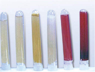
The rightmost red-colored tube most likely represents synovial fluid from a patient with:
- A) Septic arthritis
- B) Monosodium urate (gout) crystal arthritis
- C) Hemorrhagic effusion (trauma/hemophilia)
- D) Non-inflammatory osteoarthritis
✅ Answer: C - Hemorrhagic effusion Red/bloody synovial fluid = Hemorrhagic group (trauma, tumors, hemophilia, coagulation disorders, anticoagulant overdose). This is ALSO the only group with normal glucose levels. Remember: Leftmost tube (clear/pale yellow) = Normal. Red tube = Hemorrhage.
QUESTION 2 - GROSS APPEARANCE (MULTIPLE TUBES)
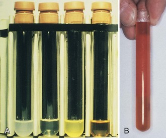
Looking at Image A (4 dark tubes) left to right, the YELLOW-GREEN turbid tube is most consistent with:
- A) Normal synovial fluid
- B) Non-inflammatory effusion
- C) Bacterial/Septic arthritis
- D) Crystal-induced (milky) arthritis
✅ Answer: C - Bacterial/Septic arthritis Per the DMSF transcript: Greenish tinge = Bacterial infection. Septic arthritis fluid = Cloudy, Yellow-green. Also confirmed by: WBC 50,000-100,000/uL, neutrophils >75%, positive culture and Gram stain.
QUESTION 3 - ABNORMAL SYNOVIAL FLUID CELLS
The image below shows a microscopic view of synovial fluid cells. The arrows point to neutrophils that have engulfed a large, rounded, homogeneous purple mass (the degenerating nucleus of a lymphocyte):
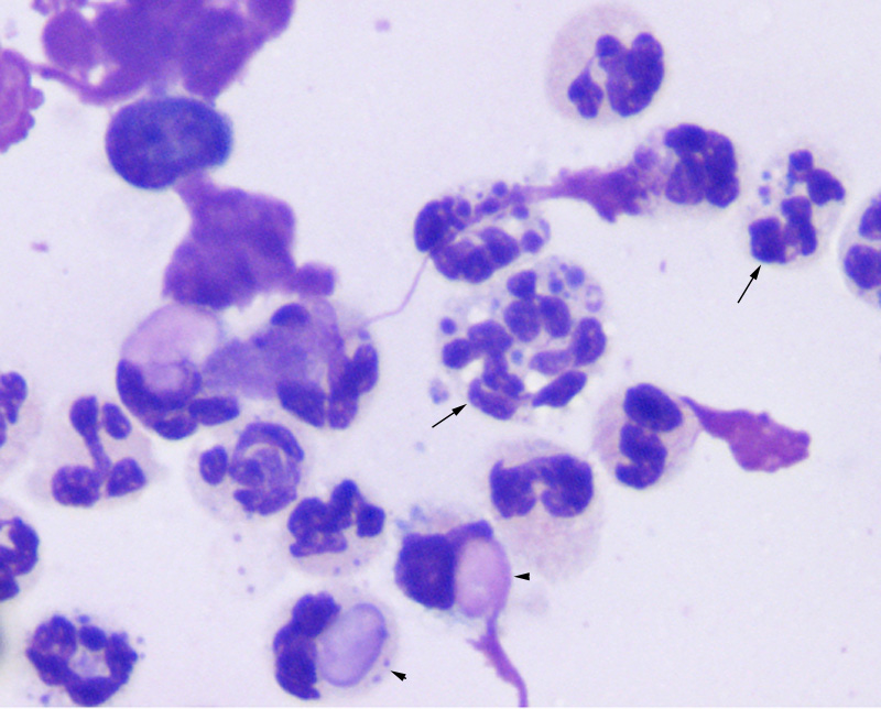
The cells indicated by the small arrowheads (▲) at the bottom of the image, which show neutrophils engulfing a rounded homogeneous nuclear mass, are called:
- A) Ragocytes (RA cells)
- B) Reiter cells
- C) LE cells (Lupus Erythematosus cells)
- D) Synovial lining cells
✅ Answer: C - LE cells LE cells = Neutrophils engulfing the degenerating nucleus of lymphocytes (rounded homogeneous purple mass). Associated with: SLE (Systemic Lupus Erythematosus). Also: ANA positive in 70% of SLE patients.⚠️ Contrast:
- LE cells = neutrophil eating lymphocyte nucleus → SLE
- Ragocytes = neutrophil with bluish RF inclusions → RA
- Reiter cells = macrophage eating a degenerating neutrophil → Reiter's syndrome
QUESTION 4 - ABNORMAL SYNOVIAL FLUID CELLS
The image below shows a macrophage (large pale cell) with an arrow pointing to an engulfed degenerating neutrophil (showing lobulated nuclear fragments inside the macrophage cytoplasm):

This cell type (macrophage engulfing a degenerating neutrophil) is called:
- A) LE cell
- B) Ragocyte
- C) Reiter cell
- D) Hemosiderin-laden macrophage
✅ Answer: C - Reiter cell Reiter cells = Macrophage engulfing degenerating neutrophil (the lobes of the neutrophil are degenerating inside the macrophage). Associated with: Reiter's syndrome.📝 Key distinction from LE cells: LE cell = NEUTROPHIL eating lymphocyte nucleus. Reiter cell = MACROPHAGE eating a neutrophil.
QUESTION 5 - CRYSTAL IDENTIFICATION
The image below shows crystals from a synovial fluid aspirate viewed under compensated polarized light microscopy. The crystals appear needle-shaped with bright birefringence (greenish/yellow discoloration):

These needle-shaped, strongly birefringent crystals are diagnostic of:
- A) Pseudogout (CPPD)
- B) Calcium oxalate crystals
- C) Urate Gout - Monosodium Urate (MSU)
- D) Cholesterol crystals
✅ Answer: C - Monosodium Urate (MSU) / Urate Gout MSU crystals = Needle-like shape + positive birefringence (greenish discoloration under polarized light). The fluid will also be: Cloudy/milky, WBC up to 100,000/uL, neutrophils >70%, decreased glucose.
QUESTION 6 - CRYSTAL IDENTIFICATION
The image below shows crystals from a synovial fluid aspirate. The crystals appear rhomboid/rectangular/4-sided in shape with weak birefringence:

These 4-sided (rhomboid/rectangular) crystals with weak birefringence are called:
- A) Monosodium urate crystals
- B) Calcium pyrophosphate dihydrate (CPPD) crystals
- C) Calcium oxalate dihydrate crystals
- D) Cholesterol crystals
✅ Answer: B - Calcium Pyrophosphate Dihydrate (CPPD) CPPD crystals = 4-sided shape (square, rhomboid, rectangle) + weak birefringence Associated disease = Pseudogout / Chondrocalcinosis (pyrophosphate gout)⚠️ Critical distinction for DMSF exam:
Feature MSU (Gout) CPPD (Pseudogout) Shape Needle-like 4-sided (rhomboid) Birefringence Strong (bright green) Weak Disease Urate Gout Pseudogout
QUESTION 7 - CRYSTAL COMPARISON
Study the crystal shapes below and match them to the correct crystal type and disease:
Crystal A:  ==================  (long, thin, needle)
Crystal B:  ▱ or ◇             (rhomboid / 4-sided)
Crystal C:  🪜 staircase/ladder shape
Crystal D:  ✚ within a square (cross/envelope)
Crystal A is to Gout as Crystal B is to:
- A) Urate gout
- B) Pseudogout (CPPD)
- C) Oxalate gout
- D) Lipid gout
✅ Answer: B - Pseudogout (CPPD)Full crystal-disease matching (DMSF must-know):
Crystal Shape Disease A - MSU Needle Urate Gout B - CPPD 4-sided (rhomboid) Pseudogout C - Cholesterol Staircase/ladder Non-specific D - Calcium oxalate Cross/envelope in square Oxalate gout
QUESTION 8 - VISCOSITY / STRING TEST
The image shows two pouring tests of synovial fluid:
Tube A: Fluid pours like water - thin, breaks immediately
Tube B: Fluid stretches into a long honey-like unbroken string
Tube A (thin, water-like fluid) most likely belongs to which condition?
- A) Normal synovial fluid
- B) Osteoarthritis (non-inflammatory)
- C) Gouty arthritis or septic arthritis (poor viscosity)
- D) Traumatic effusion
✅ Answer: C - Gout or Septic arthritis Per DMSF transcript viscosity grading:
- Thick/honey-like (long unbroken string) = Good = Normal, Osteoarthritis, Trauma
- Thin/water-like = Poor = Crystal-induced (Gout), Septic (Gonorrhea) Normal string test = 4-6 cm before breaking.
QUESTION 9 - MUCIN CLOT TEST
The image below (from the DMSF lecture) shows two mucin/Ropes test results after adding 2-5% acetic acid:
LEFT tube:   One solid, firm clot — clear surrounding fluid
RIGHT tube:  Fragmented clots with hazy/cloudy surrounding fluid
The RIGHT tube result (fragmented clot + hazy fluid) is most consistent with:
- A) Normal synovial fluid
- B) Osteoarthritis
- C) Traumatic effusion
- D) Inflammatory arthritis (RA, Lupus, Reiter's)
✅ Answer: D - Inflammatory arthritis Per DMSF transcript:
- Good (solid clot, clear fluid) = Normal, Osteoarthritis, Trauma, Bleed
- Fair (fragmented/soft clot, hazy fluid) = Lupus, RA, Reiter's syndrome
- Poor (no visible clot) = Gout, Gonorrhea
QUESTION 10 - INCLUSION BODIES
Based on the DMSF lecture description, the image below shows rice-shaped white bodies floating in synovial fluid:
[Visual: Small white oval/rice-grain shaped bodies in joint fluid]
These "rice bodies" found in synovial fluid are most associated with:
- A) Gout (crystal deposition)
- B) Rheumatoid arthritis (degenerated synovium with fibrin)
- C) Septic arthritis
- D) SLE
✅ Answer: B - Rheumatoid Arthritis Rice bodies = degenerated synovium enriched with fibrin Associated with: Rheumatoid Arthritis Compare with Ochronotic shards = debris from joint prosthesis
QUESTION 11 - CELL TYPE RECOGNITION
The image shows a large cell with columnar/cuboidal configuration, rounded nuclei, and dense chromatin pattern without prominent nucleoli:
[Cell with columnar/cuboidal shape, rounded dense nucleus]
This cell is a:
- A) Macrophage
- B) Neutrophil
- C) Synovial lining cell (synoviocyte)
- D) LE cell
✅ Answer: C - Synovial lining cell (synoviocyte) Per DMSF transcript: Synovial lining cells have columnar or cuboidal configuration with rounded nuclei and dense chromatin pattern without prominent nucleoli. These are normal primary cells seen in synovial fluid differential.
QUESTION 12 - INTEGRATED CASE (PICTURE-BASED)
A 45-year-old male presents with sudden severe pain and swelling of the first metatarsophalangeal joint. Synovial fluid aspiration is done. The findings are:
| Finding | Result |
|---|---|
| Color | Cloudy, milky white |
| Viscosity | Low |
| WBC | 85,000/uL |
| Neutrophils | 72% |
| Glucose | Decreased |
| Crystal under polarized light | Needle-shaped, strongly birefringent (greenish) |
What is the diagnosis AND what is the crystal type?
- A) Pseudogout; CPPD crystals
- B) Septic arthritis; no crystals
- C) Urate Gout; Monosodium Urate (MSU) crystals
- D) Rheumatoid arthritis; Ragocytes
✅ Answer: C - Urate Gout; MSU crystals Classic gout presentation:
- First MTP joint ("podagra")
- Milky fluid + decreased glucose + high WBC (crystal-induced = up to 100,000/uL)
- Needle-shaped birefringent crystals = MSU = Urate Gout
QUESTION 13 - INTEGRATED CASE
A 60-year-old woman with known Rheumatoid Arthritis has her knee joint aspirated. The lab reports:
| Finding | Result |
|---|---|
| Fluid color | Cloudy, yellow |
| Viscosity | Poor |
| WBC | 35,000/uL |
| Neutrophils | 60% |
| Special cells | Neutrophils with dark bluish inclusions of precipitated protein |
| Immunology | RF positive |
The "neutrophils with dark bluish inclusions of precipitated protein" are called:
- A) LE cells
- B) Reiter cells
- C) Ragocytes (RA cells)
- D) Hemosiderin-laden macrophages
✅ Answer: C - Ragocytes (RA cells) RA cells / Ragocytes = Neutrophils containing bluish inclusions composed of precipitated Rheumatoid factor Context clues: Known RA + RF positive + neutrophils with inclusions = Ragocytes Remember: Rheumatoid factor is positive in 60% of RA patients
QUESTION 14 - TUBE SELECTION
A synovial fluid specimen is obtained and needs to be distributed into the correct tubes. The lab technician needs to perform crystal examination:
- A) Sterile heparinized tube
- B) Sodium fluoride tube
- C) Plain tube - no anticoagulant
- D) Powdered anticoagulant tube
✅ Answer: C - Plain tube (or heparinized/EDTA liquid - NOT powdered) Powdered anticoagulants MUST NOT be used because they interfere with crystal examination. The DMSF rule: "Powdered anticoagulant should NOT be used - interferes with crystal examination/analysis."Correct tube assignment:
- Gram stain + Culture → Sterile heparinized tube
- Cell count → Heparinized/EDTA tube
- Other tests → Plain tube
- Glucose → Sodium fluoride tube
QUESTION 15 - FINAL INTEGRATOR
Match the image descriptions to the correct diagnosis:
| Image Finding | Match |
|---|---|
| A. Yellow-green turbid fluid, WBC 80,000, neutrophils 78%, positive Gram stain | ? |
| B. Clear pale yellow fluid, WBC 700, neutrophils 20%, good viscosity, normal glucose | ? |
| C. Milky cloudy fluid, WBC 90,000, needle-shaped birefringent crystals | ? |
| D. Red fluid, WBC equal to blood, NORMAL glucose | ? |
| E. Cloudy yellow fluid, WBC 40,000, neutrophils 55%, RF positive, Ragocytes seen | ? |
Options:
- Non-inflammatory (Osteoarthritis)
- Urate Gout
- Septic Arthritis
- Hemorrhagic Effusion
- Rheumatoid Arthritis
✅ Answers:
- A → 3 (Septic): Yellow-green + high WBC + positive Gram stain
- B → 1 (Non-inflammatory): Clear + WBC <1,000 + normal glucose + good viscosity
- C → 2 (Gout): Milky + needle crystals + WBC up to 100,000
- D → 4 (Hemorrhagic): Red fluid + normal glucose (only group!)
- E → 5 (RA): Cloudy + RF positive + Ragocytes
🎯 QUICK IMAGE RECALL CHEAT SHEET
| What you SEE in the image | What to ANSWER |
|---|---|
| Needle-shaped crystal + birefringent green | MSU = Gout |
| 4-sided/rhomboid crystal | CPPD = Pseudogout |
| Staircase/ladder crystal | Cholesterol crystal |
| Cross/envelope crystal | Calcium oxalate |
| Neutrophil eating round purple mass | LE cell = SLE |
| Neutrophil with bluish granular inclusions | Ragocyte = RA |
| Macrophage eating a neutrophil | Reiter cell = Reiter's syndrome |
| Rice-grain shaped bodies in fluid | Rice bodies = RA |
| Red/bloody fluid + normal glucose | Hemorrhagic |
| Yellow-green turbid + positive culture | Septic |
| Milky fluid + crystals | Crystal-induced (Gout/Pseudogout) |
| Honey-like unbroken string test | Good viscosity = Normal/OA |
| Water-like thin fluid | Poor viscosity = Gout/Gonorrhea |
| Solid clot (Mucin test) | Normal / OA / Trauma |
| Fragmented clot (Mucin test) | RA / Lupus / Reiter's |
This is a shared conversation. Sign in to Orris to start your own chat.