Barium studies in radiology with images and pathologies

Reading File
Finding Sources
Searching Images

barium swallow esophagus radiology

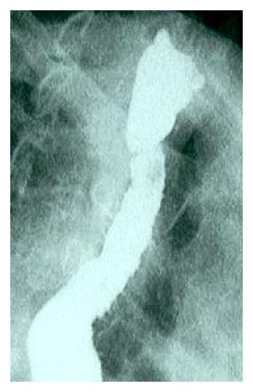
This diagnostic image is a barium swallow fluoroscopy (X-ray radiography) of the upper gastrointestinal tract, specifically the esophagus. The image reveals classic radiographic findings of achalasia. There is marked dilation of the esophageal body (megaesophagus), which appears as a widened, radiopaque column due to the retention of barium contrast. At the distal end, the esophagus exhibits a smooth, symmetrical tapering that terminates in a narrow, 'bird's beak' or 'rat-tail' appearance at the level of the lower esophageal sphincter (LES). This tapering signifies failure of the LES to relax, causing functional obstruction and a delay in contrast passage into the stomach. The clinical significance of this finding is the confirmation of achalasia, often associated with symptoms like dysphagia, regurgitation, and failure to thrive. The image provides a primary educational example of esophageal motility disorders within the field of gastroenterology and pediatric radiology.
This diagnostic image is a barium swallow fluoroscopy (X-ray radiography) of the upper gastrointestinal tract, specifically the esophagus. The image reveals classic radiographic findings of achalasia. There is marked dilation of the esophageal body (megaesophagus), which appears as a widened, radiopaque column due to the retention of barium contrast. At the distal end, the esophagus exhibits a smooth, symmetrical tapering that terminates in a narrow, 'bird's beak' or 'rat-tail' appearance at the level of the lower esophageal sphincter (LES). This tapering signifies failure of the LES to relax, causing functional obstruction and a delay in contrast passage into the stomach. The clinical significance of this finding is the confirmation of achalasia, often associated with symptoms like dysphagia, regurgitation, and failure to thrive. The image provides a primary educational example of esophageal motility disorders within the field of gastroenterology and pediatric radiology.

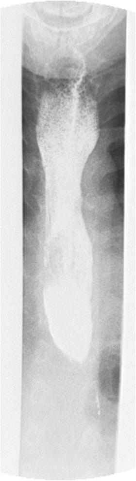
This diagnostic image is an esophageal barium swallow (contrast radiography) demonstrating classic features of esophageal intramural pseudodiverticulosis (EIP). The radiopaque contrast material highlights a significant focal narrowing in the upper esophagus, consistent with a proximal esophageal stricture. Associated with this stricture, multiple small, flask-shaped, or linear collections of contrast are visible running parallel to the esophageal wall. These represent dilated excretory ducts of the deep esophageal mucosal glands that have filled with barium, creating a characteristic 'dotted line' or 'pseudodiverticula' appearance. The image serves as a clinical example of EIP, a condition often associated with chronic inflammatory processes such as Candida esophagitis or chronic gastroesophageal reflux. This imaging is crucial for gastroenterology and radiology education, illustrating the typical radiographic morphology used to distinguish intramural pseudodiverticula from true esophageal diverticula.
This diagnostic image is an esophageal barium swallow (contrast radiography) demonstrating classic features of esophageal intramural pseudodiverticulosis (EIP). The radiopaque contrast material highlights a significant focal narrowing in the upper esophagus, consistent with a proximal esophageal stricture. Associated with this stricture, multiple small, flask-shaped, or linear collections of contrast are visible running parallel to the esophageal wall. These represent dilated excretory ducts of the deep esophageal mucosal glands that have filled with barium, creating a characteristic 'dotted line' or 'pseudodiverticula' appearance. The image serves as a clinical example of EIP, a condition often associated with chronic inflammatory processes such as Candida esophagitis or chronic gastroesophageal reflux. This imaging is crucial for gastroenterology and radiology education, illustrating the typical radiographic morphology used to distinguish intramural pseudodiverticula from true esophageal diverticula.

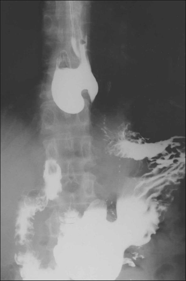
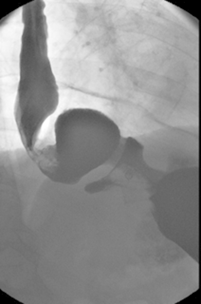
This diagnostic image is a barium swallow (upper gastrointestinal series) radiograph of the esophagus and stomach. The image demonstrates a prominent epiphrenic diverticulum, which appears as a large, rounded, radiopaque outpouching filled with contrast medium in the distal thoracic esophagus, just above the diaphragm. The esophagus itself shows significant architectural abnormalities, including distal dilatation and a non-straight, tortuous contour suggestive of a motility disorder such as achalasia. Contrast material is visible passing through the gastroesophageal junction into the stomach, where mucosal folds are outlined. The presence of this pulsion diverticulum in the lower third of the esophagus is a key finding often associated with increased intraluminal pressure. This educational material is relevant for gastroenterology and radiology specialties, focusing on structural esophageal pathologies and diagnostic fluoroscopy.
This diagnostic image is a barium swallow (upper gastrointestinal series) radiograph of the esophagus and stomach. The image demonstrates a prominent epiphrenic diverticulum, which appears as a large, rounded, radiopaque outpouching filled with contrast medium in the distal thoracic esophagus, just above the diaphragm. The esophagus itself shows significant architectural abnormalities, including distal dilatation and a non-straight, tortuous contour suggestive of a motility disorder such as achalasia. Contrast material is visible passing through the gastroesophageal junction into the stomach, where mucosal folds are outlined. The presence of this pulsion diverticulum in the lower third of the esophagus is a key finding often associated with increased intraluminal pressure. This educational material is relevant for gastroenterology and radiology specialties, focusing on structural esophageal pathologies and diagnostic fluoroscopy.

This diagnostic image is a barium swallow (barium meal) radiograph demonstrating classic features of advanced achalasia. The esophagus is severely dilated with a tortuous, 'sigmoid' morphology, indicative of chronic obstruction and loss of esophageal peristalsis (megaesophagus). At the distal end, the esophagus terminates in a smooth, tapered narrowing at the gastroesophageal junction, creating the characteristic 'bird's beak' appearance. This finding represents a failure of the lower esophageal sphincter (LES) to relax, leading to the pooling of contrast material and food within the thoracic esophagus. The image illustrates the anatomical and functional changes associated with esophageal motility disorders, specifically the late stages of achalasia where the organ becomes decompensated and redundant. It serves as a key clinical example for medical students and residents in gastroenterology and radiology for identifying esophageal outflow obstruction.
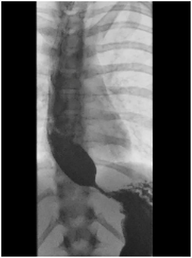
This diagnostic image is a barium swallow (barium meal) radiograph demonstrating classic features of advanced achalasia. The esophagus is severely dilated with a tortuous, 'sigmoid' morphology, indicative of chronic obstruction and loss of esophageal peristalsis (megaesophagus). At the distal end, the esophagus terminates in a smooth, tapered narrowing at the gastroesophageal junction, creating the characteristic 'bird's beak' appearance. This finding represents a failure of the lower esophageal sphincter (LES) to relax, leading to the pooling of contrast material and food within the thoracic esophagus. The image illustrates the anatomical and functional changes associated with esophageal motility disorders, specifically the late stages of achalasia where the organ becomes decompensated and redundant. It serves as a key clinical example for medical students and residents in gastroenterology and radiology for identifying esophageal outflow obstruction.

This diagnostic image is a barium swallow fluoroscopy (upper gastrointestinal series) radiograph of the chest and upper abdomen, demonstrating a classic 'bird-beak' appearance of the distal esophagus. The image shows significant proximal esophageal dilation filled with radiopaque barium contrast material. At the level of the gastroesophageal junction, there is a smooth, tapered, and symmetrical narrowing where the contrast terminates in a sharp point before entering the stomach. This visual sign is highly characteristic of achalasia, a primary esophageal motility disorder characterized by failure of the lower esophageal sphincter (LES) to relax and absent peristalsis. The background anatomy includes the thoracic vertebral column and ribs, which are positioned posterior to the dilated esophagus. The image serves as a definitive educational example of esophageal stasis and obstructive pathophysiology at the LES, often used for training in gastroenterology and radiology for the diagnosis of Type I or Type II achalasia.
This diagnostic image is a barium swallow fluoroscopy (upper gastrointestinal series) radiograph of the chest and upper abdomen, demonstrating a classic 'bird-beak' appearance of the distal esophagus. The image shows significant proximal esophageal dilation filled with radiopaque barium contrast material. At the level of the gastroesophageal junction, there is a smooth, tapered, and symmetrical narrowing where the contrast terminates in a sharp point before entering the stomach. This visual sign is highly characteristic of achalasia, a primary esophageal motility disorder characterized by failure of the lower esophageal sphincter (LES) to relax and absent peristalsis. The background anatomy includes the thoracic vertebral column and ribs, which are positioned posterior to the dilated esophagus. The image serves as a definitive educational example of esophageal stasis and obstructive pathophysiology at the LES, often used for training in gastroenterology and radiology for the diagnosis of Type I or Type II achalasia.

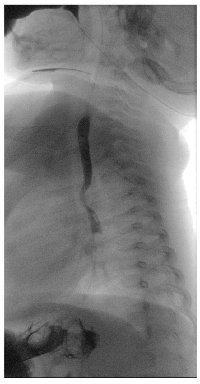
This lateral-view diagnostic image demonstrates a pediatric barium swallow test (contrast esophagram) in a 5-month-old patient. The esophagus is opacified with contrast, appearing as a dark, vertical column against the radiolucent thoracic cavity and the radiopaque vertebral column. A significant pathological finding is visible in the proximal thoracic region: a focal posterior indentation of the esophageal wall. This indentation causes localized narrowing of the contrast column and anterior displacement, which is a classic radiographic sign of extrinsic compression. In the clinical context of dysphagia and failure to thrive, such a finding is highly suggestive of a vascular ring, such as a right aortic arch with an aberrant left subclavian artery and Kommerell's diverticulum. The remainder of the esophagus distal to the compression appears to have a normal caliber. This image serves as a critical diagnostic step in pediatric radiology for identifying congenital anatomical anomalies affecting the upper gastrointestinal tract.

This lateral-view diagnostic image demonstrates a pediatric barium swallow test (contrast esophagram) in a 5-month-old patient. The esophagus is opacified with contrast, appearing as a dark, vertical column against the radiolucent thoracic cavity and the radiopaque vertebral column. A significant pathological finding is visible in the proximal thoracic region: a focal posterior indentation of the esophageal wall. This indentation causes localized narrowing of the contrast column and anterior displacement, which is a classic radiographic sign of extrinsic compression. In the clinical context of dysphagia and failure to thrive, such a finding is highly suggestive of a vascular ring, such as a right aortic arch with an aberrant left subclavian artery and Kommerell's diverticulum. The remainder of the esophagus distal to the compression appears to have a normal caliber. This image serves as a critical diagnostic step in pediatric radiology for identifying congenital anatomical anomalies affecting the upper gastrointestinal tract.

Reading File
Reading File
Searching Images

barium enema colon pathology radiology
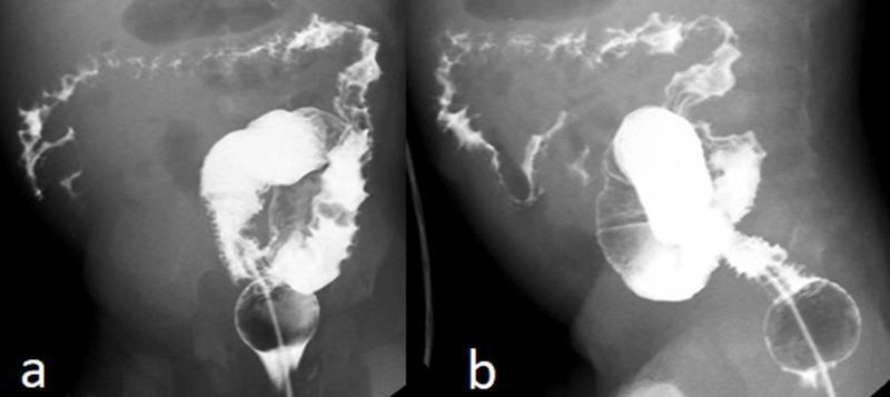
This diagnostic fluoroscopic image displays a barium enema examination of the colon in an infant, presented in two views (a and b). The primary findings include a significantly reduced caliber of the colonic lumen and a strikingly irregular mucosal contour. A classic radiological 'thumbprinting' sign is evident, characterized by rounded, smooth indentations along the colonic wall. These features represent submucosal edema, hemorrhage, or intraluminal masses, which in this clinical context correspond to gastrointestinal hemangiomatosis. The distribution of the pathology is extensive, involving the ascending, transverse, and descending segments of the colon, while the sigmoid colon appears relatively spared with a more preserved caliber. The image demonstrates key signs of diffuse inflammatory or vascular colonic disease, essential for pediatric radiology and gastroenterology education. The balloon of the enema catheter is visible in the rectum, marking the distal extent of the study.
This diagnostic fluoroscopic image displays a barium enema examination of the colon in an infant, presented in two views (a and b). The primary findings include a significantly reduced caliber of the colonic lumen and a strikingly irregular mucosal contour. A classic radiological 'thumbprinting' sign is evident, characterized by rounded, smooth indentations along the colonic wall. These features represent submucosal edema, hemorrhage, or intraluminal masses, which in this clinical context correspond to gastrointestinal hemangiomatosis. The distribution of the pathology is extensive, involving the ascending, transverse, and descending segments of the colon, while the sigmoid colon appears relatively spared with a more preserved caliber. The image demonstrates key signs of diffuse inflammatory or vascular colonic disease, essential for pediatric radiology and gastroenterology education. The balloon of the enema catheter is visible in the rectum, marking the distal extent of the study.

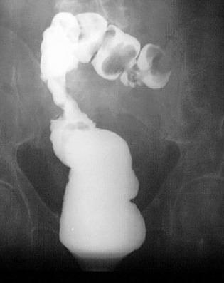
This diagnostic image is an irigography (barium enema radiography) of the colon, demonstrating a significant pathology in the region of the hepatic flexure. The lower segment of the contrast-filled colon appears smooth and normal in caliber. As the contrast ascends, there is a clear transition zone marked by a sudden, severe circumferential constriction (stenosis). Superior to this constriction, the colon exhibits massive dilation and an irregular, lobulated appearance with multiple bulbous segments. The contrast within these dilated lobes shows heterogeneous density, including lucent areas likely representing gas or displacement by a large mass. The morphology is highly suggestive of an obstructing tumor at the hepatic flexure, showing the classic 'apple-core' sign or shelf-like transition typical of malignancy. This image is used in gastroenterology and radiology curricula to teach the visual manifestations of colorectal cancer and the utility of fluoroscopic contrast studies in identifying bowel obstructions and anatomical abnormalities.
This diagnostic image is an irigography (barium enema radiography) of the colon, demonstrating a significant pathology in the region of the hepatic flexure. The lower segment of the contrast-filled colon appears smooth and normal in caliber. As the contrast ascends, there is a clear transition zone marked by a sudden, severe circumferential constriction (stenosis). Superior to this constriction, the colon exhibits massive dilation and an irregular, lobulated appearance with multiple bulbous segments. The contrast within these dilated lobes shows heterogeneous density, including lucent areas likely representing gas or displacement by a large mass. The morphology is highly suggestive of an obstructing tumor at the hepatic flexure, showing the classic 'apple-core' sign or shelf-like transition typical of malignancy. This image is used in gastroenterology and radiology curricula to teach the visual manifestations of colorectal cancer and the utility of fluoroscopic contrast studies in identifying bowel obstructions and anatomical abnormalities.

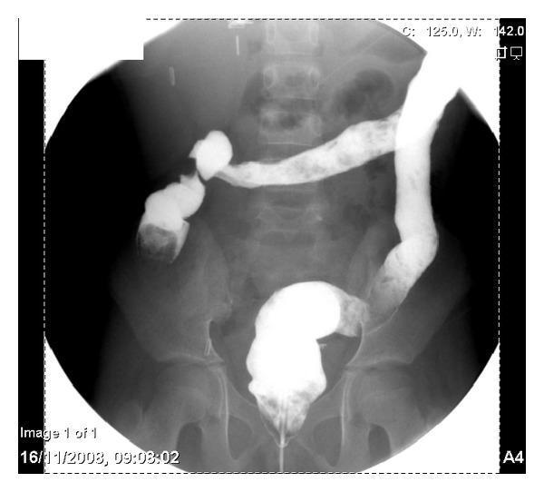
A diagnostic image showing a barium enema examination of the lower gastrointestinal tract. The radiograph displays a significant luminal narrowing, or stricture, located in the rectosigmoid region of the colon. The barium contrast agent clearly outlines a transition from a normal, dilated colonic diameter to an abruptly constricted, irregular channel where the lumen is markedly reduced. This finding suggests a partial obstruction. The surrounding anatomical context includes the lumbar vertebrae and the sacrum, visible in the background. The image is used for clinical education in gastroenterology and radiology to demonstrate the radiographic presentation of colonic strictures, which may be associated with inflammatory bowel disease, tuberculosis, or primary malignancies such as plasmacytoma of the colon.
A diagnostic image showing a barium enema examination of the lower gastrointestinal tract. The radiograph displays a significant luminal narrowing, or stricture, located in the rectosigmoid region of the colon. The barium contrast agent clearly outlines a transition from a normal, dilated colonic diameter to an abruptly constricted, irregular channel where the lumen is markedly reduced. This finding suggests a partial obstruction. The surrounding anatomical context includes the lumbar vertebrae and the sacrum, visible in the background. The image is used for clinical education in gastroenterology and radiology to demonstrate the radiographic presentation of colonic strictures, which may be associated with inflammatory bowel disease, tuberculosis, or primary malignancies such as plasmacytoma of the colon.

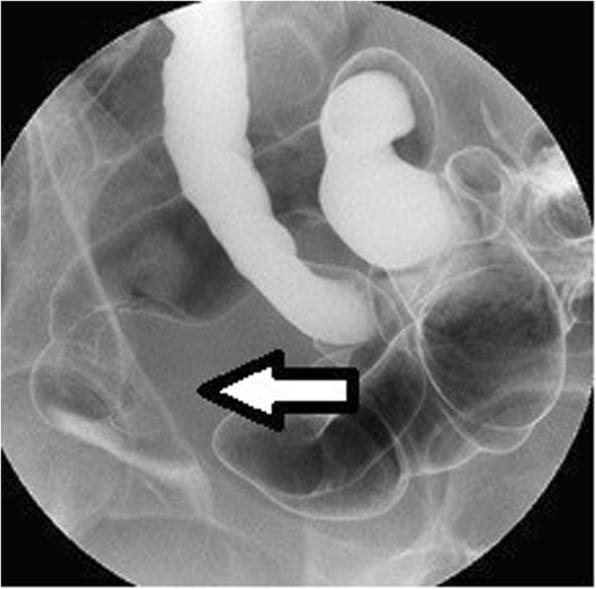
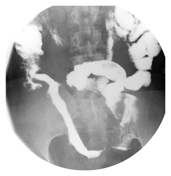
This diagnostic image is a barium enema (single-contrast radiography) of the pelvic region focusing on the distal large intestine. The radiograph displays the rectum and sigmoid colon filled with radiopaque barium contrast. A prominent finding is a 40-mm extramural mass located in the rectum, indicated by a white arrow. This mass manifests as a smooth, rounded indentation and outward distortion of the rectal wall contour, suggesting an external compressive force rather than an intraluminal mucosal lesion. The surrounding anatomical structures are visible through faint gas and contrast shadows. Such findings are clinically significant in the diagnostic workup of extrinsic pelvic masses, including intestinal endometriosis or gastrointestinal stromal tumors (GIST), particularly when mucosal findings on colonoscopy are absent. This material is suitable for intermediate to advanced medical learners studying gastrointestinal radiology and surgical pathology.

This diagnostic image is a barium enema (single-contrast radiography) of the pelvic region focusing on the distal large intestine. The radiograph displays the rectum and sigmoid colon filled with radiopaque barium contrast. A prominent finding is a 40-mm extramural mass located in the rectum, indicated by a white arrow. This mass manifests as a smooth, rounded indentation and outward distortion of the rectal wall contour, suggesting an external compressive force rather than an intraluminal mucosal lesion. The surrounding anatomical structures are visible through faint gas and contrast shadows. Such findings are clinically significant in the diagnostic workup of extrinsic pelvic masses, including intestinal endometriosis or gastrointestinal stromal tumors (GIST), particularly when mucosal findings on colonoscopy are absent. This material is suitable for intermediate to advanced medical learners studying gastrointestinal radiology and surgical pathology.

Searching Images

barium meal stomach duodenum ulcer radiology
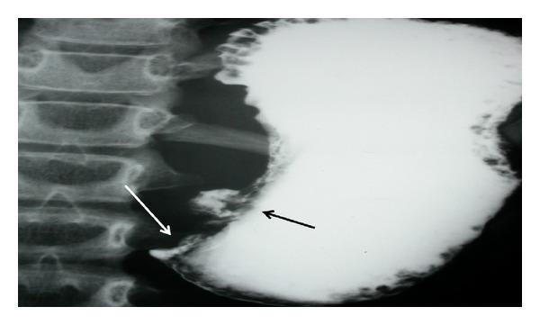
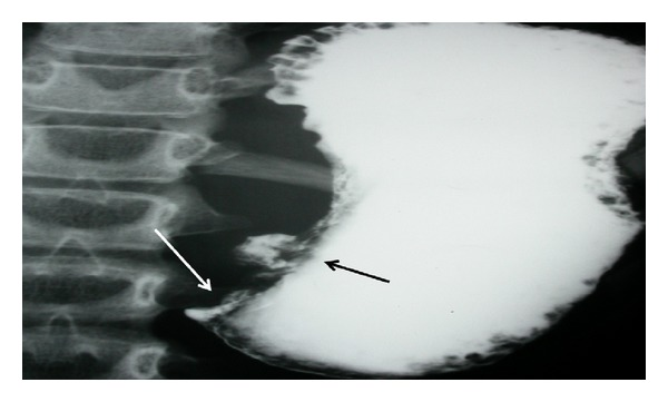
This diagnostic image is an upper gastrointestinal barium meal radiograph (X-ray) focusing on the stomach and pyloric region. The contrast study reveals a significantly distended stomach filled with radiopaque barium. Two key pathological findings are annotated: a black arrow indicates a localized, irregular projection of contrast along the lesser curvature, representing an ulcer crater. A white arrow points to the distal antrum/pylorus, where there is an abrupt, tapered termination of the contrast column with no visible passage into the duodenum, characteristic of a high-grade gastric outlet obstruction. The vertebral column is visible to the left of the gastric shadow. This radiographic appearance is clinically significant for diagnosing mechanical obstruction and mucosal lesions, such as those caused by peptic ulcer disease or gastric malignancy. The image is intended for intermediate to advanced medical learners studying gastrointestinal radiology and surgical pathology.
This diagnostic image is an upper gastrointestinal barium meal radiograph (X-ray) focusing on the stomach and pyloric region. The contrast study reveals a significantly distended stomach filled with radiopaque barium. Two key pathological findings are annotated: a black arrow indicates a localized, irregular projection of contrast along the lesser curvature, representing an ulcer crater. A white arrow points to the distal antrum/pylorus, where there is an abrupt, tapered termination of the contrast column with no visible passage into the duodenum, characteristic of a high-grade gastric outlet obstruction. The vertebral column is visible to the left of the gastric shadow. This radiographic appearance is clinically significant for diagnosing mechanical obstruction and mucosal lesions, such as those caused by peptic ulcer disease or gastric malignancy. The image is intended for intermediate to advanced medical learners studying gastrointestinal radiology and surgical pathology.

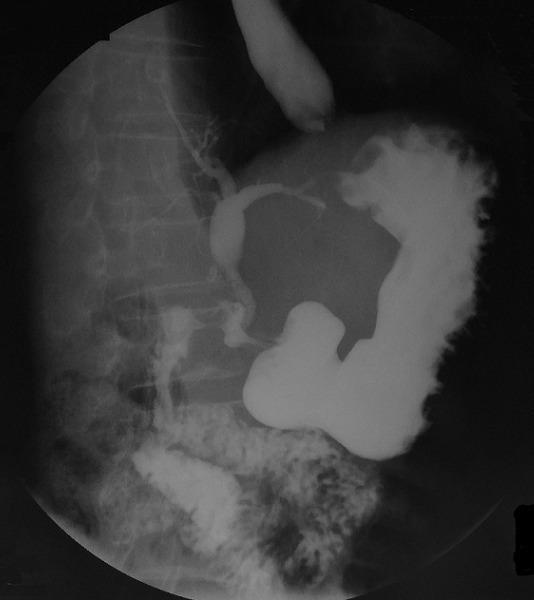
This diagnostic fluoroscopic image is a barium meal study (upper gastrointestinal series) demonstrating a choledochoduodenal fistula (CDF). The image shows radiopaque contrast material opacifying the esophagus, stomach, and a deformed duodenal bulb. Critically, there is clear evidence of contrast reflux from the duodenum into the biliary tract, resulting in the opacification of the common bile duct and intrahepatic bile ducts. This retrograde filling of the biliary tree is a pathological finding indicating an abnormal communication between the gastrointestinal and biliary systems. The duodenal bulb appears distorted, consistent with chronic peptic ulcer disease which can lead to fistula formation. This radiographic study is highly characteristic for diagnosing spontaneous internal biliary fistulas, where contrast or air (pneumobilia) occupies the biliary tree. This material is useful for teaching radiology and gastroenterology students about the complications of severe duodenal ulcers and the application of barium studies in identifying hepatobiliary anomalies.
This diagnostic fluoroscopic image is a barium meal study (upper gastrointestinal series) demonstrating a choledochoduodenal fistula (CDF). The image shows radiopaque contrast material opacifying the esophagus, stomach, and a deformed duodenal bulb. Critically, there is clear evidence of contrast reflux from the duodenum into the biliary tract, resulting in the opacification of the common bile duct and intrahepatic bile ducts. This retrograde filling of the biliary tree is a pathological finding indicating an abnormal communication between the gastrointestinal and biliary systems. The duodenal bulb appears distorted, consistent with chronic peptic ulcer disease which can lead to fistula formation. This radiographic study is highly characteristic for diagnosing spontaneous internal biliary fistulas, where contrast or air (pneumobilia) occupies the biliary tree. This material is useful for teaching radiology and gastroenterology students about the complications of severe duodenal ulcers and the application of barium studies in identifying hepatobiliary anomalies.

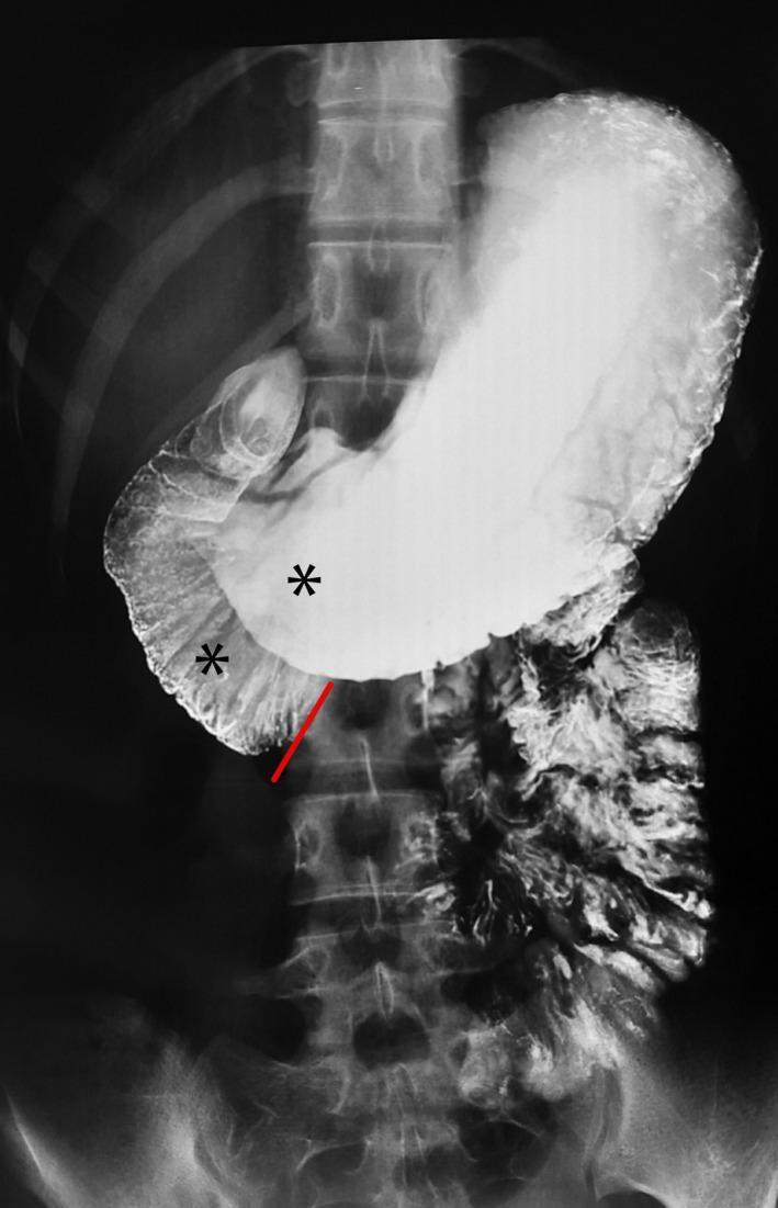
This diagnostic image is a barium meal radiograph (upper gastrointestinal series) showing the stomach and proximal duodenum. The image demonstrates a significantly distended, contrast-filled stomach and marked dilatation of the first and second parts of the duodenum (indicated by asterisks). A clear, linear, abrupt cutoff of the barium column is visible at the third (transverse) part of the duodenum, highlighted by a red annotation line. This 'cutoff sign' suggests an extrinsic compression or obstruction at the level where the duodenum crosses the midline. The spinal column and ribs provide anatomical landmarks for the upper abdomen. The visual findings are classic radiological indicators of Superior Mesenteric Artery (SMA) syndrome, where the third part of the duodenum is compressed between the aorta and the overlying SMA. This material is designed for intermediate to advanced medical learners studying gastrointestinal radiology and surgical pathology.
This diagnostic image is a barium meal radiograph (upper gastrointestinal series) showing the stomach and proximal duodenum. The image demonstrates a significantly distended, contrast-filled stomach and marked dilatation of the first and second parts of the duodenum (indicated by asterisks). A clear, linear, abrupt cutoff of the barium column is visible at the third (transverse) part of the duodenum, highlighted by a red annotation line. This 'cutoff sign' suggests an extrinsic compression or obstruction at the level where the duodenum crosses the midline. The spinal column and ribs provide anatomical landmarks for the upper abdomen. The visual findings are classic radiological indicators of Superior Mesenteric Artery (SMA) syndrome, where the third part of the duodenum is compressed between the aorta and the overlying SMA. This material is designed for intermediate to advanced medical learners studying gastrointestinal radiology and surgical pathology.

**Imaging Modality:** Barium meal (upper gastrointestinal fluoroscopy with oral contrast).

**Anatomical Region:** Distal stomach, specifically the antrum and pyloric region.

**Observed Pathology:** The image demonstrates a significant gastric outlet obstruction. A prominent barium-filled collection, characteristic of an ulcer crater, is visible along the lesser curvature of the stomach (indicated by the black arrow). Distal to this, there is a marked narrowing and abrupt termination of the contrast column at the pylorus (indicated by the white arrow), suggesting luminal obstruction.

**Characteristic Visual Features:**
*   **Ulcer Niche:** An out-pouching of contrast material representing a localized mucosal defect or crater.
*   **Luminal Narrowing:** Severe attenuation of the gastric lumen in the prepyloric region.
*   **Contrast Retention:** Significant pooling of barium within a dilated gastric body and antrum, with minimal to no contrast passing into the duodenum.

**Clinical Significance:** These findings are highly suggestive of chronic peptic ulcer disease leading to cicatricial pyloric stenosis or acute inflammatory edema resulting in gastric outlet obstruction. This description is optimized for retrieval in databases concerning gastrointestinal radiology and obstructive gastric pathologies.

**Imaging Modality:** Barium meal (upper gastrointestinal fluoroscopy with oral contrast). **Anatomical Region:** Distal stomach, specifically the antrum and pyloric region. **Observed Pathology:** The image demonstrates a significant gastric outlet obstruction. A prominent barium-filled collection, characteristic of an ulcer crater, is visible along the lesser curvature of the stomach (indicated by the black arrow). Distal to this, there is a marked narrowing and abrupt termination of the contrast column at the pylorus (indicated by the white arrow), suggesting luminal obstruction. **Characteristic Visual Features:** * **Ulcer Niche:** An out-pouching of contrast material representing a localized mucosal defect or crater. * **Luminal Narrowing:** Severe attenuation of the gastric lumen in the prepyloric region. * **Contrast Retention:** Significant pooling of barium within a dilated gastric body and antrum, with minimal to no contrast passing into the duodenum. **Clinical Significance:** These findings are highly suggestive of chronic peptic ulcer disease leading to cicatricial pyloric stenosis or acute inflammatory edema resulting in gastric outlet obstruction. This description is optimized for retrieval in databases concerning gastrointestinal radiology and obstructive gastric pathologies.

Finding Sources
Searching Images

small bowel follow through Crohn's disease barium
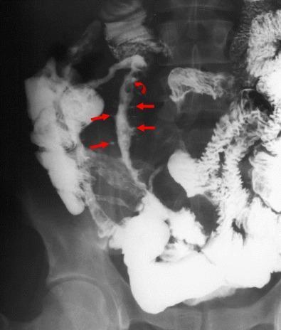
This diagnostic image is a barium follow-through radiograph showing a patient with recurrent active Crohn’s disease in the neo-terminal ileum following prior surgery. The imaging demonstrates severe, irregular luminal narrowing (stricture) of the neo-terminal ileum. Key pathological features include a characteristic 'cobblestoning' mucosal pattern, indicated by a curved red arrow, resulting from interlacing longitudinal and transverse ulcerations. Multiple linear, deep 'rose-thorn' ulcers are highlighted by straight red arrows, appearing as dark indentations within the barium column. The affected ileal segment shows significant separation from adjacent bowel loops, a finding suggestive of transmural inflammation and mesenteric thickening. This case illustrates the classic radiological manifestations of inflammatory bowel disease (IBD) recurrence at the surgical anastomosis, emphasizing the utility of small bowel follow-through for detecting fine mucosal details and structural changes like strictures and deep ulcerations in a clinical setting.
This diagnostic image is a barium follow-through radiograph showing a patient with recurrent active Crohn’s disease in the neo-terminal ileum following prior surgery. The imaging demonstrates severe, irregular luminal narrowing (stricture) of the neo-terminal ileum. Key pathological features include a characteristic 'cobblestoning' mucosal pattern, indicated by a curved red arrow, resulting from interlacing longitudinal and transverse ulcerations. Multiple linear, deep 'rose-thorn' ulcers are highlighted by straight red arrows, appearing as dark indentations within the barium column. The affected ileal segment shows significant separation from adjacent bowel loops, a finding suggestive of transmural inflammation and mesenteric thickening. This case illustrates the classic radiological manifestations of inflammatory bowel disease (IBD) recurrence at the surgical anastomosis, emphasizing the utility of small bowel follow-through for detecting fine mucosal details and structural changes like strictures and deep ulcerations in a clinical setting.

**Imaging Modality:** Fluoroscopic barium small bowel follow-through (radiogram).

**Anatomical Region:** Lower abdomen and pelvis, specifically the distal small intestine and terminal ileum.

**Observed Pathology:** Segmental mural thickening and mass effect involving a significant portion of the distal small bowel/terminal ileum. The affected loop demonstrates significant separation and displacement from adjacent intestinal segments, suggesting transmural inflammation or mesenteric thickening.

**Characteristic Visual Features:**
- **Contrast Distribution:** Positive oral contrast (barium) opacifies the lumen of several small bowel loops.
- **Mural Appearance:** There is a notable "string-sign-like" appearance or narrowing of the terminal ileum leading to the cecum, though the mucosal surface remains relatively smooth without overt ulceration or cobblestoning in this view.
- **Spatial Arrangement:** A long segment of the distal ileum is isolated from surrounding loops, a radiologic indicator of significant bowel wall thickening or fibrofatty proliferation (creeping fat) often associated with chronic inflammatory processes.
- **Negative Findings:** Absence of pneumoperitoneum, high-grade mechanical obstruction, or extraluminal contrast extravasation.

**Clinical Context:** Radiologic features are consistent with the terminal ileal involvement of Crohn’s disease, characterized by bowel wall thickening and mesenteric separation.
**Imaging Modality:** Fluoroscopic barium small bowel follow-through (radiogram). **Anatomical Region:** Lower abdomen and pelvis, specifically the distal small intestine and terminal ileum. **Observed Pathology:** Segmental mural thickening and mass effect involving a significant portion of the distal small bowel/terminal ileum. The affected loop demonstrates significant separation and displacement from adjacent intestinal segments, suggesting transmural inflammation or mesenteric thickening. **Characteristic Visual Features:** - **Contrast Distribution:** Positive oral contrast (barium) opacifies the lumen of several small bowel loops. - **Mural Appearance:** There is a notable "string-sign-like" appearance or narrowing of the terminal ileum leading to the cecum, though the mucosal surface remains relatively smooth without overt ulceration or cobblestoning in this view. - **Spatial Arrangement:** A long segment of the distal ileum is isolated from surrounding loops, a radiologic indicator of significant bowel wall thickening or fibrofatty proliferation (creeping fat) often associated with chronic inflammatory processes. - **Negative Findings:** Absence of pneumoperitoneum, high-grade mechanical obstruction, or extraluminal contrast extravasation. **Clinical Context:** Radiologic features are consistent with the terminal ileal involvement of Crohn’s disease, characterized by bowel wall thickening and mesenteric separation.

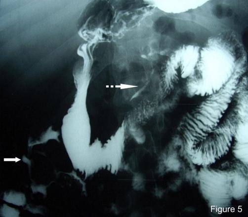
This diagnostic fluoroscopic image captures a small bowel barium follow-through study, a common radiographic procedure for evaluating the gastrointestinal tract. The image highlights significant pathology through the abnormal distribution of radiopaque barium contrast. A continuous white arrow identifies a coloduodenal fistula, showing a direct connection between the ascending colon and the second portion of the duodenum. A dotted white arrow points to multiple fistulous tracts originating from the jejunum and extending into the lesser sac (omental bursa). The small bowel loops demonstrate characteristic valvulae conniventes, while the barium-filled fistulae appear as narrow, linear channels of contrast escaping the normal intestinal lumen. This study is clinically significant for diagnosing penetrating Crohn's disease or other inflammatory/infectious processes that lead to enteric fistula formation. The image provides critical visual evidence for surgical planning and disease staging by mapping complex anatomical communications between adjacent organs and peritoneal spaces.

This diagnostic fluoroscopic image captures a small bowel barium follow-through study, a common radiographic procedure for evaluating the gastrointestinal tract. The image highlights significant pathology through the abnormal distribution of radiopaque barium contrast. A continuous white arrow identifies a coloduodenal fistula, showing a direct connection between the ascending colon and the second portion of the duodenum. A dotted white arrow points to multiple fistulous tracts originating from the jejunum and extending into the lesser sac (omental bursa). The small bowel loops demonstrate characteristic valvulae conniventes, while the barium-filled fistulae appear as narrow, linear channels of contrast escaping the normal intestinal lumen. This study is clinically significant for diagnosing penetrating Crohn's disease or other inflammatory/infectious processes that lead to enteric fistula formation. The image provides critical visual evidence for surgical planning and disease staging by mapping complex anatomical communications between adjacent organs and peritoneal spaces.

Searching Images

barium enema ulcerative colitis lead pipe appearance
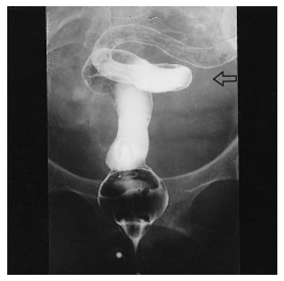
**Imaging Modality:** Fluoroscopic Barium Enema (single-contrast study).

**Anatomical Region:** Lower gastrointestinal tract, specifically the rectum and sigmoid colon.

**Observed Pathology:** Chronic inflammatory bowel disease, consistent with ulcerative colitis.

**Characteristic Visual Features:**
*   **Lead Pipe Appearance:** There is a marked loss of normal haustral markings throughout the visualized segments of the distal colon, resulting in a smooth, featureless, and tubular appearance of the bowel wall.
*   **Luminal Narrowing:** Diffuse and symmetric narrowing of the colonic lumen is evident in the rectum and sigmoid colon.
*   **Shortening:** The affected segments appear shortened and rigid.
*   **Landmarks:** A rectal catheter with an inflated retention balloon is visible at the inferior aspect of the image. An open arrow points to the featureless descending/sigmoid junction where haustral loss is prominent.

**Key Diagnostic Features:** The combination of diffuse haustral effacement and luminal narrowing ("lead pipe" sign) is a classic radiologic indicator of chronic ulcerative colitis, distinguishing it from the skip lesions and focal strictures typically seen in Crohn's disease.
**Imaging Modality:** Fluoroscopic Barium Enema (single-contrast study). **Anatomical Region:** Lower gastrointestinal tract, specifically the rectum and sigmoid colon. **Observed Pathology:** Chronic inflammatory bowel disease, consistent with ulcerative colitis. **Characteristic Visual Features:** * **Lead Pipe Appearance:** There is a marked loss of normal haustral markings throughout the visualized segments of the distal colon, resulting in a smooth, featureless, and tubular appearance of the bowel wall. * **Luminal Narrowing:** Diffuse and symmetric narrowing of the colonic lumen is evident in the rectum and sigmoid colon. * **Shortening:** The affected segments appear shortened and rigid. * **Landmarks:** A rectal catheter with an inflated retention balloon is visible at the inferior aspect of the image. An open arrow points to the featureless descending/sigmoid junction where haustral loss is prominent. **Key Diagnostic Features:** The combination of diffuse haustral effacement and luminal narrowing ("lead pipe" sign) is a classic radiologic indicator of chronic ulcerative colitis, distinguishing it from the skip lesions and focal strictures typically seen in Crohn's disease.

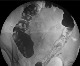
**Modality:** Single-contrast fluoroscopic barium enema.

**Anatomic Region:** Abdomen and pelvis, specifically the lower gastrointestinal tract including the rectum and sigmoid colon.

**Observed Findings:**
The image demonstrates a significant contrast-enhanced segment of the distal colon. The rectum and distal sigmoid colon exhibit a diffuse, "lead-pipe" appearance characterized by a complete loss of normal haustral markings. The mucosal surface appears irregular with fine ulcerations or a "granular" texture, suggestive of chronic inflammatory changes. In contrast, the more proximal colonic segments (visible in the upper left) retain more recognizable haustral patterns but appear distended. There is no evidence of immediate mechanical obstruction or gross perforation in this view.

**Diagnostic Features:**
*   **Ahaustral Colon:** Loss of haustrations in the descending/sigmoid region, a classic sign of chronic ulcerative colitis.
*   **Mucosal Granularity:** Suggestive of active inflammatory bowel disease (IBD).
*   **Tubular Configuration:** Narrowing and shortening of the bowel segment.

**Clinical Significance:** These radiologic findings are highly characteristic of chronic ulcerative colitis with distal involvement. The image is suitable for indexing under inflammatory bowel disease (IBD), barium enema findings, and chronic colonic inflammation.
**Modality:** Single-contrast fluoroscopic barium enema. **Anatomic Region:** Abdomen and pelvis, specifically the lower gastrointestinal tract including the rectum and sigmoid colon. **Observed Findings:** The image demonstrates a significant contrast-enhanced segment of the distal colon. The rectum and distal sigmoid colon exhibit a diffuse, "lead-pipe" appearance characterized by a complete loss of normal haustral markings. The mucosal surface appears irregular with fine ulcerations or a "granular" texture, suggestive of chronic inflammatory changes. In contrast, the more proximal colonic segments (visible in the upper left) retain more recognizable haustral patterns but appear distended. There is no evidence of immediate mechanical obstruction or gross perforation in this view. **Diagnostic Features:** * **Ahaustral Colon:** Loss of haustrations in the descending/sigmoid region, a classic sign of chronic ulcerative colitis. * **Mucosal Granularity:** Suggestive of active inflammatory bowel disease (IBD). * **Tubular Configuration:** Narrowing and shortening of the bowel segment. **Clinical Significance:** These radiologic findings are highly characteristic of chronic ulcerative colitis with distal involvement. The image is suitable for indexing under inflammatory bowel disease (IBD), barium enema findings, and chronic colonic inflammation.

This abdominal diagnostic image is an X-ray radiography showing a Gastrografin enema examination of the large intestine. The contrast study reveals a significant loss of normal haustral folds throughout the visualized segments of the colon, resulting in a smooth, tubular appearance often referred to as a 'lead pipe' appearance. There is evidence of mucosal thickening and fine contour irregularities consistent with small ulcerations. The primary educational focus is to demonstrate the radiological features of chronic inflammation or pancolitis, where the loss of haustration indicates severe or long-standing inflammatory bowel disease. Anatomical landmarks include the rectum, descending colon, transverse colon, and ascending colon, all of which appear narrow and devoid of their typical segmented structure. This visual is characteristic of conditions such as Ulcerative Colitis, where continuous mucosal inflammation leads to structural remodeling of the colonic wall.

This abdominal diagnostic image is an X-ray radiography showing a Gastrografin enema examination of the large intestine. The contrast study reveals a significant loss of normal haustral folds throughout the visualized segments of the colon, resulting in a smooth, tubular appearance often referred to as a 'lead pipe' appearance. There is evidence of mucosal thickening and fine contour irregularities consistent with small ulcerations. The primary educational focus is to demonstrate the radiological features of chronic inflammation or pancolitis, where the loss of haustration indicates severe or long-standing inflammatory bowel disease. Anatomical landmarks include the rectum, descending colon, transverse colon, and ascending colon, all of which appear narrow and devoid of their typical segmented structure. This visual is characteristic of conditions such as Ulcerative Colitis, where continuous mucosal inflammation leads to structural remodeling of the colonic wall.

Reading File
Reading File
Searching Images

barium follow through normal small bowel enteroclysis
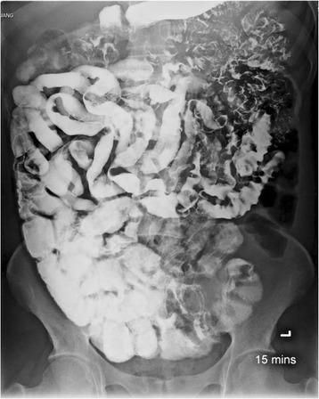
This abdominal X-ray demonstrates a small bowel follow-through (SBFT) study using oral barium contrast. The image reveals significant radiological abnormalities throughout the small intestine, specifically involving the jejunum and ileum. Key findings include a continuous long segment of diffuse mucosal fold thickening and areas of fold effacement, where the normal valvulae conniventes appear flattened or absent. There is evidence of increased luminal fluid, which contributes to the dilution and flocculation of the barium contrast, creating a characteristic 'clumping' or irregular distribution. Morphologically, the bowel loops show varying degrees of luminal narrowing and focal dilatation. These radiographic features are characteristic of malabsorption syndromes and inflammatory processes, specifically associated here with intestinal capillariasis. The 15-minute time marker suggests rapid transit of the contrast through the small bowel, a common feature in protein-losing enteropathies and chronic parasitic infections.

This abdominal X-ray demonstrates a small bowel follow-through (SBFT) study using oral barium contrast. The image reveals significant radiological abnormalities throughout the small intestine, specifically involving the jejunum and ileum. Key findings include a continuous long segment of diffuse mucosal fold thickening and areas of fold effacement, where the normal valvulae conniventes appear flattened or absent. There is evidence of increased luminal fluid, which contributes to the dilution and flocculation of the barium contrast, creating a characteristic 'clumping' or irregular distribution. Morphologically, the bowel loops show varying degrees of luminal narrowing and focal dilatation. These radiographic features are characteristic of malabsorption syndromes and inflammatory processes, specifically associated here with intestinal capillariasis. The 15-minute time marker suggests rapid transit of the contrast through the small bowel, a common feature in protein-losing enteropathies and chronic parasitic infections.

This diagnostic fluoroscopic image captures a small bowel barium follow-through study, a common radiographic procedure for evaluating the gastrointestinal tract. The image highlights significant pathology through the abnormal distribution of radiopaque barium contrast. A continuous white arrow identifies a coloduodenal fistula, showing a direct connection between the ascending colon and the second portion of the duodenum. A dotted white arrow points to multiple fistulous tracts originating from the jejunum and extending into the lesser sac (omental bursa). The small bowel loops demonstrate characteristic valvulae conniventes, while the barium-filled fistulae appear as narrow, linear channels of contrast escaping the normal intestinal lumen. This study is clinically significant for diagnosing penetrating Crohn's disease or other inflammatory/infectious processes that lead to enteric fistula formation. The image provides critical visual evidence for surgical planning and disease staging by mapping complex anatomical communications between adjacent organs and peritoneal spaces.

This diagnostic fluoroscopic image captures a small bowel barium follow-through study, a common radiographic procedure for evaluating the gastrointestinal tract. The image highlights significant pathology through the abnormal distribution of radiopaque barium contrast. A continuous white arrow identifies a coloduodenal fistula, showing a direct connection between the ascending colon and the second portion of the duodenum. A dotted white arrow points to multiple fistulous tracts originating from the jejunum and extending into the lesser sac (omental bursa). The small bowel loops demonstrate characteristic valvulae conniventes, while the barium-filled fistulae appear as narrow, linear channels of contrast escaping the normal intestinal lumen. This study is clinically significant for diagnosing penetrating Crohn's disease or other inflammatory/infectious processes that lead to enteric fistula formation. The image provides critical visual evidence for surgical planning and disease staging by mapping complex anatomical communications between adjacent organs and peritoneal spaces.

Searching Images

barium esophagram Zenker diverticulum stricture cancer
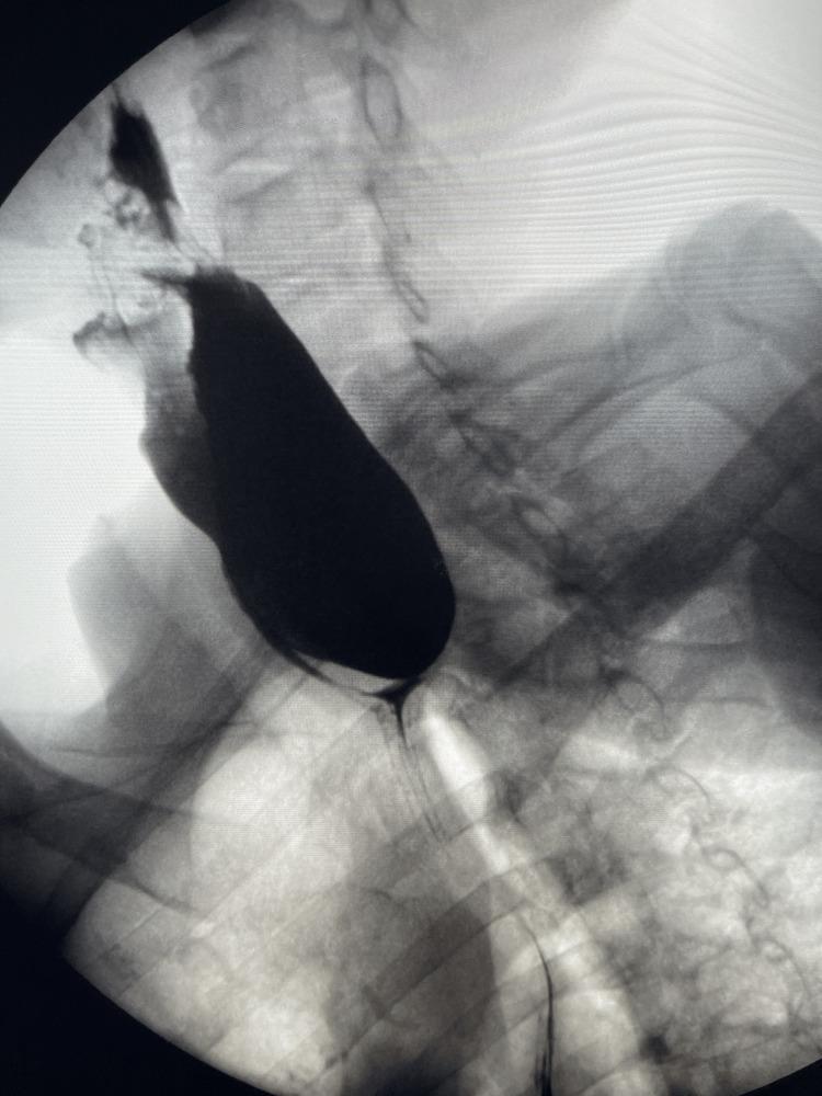
This lateral barium swallow radiograph (contrast esophagram) of the cervical and upper thoracic region demonstrates a large Zenker's diverticulum. A significant, well-defined, radiopaque, sac-like outpouching is visible originating from the posterior wall of the pharyngoesophageal junction, specifically at Killian's dehiscence above the cricopharyngeal muscle. The diverticulum is filled with barium contrast, appearing as an elongated, dependent sac approximately 7 cm in length that displaces the esophagus anteriorly. There is a clear narrowing or 'neck' at the proximal connection point where the diverticulum communicates with the esophageal lumen. The image highlights significant barium retention within the pouch, a hallmark finding of this false diverticulum. This diagnostic image is intended for medical education regarding upper gastrointestinal pathologies, specifically pharyngeal pouch disorders and causes of cervical dysphagia and regurgitation.
This lateral barium swallow radiograph (contrast esophagram) of the cervical and upper thoracic region demonstrates a large Zenker's diverticulum. A significant, well-defined, radiopaque, sac-like outpouching is visible originating from the posterior wall of the pharyngoesophageal junction, specifically at Killian's dehiscence above the cricopharyngeal muscle. The diverticulum is filled with barium contrast, appearing as an elongated, dependent sac approximately 7 cm in length that displaces the esophagus anteriorly. There is a clear narrowing or 'neck' at the proximal connection point where the diverticulum communicates with the esophageal lumen. The image highlights significant barium retention within the pouch, a hallmark finding of this false diverticulum. This diagnostic image is intended for medical education regarding upper gastrointestinal pathologies, specifically pharyngeal pouch disorders and causes of cervical dysphagia and regurgitation.

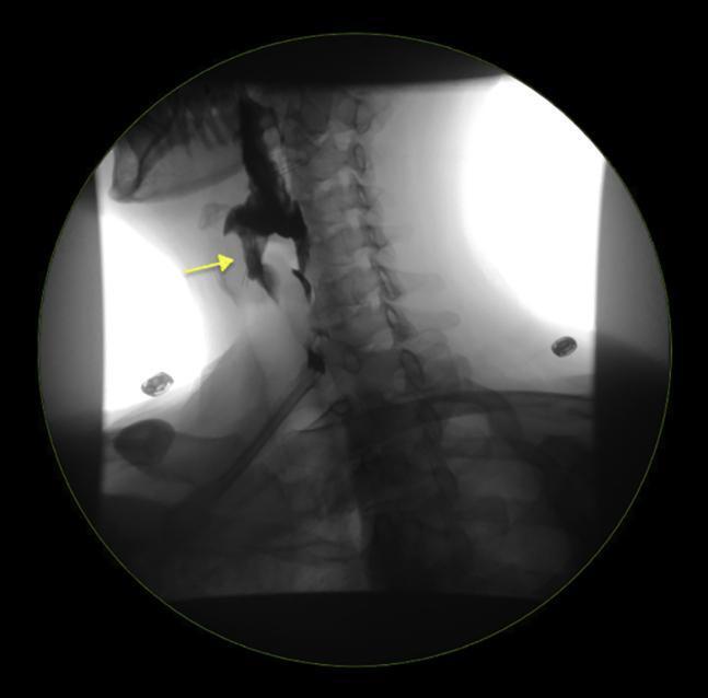
This diagnostic image is a lateral-view contrast esophagram (barium swallow) focusing on the cervical and upper thoracic regions. The radiographic study clearly demonstrates a Zenker's diverticulum, appearing as a contrast-filled outpouching projecting posteriorly from the pharyngoesophageal junction, specifically through Killian's dehiscence above the cricopharyngeal muscle. A yellow arrow points to this prominent sac-like structure, which is located dorsal to the esophageal lumen and anterior to the cervical vertebral column. The contrast agent outlines the normal esophageal lumen distal to the diverticulum, although the diverticulum itself retains a significant volume of the material. Anatomical landmarks visualized include the cervical vertebrae, the mandible, and the soft tissues of the neck. This finding is a classic representation of a pulsion diverticulum, clinically significant for causing dysphagia, regurgitation, and halitosis. It is an essential teaching image for gastroenterology and radiology regarding upper gastrointestinal motility disorders and anatomical defects.
This diagnostic image is a lateral-view contrast esophagram (barium swallow) focusing on the cervical and upper thoracic regions. The radiographic study clearly demonstrates a Zenker's diverticulum, appearing as a contrast-filled outpouching projecting posteriorly from the pharyngoesophageal junction, specifically through Killian's dehiscence above the cricopharyngeal muscle. A yellow arrow points to this prominent sac-like structure, which is located dorsal to the esophageal lumen and anterior to the cervical vertebral column. The contrast agent outlines the normal esophageal lumen distal to the diverticulum, although the diverticulum itself retains a significant volume of the material. Anatomical landmarks visualized include the cervical vertebrae, the mandible, and the soft tissues of the neck. This finding is a classic representation of a pulsion diverticulum, clinically significant for causing dysphagia, regurgitation, and halitosis. It is an essential teaching image for gastroenterology and radiology regarding upper gastrointestinal motility disorders and anatomical defects.

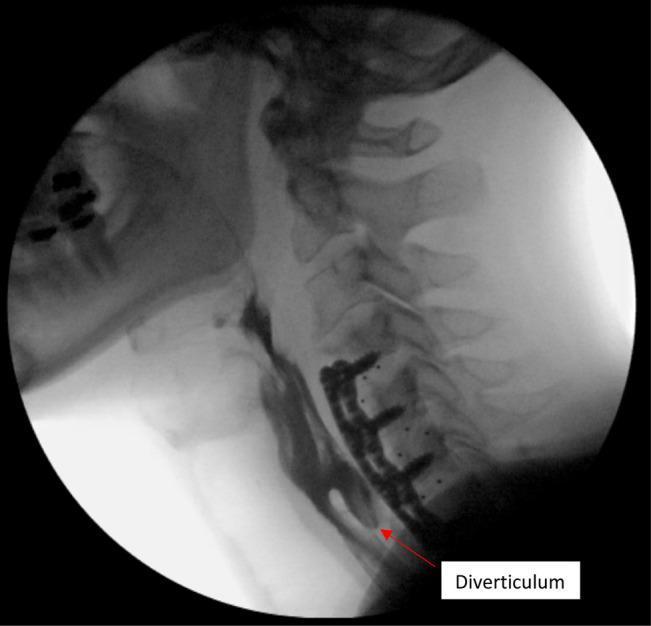
This lateral view barium swallow (esophagram) demonstrates the cervical spine and esophagus in a patient with a history of anterior cervical discectomy and fusion (ACDF). The image shows radiopaque orthopedic hardware consisting of a plate and multiple screws extending from C4 to C7. The contrast-filled esophagus is visible posterior to the hardware. A prominent outpouching or sac, labeled as a 'Diverticulum', is seen originating from the anterior aspect of the esophagus near the lower margin of the cervical plate. The diverticulum is filled with barium and shows smooth, rounded borders. While the location and appearance may mimic a Zenker diverticulum, the clinical context and visual proximity to the instrumentation suggest an ACDF-related diverticulum or an acquired traction diverticulum possibly related to hardware irritation or chronic esophageal wall compromise. The study highlights the anatomical relationship between spinal instrumentation and the aerodigestive tract, relevant for evaluating postoperative dysphagia.

This lateral view barium swallow (esophagram) demonstrates the cervical spine and esophagus in a patient with a history of anterior cervical discectomy and fusion (ACDF). The image shows radiopaque orthopedic hardware consisting of a plate and multiple screws extending from C4 to C7. The contrast-filled esophagus is visible posterior to the hardware. A prominent outpouching or sac, labeled as a 'Diverticulum', is seen originating from the anterior aspect of the esophagus near the lower margin of the cervical plate. The diverticulum is filled with barium and shows smooth, rounded borders. While the location and appearance may mimic a Zenker diverticulum, the clinical context and visual proximity to the instrumentation suggest an ACDF-related diverticulum or an acquired traction diverticulum possibly related to hardware irritation or chronic esophageal wall compromise. The study highlights the anatomical relationship between spinal instrumentation and the aerodigestive tract, relevant for evaluating postoperative dysphagia.

Finding Sources
Searching Images

hiatus hernia barium meal gastroesophageal reflux
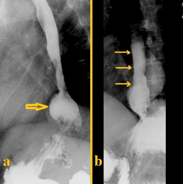
This composite diagnostic image consists of two panels (a and b) showing a barium esophagogram, a radiographic study used to evaluate the upper gastrointestinal tract. Panel (a) illustrates a sliding hiatal hernia, indicated by a yellow arrow pointing to a saccular outpouching of barium-filled gastric tissue protruding through the esophageal hiatus into the thoracic cavity. The gastric mucosal folds are visible above the level of the diaphragm. Panel (b) demonstrates gastroesophageal reflux disease (GERD), with three yellow arrows highlighting the retrograde flow of the radio-opaque barium contrast material from the stomach upwards into the distal and mid-esophagus. These images provide key clinical evidence for structural abnormalities (hiatal hernia) and functional impairments (reflux) of the gastroesophageal junction. The study is typical of those used in gastroenterology and radiology to diagnose causes of dysphagia, chronic heartburn, or regurgitation.
This composite diagnostic image consists of two panels (a and b) showing a barium esophagogram, a radiographic study used to evaluate the upper gastrointestinal tract. Panel (a) illustrates a sliding hiatal hernia, indicated by a yellow arrow pointing to a saccular outpouching of barium-filled gastric tissue protruding through the esophageal hiatus into the thoracic cavity. The gastric mucosal folds are visible above the level of the diaphragm. Panel (b) demonstrates gastroesophageal reflux disease (GERD), with three yellow arrows highlighting the retrograde flow of the radio-opaque barium contrast material from the stomach upwards into the distal and mid-esophagus. These images provide key clinical evidence for structural abnormalities (hiatal hernia) and functional impairments (reflux) of the gastroesophageal junction. The study is typical of those used in gastroenterology and radiology to diagnose causes of dysphagia, chronic heartburn, or regurgitation.

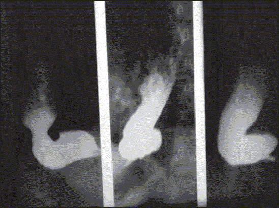
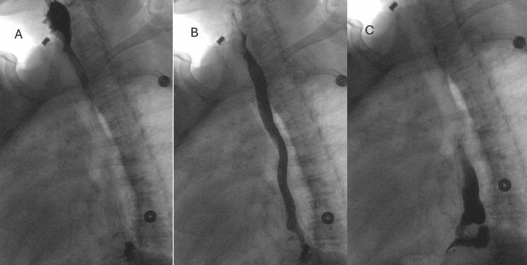
A three-panel figure (A, B, C) showing an upper gastrointestinal (GI) series using fluoroscopic barium contrast. Panel A demonstrates a moderate paraesophageal hernia, characterized by the pooling of radio-opaque barium within a herniated portion of the gastric fundus located above the diaphragmatic hiatus. Panel B illustrates the esophagus filled with barium, showing evidence of mild dysmotility and a small outpouching suspicious for a pulsion diverticulum in the mid-to-distal esophageal segment. Panel C captures an instance of gastroesophageal reflux, where barium contrast is seen retrograde in the distal esophagus above the gastroesophageal junction. The images provide clinical evidence of anatomical and functional abnormalities of the upper GI tract, including hiatal herniation and impaired sphincter or motility function. External cardiac monitoring leads are visible as circular radio-opaque markers on the thorax.
A three-panel figure (A, B, C) showing an upper gastrointestinal (GI) series using fluoroscopic barium contrast. Panel A demonstrates a moderate paraesophageal hernia, characterized by the pooling of radio-opaque barium within a herniated portion of the gastric fundus located above the diaphragmatic hiatus. Panel B illustrates the esophagus filled with barium, showing evidence of mild dysmotility and a small outpouching suspicious for a pulsion diverticulum in the mid-to-distal esophageal segment. Panel C captures an instance of gastroesophageal reflux, where barium contrast is seen retrograde in the distal esophagus above the gastroesophageal junction. The images provide clinical evidence of anatomical and functional abnormalities of the upper GI tract, including hiatal herniation and impaired sphincter or motility function. External cardiac monitoring leads are visible as circular radio-opaque markers on the thorax.

**Imaging Modality:** Barium Swallow (Fluoroscopy/Upper Gastrointestinal Series).

**Anatomical Region:** Thoraco-abdominal junction, including the distal esophagus, diaphragm, and stomach.

**Observed Pathology:** Large sliding hiatus hernia and gastroesophageal reflux.

**Characteristic Visual Features:** 
The image demonstrates a significant herniation of the proximal stomach (gastric cardia and fundus) through the esophageal hiatus into the posterior mediastinum, situated above the level of the hemidiaphragm. The distal esophagus appears dilated and enters the herniated gastric pouch superiorly. Barium contrast is visible within both the esophageal lumen and the intrathoracic portion of the stomach. There is evidence of retrograde flow of contrast from the stomach back into the esophagus, indicating active gastroesophageal reflux. 

**Key Diagnostic Features:**
- Presence of gastric mucosal folds above the diaphragmatic level.
- Widening of the diaphragmatic hiatus.
- Direct visualization of contrast reflux into the mid-to-distal esophagus.
- Displacement of the gastroesophageal junction into the thoracic cavity.

**Imaging Modality:** Barium Swallow (Fluoroscopy/Upper Gastrointestinal Series). **Anatomical Region:** Thoraco-abdominal junction, including the distal esophagus, diaphragm, and stomach. **Observed Pathology:** Large sliding hiatus hernia and gastroesophageal reflux. **Characteristic Visual Features:** The image demonstrates a significant herniation of the proximal stomach (gastric cardia and fundus) through the esophageal hiatus into the posterior mediastinum, situated above the level of the hemidiaphragm. The distal esophagus appears dilated and enters the herniated gastric pouch superiorly. Barium contrast is visible within both the esophageal lumen and the intrathoracic portion of the stomach. There is evidence of retrograde flow of contrast from the stomach back into the esophagus, indicating active gastroesophageal reflux. **Key Diagnostic Features:** - Presence of gastric mucosal folds above the diaphragmatic level. - Widening of the diaphragmatic hiatus. - Direct visualization of contrast reflux into the mid-to-distal esophagus. - Displacement of the gastroesophageal junction into the thoracic cavity.

Loading Image
Finding Sources
I now have a very rich collection of textbook content and images. Let me compile the comprehensive response.

Barium Studies in Radiology

Barium studies are fluoroscopic contrast examinations of the gastrointestinal (GI) tract using barium sulfate (BaSO₄), an inert, radio-opaque compound that coats the mucosal lining and permits real-time visualization of luminal anatomy, mucosal detail, and motility. They remain cost-effective and offer superior spatial and mucosal resolution compared to CT or MRI, despite increasing competition from cross-sectional imaging. - Grainger & Allison's Diagnostic Radiology

Barium Sulfate: The Contrast Agent

  • Chemical: BaSO₄ suspension (water-insoluble, biologically inert)
  • Technique variants:
    • Single contrast: barium alone - fills and coats the lumen
    • Double contrast (air contrast): barium + air/gas insufflation - outlines mucosal surface with high detail
  • Contraindication: Suspected perforation - use water-soluble contrast (Gastrografin) instead, as barium in the peritoneum causes fatal chemical peritonitis

Types of Barium Studies

1. Barium Swallow (Esophagogram)

Purpose: Evaluates pharynx, upper esophageal sphincter (UES), esophageal body, lower esophageal sphincter (LES), and gastroesophageal junction (GEJ).
Technique: Patient swallows a large bolus of barium; dynamic fluoroscopy captures transit in real time. A modified barium swallow (videofluoroscopic swallowing study) uses multiple consistencies (thin liquid, paste, solid) and is the gold standard for evaluating oropharyngeal dysphagia.
According to Yamada's Textbook of Gastroenterology, the barium esophagogram is uniquely able to detect subtle esophageal dilation or stenosis better than endoscopy, and provides functional transit information that endoscopy cannot.

Key Esophageal Pathologies

Achalasia - "Bird's Beak" / "Rat-Tail" Sign
The most classic barium swallow finding: smooth, symmetrical tapering of the distal esophagus at the LES with massive proximal dilation (megaesophagus). In advanced (sigmoid) achalasia, the esophagus becomes tortuous and S-shaped.
Achalasia - Bird's Beak appearance on barium swallow
Classic bird's beak tapering at the LES with dilated proximal esophagus - hallmark of achalasia
Advanced sigmoid achalasia
Late-stage achalasia: massively dilated, tortuous ('sigmoid') esophagus with bird's beak at GEJ

Zenker's Diverticulum
A pulsion diverticulum arising from Killian's dehiscence (posterior wall of the pharyngoesophageal junction, above the cricopharyngeal muscle). Barium fills the dependent sac, which projects posteriorly and displaces the esophagus anteriorly. Causes dysphagia, regurgitation of undigested food, and halitosis.
Zenker's diverticulum on lateral barium swallow
Large Zenker's diverticulum: barium-filled posterior sac at the pharyngoesophageal junction

Epiphrenic Diverticulum
Pulsion diverticulum in the distal 10 cm of the esophagus, just above the diaphragm - often associated with underlying esophageal motility disorder (achalasia, diffuse esophageal spasm). Appears as a rounded outpouching filled with barium on the right side of the distal thoracic esophagus.
Epiphrenic diverticulum with distal esophageal dilation
Epiphrenic diverticulum (right-sided outpouching in distal thoracic esophagus) with associated esophageal motility disorder

Intramural Pseudodiverticulosis
Multiple tiny, flask-shaped collections of contrast running parallel to the esophageal wall - represent dilated excretory ducts of deep esophageal mucosal glands. Often associated with a proximal stricture. Classic "dotted line" appearance.
Esophageal intramural pseudodiverticulosis
Intramural pseudodiverticulosis: multiple flask-shaped contrast collections along the esophageal wall with proximal stricture

Sliding Hiatus Hernia & GERD
Barium swallow demonstrates gastric mucosal folds herniating above the diaphragmatic hiatus. Active reflux shows retrograde flow of barium from stomach into the distal esophagus.
Sliding hiatus hernia and GERD on barium
Left panel: sliding hiatal hernia (gastric tissue above diaphragm). Right panel: active gastroesophageal reflux shown by retrograde barium flow

Vascular Ring (Pediatric)
In infants with dysphagia, a posterior indentation of the contrast-filled esophagus on lateral view suggests extrinsic compression by a vascular ring (e.g., right aortic arch with aberrant left subclavian artery).
Pediatric vascular ring - posterior esophageal indentation
Pediatric barium swallow: posterior indentation of the esophagus in a 5-month-old suggesting vascular ring

Esophageal Carcinoma
On barium study, features include mucosal irregularity, shouldering, luminal narrowing, and proximal esophageal dilation. Tortuosity, axis deviation, sinus formation, and fistulization to the bronchial tree indicate advanced disease. According to Maingot's Abdominal Operations, barium studies are becoming less essential with modern staging modalities available, but still show the classic findings of: mucosal irregularity, shouldering, luminal narrowing, and proximal dilation.

2. Barium Meal (Upper GI Series)

Purpose: Evaluates the stomach and duodenum (1st and 2nd parts). Can include the esophagus through to the duodenojejunal flexure.

Key Pathologies

Peptic Ulcer Disease - Gastric Outlet Obstruction
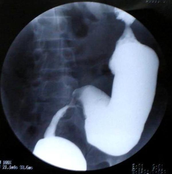
Barium meal shows a large, distended stomach with barium retention, an ulcer niche (out-pouching of contrast) along the lesser curvature, and severe narrowing/abrupt termination of contrast at the pylorus.
Gastric ulcer with pyloric obstruction on barium meal
Barium meal: ulcer niche at lesser curvature (black arrow) with severe pyloric obstruction and barium retention (white arrow)

Superior Mesenteric Artery (SMA) Syndrome
Barium meal shows distension of the stomach, 1st and 2nd parts of the duodenum, with an abrupt linear "cut-off" at the 3rd (transverse) part where the duodenum is compressed between the aorta and the SMA.
SMA syndrome on barium meal
Barium meal: marked duodenal dilation with abrupt cutoff at D3 - classic for SMA syndrome

Choledochoduodenal Fistula
Barium meal shows retrograde flow of contrast from a deformed duodenal bulb (chronic peptic ulcer disease) into the common bile duct and intrahepatic bile ducts - a rare complication of severe duodenal ulcers.
Choledochoduodenal fistula on barium meal
Barium meal: retrograde filling of biliary tree from a deformed duodenal bulb in penetrating duodenal ulcer disease

Gastric Carcinoma / GEJ Tumors
Barium contrast study of a Siewert Type II cardia cancer shows mucosal irregularity, filling defect, and luminal narrowing at the GEJ. - Maingot's Abdominal Operations

3. Small Bowel Follow-Through (SBFT) and Enteroclysis

Purpose: Evaluates the small bowel from the duodenojejunal flexure to the terminal ileum.
  • SBFT: Patient ingests a large volume of barium orally; serial abdominal films taken until contrast reaches the colon (typically 30 min - 4 hours)
  • Enteroclysis: Naso-intestinal tube placed past the ligament of Treitz; barium infused under pressure with air (double-contrast) for superior mucosal detail
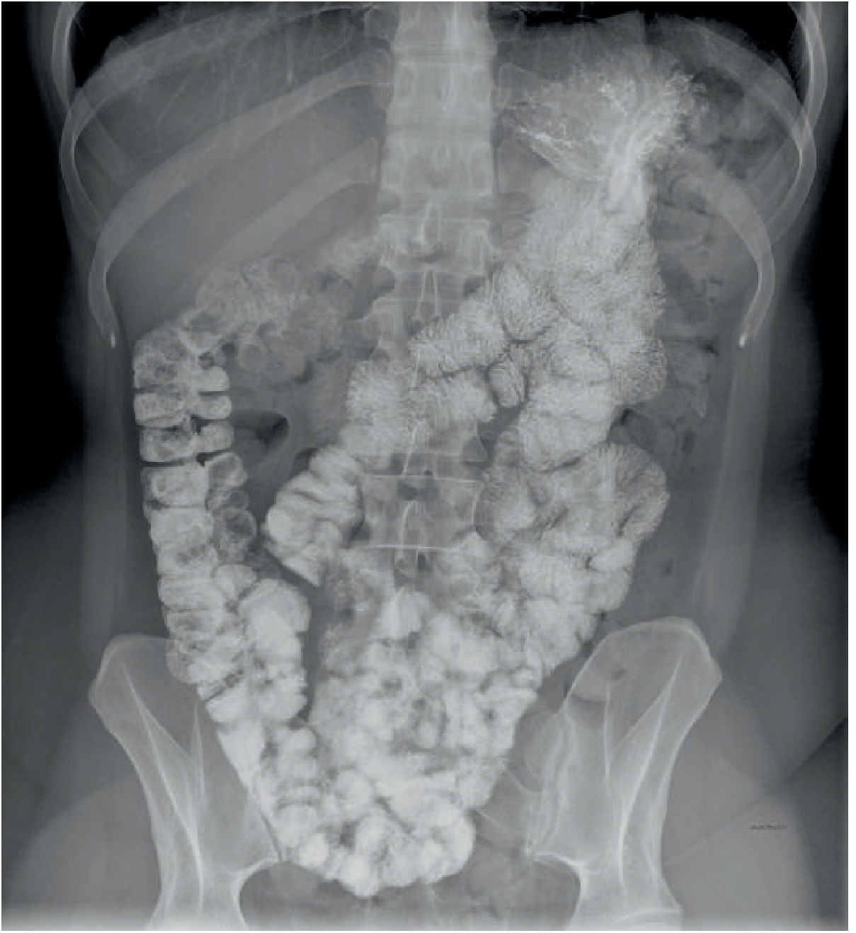
Grainger & Allison's notes that CT/MR enterography has largely replaced barium enteroclysis in resource-rich settings, but barium studies still offer superior mucosal detail and are widely used where CT/MRI availability is limited.
Normal Enteroclysis:
Normal enteroclysis - full small bowel examination
Normal enteroclysis: well-distended loops of small bowel with clear valvulae conniventes visible throughout the abdomen and pelvis

Key Small Bowel Pathologies

Crohn's Disease
Classic findings on barium SBFT / enteroclysis:
  • String sign of Kantor: narrow, smooth, thread-like column of contrast in the terminal ileum - due to spasm and mural edema or fibrotic stricture. - S. Das Manual on Clinical Surgery
  • Cobblestone pattern: interlacing longitudinal and transverse ulcers creating a cobblestone mucosal appearance
  • Rose-thorn ulcers: deep, linear ulcerations appearing as dark indentations within the barium column
  • Skip lesions: alternating normal and diseased bowel segments
  • Bowel wall thickening and loop separation: due to transmural inflammation and mesenteric fat wrapping
Crohn's disease - cobblestone pattern and rose-thorn ulcers on barium follow-through
Barium follow-through in recurrent Crohn's disease: cobblestone mucosa (curved arrow) and rose-thorn ulcers (straight arrows) with luminal narrowing at neo-terminal ileum
Crohn's fistulae: Barium can track into fistulous tracts between bowel loops, or into adjacent organs (coloduodenal fistula, enterovesical, etc.).
Crohn's disease fistulae on barium follow-through
Penetrating Crohn's disease: coloduodenal fistula and multiple jejunal fistulous tracts visible as linear contrast channels

Malabsorption (e.g., Intestinal Capillariasis)
Shows fold thickening, fold effacement (flattening of valvulae conniventes), increased intraluminal fluid diluting barium ("moulage sign"), and rapid transit.

4. Barium Enema (Large Bowel Enema)

Purpose: Evaluates the rectum, colon, and (sometimes) terminal ileum.
  • Single contrast: barium only - good for gross lesions and obstruction
  • Double contrast (air contrast): barium + air insufflation - superior for mucosal detail, polyps, and early carcinoma
Bowel preparation (clean-out) is mandatory. Contraindicated in suspected perforation and toxic megacolon.

Key Colonic Pathologies

Colorectal Carcinoma - "Apple-Core" Lesion
The most classic barium enema sign. An obstructing annular carcinoma shows an abrupt, shouldered, "apple-core" or "napkin ring" constriction of the lumen with mucosal irregularity. According to Sleisenger and Fordtran's and Swanson's Family Medicine Review, this appearance is best demonstrated on air-contrast barium enema.
Apple-core carcinoma at hepatic flexure on barium enema
Barium enema: obstructing carcinoma at the hepatic flexure showing the classic apple-core/shelf sign with massive proximal colonic dilation

Ulcerative Colitis - "Lead Pipe" Appearance
In chronic UC, continuous mucosal inflammation causes:
  • Loss of normal haustral folds throughout the colon
  • Smooth, tubular, "lead-pipe" featureless appearance
  • Diffuse luminal narrowing and colonic shortening
  • Mucosal granularity in active disease
Characteristically starts at the rectum and extends proximally in a continuous, non-skip pattern.
Lead pipe appearance of chronic ulcerative colitis on barium enema
Barium enema: lead-pipe appearance - complete loss of haustral markings in the rectosigmoid with luminal narrowing in chronic ulcerative colitis
Ulcerative colitis pancolitis - barium enema
Gastrografin enema: pancolitis with diffuse haustral effacement throughout the entire colon in UC

Colonic Stricture
Can be due to malignancy, IBD (TB, Crohn's), ischemia, or extrinsic compression. Shows abrupt transition from normal-caliber to narrowed lumen.
Rectosigmoid stricture on barium enema
Barium enema: abrupt stricture at the rectosigmoid junction - differential includes malignancy, IBD, TB

Extrinsic Rectal Compression
Smooth, rounded extramural indentation on the rectal wall - no mucosal abnormality. Suggests extrinsic pelvic mass (endometriosis, GIST, lymph node, etc.).
Extrinsic rectal mass on barium enema
Barium enema: smooth extramural indentation of the rectum (arrow) from an extrinsic pelvic mass

Thumbprinting Sign (Infantile GI Hemangiomatosis / Ischemia)
Rounded, smooth indentations ("thumbprints") along the colonic wall due to submucosal edema, hemorrhage, or intramural masses - seen in ischemic colitis, angioedema, intramural hematoma, or hemangiomatosis.
Thumbprinting sign on barium enema
Barium enema in infant: thumbprinting throughout the colon due to submucosal hemangiomatosis

Hirschsprung Disease
Barium enema shows a transitional zone between the narrow aganglionic segment (rectum) and dilated proximal colon. Manometry and rectal biopsy confirm the diagnosis. - Yamada's Textbook of Gastroenterology

Summary Table: Barium Signs and Their Pathologies

Sign / AppearanceStudyPathology
Bird's beak / Rat-tailBarium swallowAchalasia
Sigmoid/tortuous esophagusBarium swallowAdvanced achalasia
Posterior pouch at pharyngoesophageal junctionBarium swallowZenker's diverticulum
Right-sided lower esophageal outpouchingBarium swallowEpiphrenic diverticulum
Flask-shaped mucosal collectionsBarium swallowIntramural pseudodiverticulosis
Mucosal irregularity + shouldering + strictureBarium swallowEsophageal carcinoma
Gastric mucosal folds above diaphragmBarium swallow/mealSliding hiatus hernia
Retrograde barium flowBarium swallow/mealGERD
Ulcer niche + pyloric obstructionBarium mealPeptic ulcer / pyloric stenosis
D3 cutoff signBarium mealSMA syndrome
Biliary opacification via duodenumBarium mealCholedochoduodenal fistula
String sign of KantorSBFTCrohn's disease (terminal ileum)
Cobblestone pattern + rose-thorn ulcersSBFT/enteroclysisCrohn's disease
Skip lesionsSBFTCrohn's disease
Fold thickening + flocculationSBFTMalabsorption syndromes
Apple-core lesion / napkin ringBarium enemaColorectal carcinoma
Lead-pipe colonBarium enemaChronic ulcerative colitis
ThumbprintingBarium enemaIschemic colitis, intramural hematoma, hemangiomatosis
Transitional zone (narrow rectum → dilated colon)Barium enemaHirschsprung disease
Smooth extramural indentationBarium enemaExtrinsic pelvic mass

Contraindications and Precautions

  • Suspected bowel perforation: use water-soluble contrast (Gastrografin), NOT barium
  • Toxic megacolon: barium enema is absolutely contraindicated (risk of perforation)
  • Near-complete obstruction: barium can solidify proximal to obstruction
  • Aspiration risk (swallow studies): modified barium swallow should be performed with aspiration precautions
  • Radiation exposure: significant for fluoroscopic studies, particularly enteroclysis

Sources: Grainger & Allison's Diagnostic Radiology | Sleisenger and Fordtran's GI and Liver Disease | Yamada's Textbook of Gastroenterology | Maingot's Abdominal Operations | S. Das Manual on Clinical Surgery | Swanson's Family Medicine Review
This is a shared conversation. Sign in to Orris to start your own chat.