PROTEIN IN URINE (PROTEINURIA) Normal Protein Excretion * Normal urinary protein excretion = ≤150 mg/day * Proteins with MW <50,000–60,000 Da pass through GBM and are reabsorbed by proximal tubules * Only very small amount of albumin (MW 69,000 Da) is filtered * Tamm-Horsfall glycoprotein (uromucoid) * Secreted by: * Distal tubular cells * Ascending loop of Henle * Forms ≥1/3 of normal urinary protein ⸻ Proteinuria Definition * Presence of detectable protein in urine * Indicates renal disease, especially glomerular injury * If urine is turbid → filter or centrifuge before testing ⸻ Methods for Detection of Proteinuria 1. Reagent Strip Method 2. Heat & Acetic Acid Test 3. Sulfosalicylic Acid Test 4. Heller’s Test Note * Reagent strip → mainly detects albumin * Acid precipitation methods → detect all proteins ⸻ 1. Reagent Strip Method Principle * Based on protein error of pH indicators * Proteins carry charge at physiologic pH → produce pH change * Color change of indicator indicates presence of protein Reagent * Tetrabromophenol blue OR * Tetrachlorophenol-tetrabromosulfophthalein * Buffered at pH 3.0 Reaction * No protein → strip remains yellow * Protein present → indicator-protein complex forms * Color changes to green/bluish-green * Shade depends on type & concentration of protein Result * Color develops in 30–60 seconds * Reported as: * Negative * Trace * 1+ * 2+ * 3+ * 4+ Sensitivity * Detects 5–20 mg albumin/dL ⸻ 2. Heat & Acetic Acid Test Principle * Heat causes protein coagulation & precipitation * Acetic acid enhances coagulation Procedure * Take 5 mL test tube * Fill 2/3 with urine * If urine alkaline → add few drops of 3% acetic acid * Boil upper portion for 2 minutes * Lower portion acts as control * If turbidity appears → add few drops of 3% acetic acid Interpretation * Turbidity disappears after acetic acid → Phosphates present * Turbidity persists after acetic acid → Proteins present Nature of Test * Semiquantitative * Graded from: * Trace * 1+ * 2+ * 3+ * 4+ ⸻ Acetic Acid Test for Bence-Jones Protein Procedure * 5 mL clear urine * Add 1 mL acetate buffer * Heat in water bath with thermometer Characteristic Finding * Starts precipitating at 40°C * Complete precipitation at 58°C * Dissolves when heated >60°C * Reappears on cooling to 40–60°C Best Detection Method * Protein electrophoresis ⸻ 3. Sulfosalicylic Acid Test Detects * Albumin * Globulin * Glycoproteins * Bence-Jones proteins Principle * Cold precipitation of proteins by strong acid Advantage * More sensitive & reliable than heat-acetic acid test Procedure * Take 2.5 mL urine * Add 2.5 mL of 3% sulfosalicylic acid slowly * Wait 10 minutes Interpretation * Cloudy precipitate → proteins present Limitation * Also precipitates: * Mucus * Bence-Jones proteins ⸻ 4. Heller’s Test Principle * Cold precipitation of proteins by strong acid Procedure * Take 5 mL urine * Add few drops of concentrated nitric acid Interpretation * White ring at urine-acid junction → Protein present Precaution * Filter turbid urine before testing ⸻ Quantitative Estimation of Urinary Protein Esbach’s Albuminometer Indications * Nephrotic syndrome * Protein >3.5 g/24 hr * Orthostatic/Postural proteinuria Principle * Cold precipitation of proteins by strong acid Procedure * Fill urine up to mark U * Add Esbach’s reagent up to mark R * Contains: * Picric acid * Citric acid * Stopper and mix * Keep for 24 hours * Read precipitate level * Divide reading by 10 Result * Gives percentage of total urinary proteins ⸻ Categories of Proteinuria According to Amount Heavy * 4 g/day Moderate * 1–4 g/day Mild * <1 g/day ⸻ According to Site/Cause 1. Renal * Glomerular * Tubular 2. Prerenal 3. Postrenal ⸻ Types of Glomerular Proteinuria Selective Proteinuria * Leakage of intermediate-sized proteins (<100 kDa) * Examples: * Albumin * Transferrin Nonselective Proteinuria * Leakage of proteins of different sizes * Includes larger proteins * Example: * Immunoglobulins ⸻ Bence-Jones Protein Definition * Light chains of immunoglobulins Seen In * Multiple myeloma * Macroglobulinemia * Lymphoma Characteristic Property * Precipitates at 40–60°C * Dissolves near 100°C * Reappears on cooling to 40–60°C ⸻ Microalbuminuria Definition Albumin in urine: * Above normal level * Below detection limit of routine tests Diagnostic Criteria Persistent urinary albumin excretion: * 20–200 mg/L OR * 20–200 µg/min * In early morning urine sample Significance * Indicates early and potentially reversible glomerular damage Detection Methods * Immunologic methods (antibodies) * Nephelometry * Immunoassay * Protein strip electrophoresis * HPLC ⸻ Causes of Microalbuminuria Diabetes Mellitus * Associated with ↑ cardiovascular mortality * Risk factor for renal mortality * Early predictor of diabetic nephropathy Essential Hypertension * Predicts cardiovascular morbidity * Predicts cardiovascular mortality ⸻ Causes of Proteinuria 1. Renal Causes A. Glomerular Damage Mechanism * Increased permeability of glomerular capillary wall Heavy Proteinuria * Nephrotic syndrome Moderate–Heavy Proteinuria * Rapidly progressive glomerulonephritis * Chronic glomerulonephritis * Malignant hypertension * SLE Mild–Moderate Proteinuria * Acute glomerulonephritis * Nephrosclerosis ⸻ B. Tubular Damage Mechanism * Failure of tubules to reabsorb low-MW proteins Moderate Proteinuria * Heavy metal intoxication * Vitamin D intoxication Mild Proteinuria * Acute pyelonephritis * Chronic pyelonephritis * Acute tubular necrosis * Polycystic kidney disease * Hypokalemia ⸻ 2. Prerenal Causes A. Overflow Proteinuria Mechanism * Excess protein in blood exceeds tubular reabsorptive capacity Causes Immunoglobulins * Plasma cell dyscrasias * Bence-Jones proteinuria Hemoglobin * Intravascular hemolysis Myoglobin * Skeletal muscle trauma Lysozyme * Acute myeloid leukemia (M4, M5) ⸻ B. Hemodynamic Proteinuria Mechanism * Altered glomerular blood flow Causes * Excessive exercise * Postural (orthostatic) albuminuria * Congestive cardiac failure * High fever * Exposure to cold * Dehydration Usually * Mild * Asymptomatic ⸻ 3. Postrenal Causes Due To Inflammatory or neoplastic lesions of: * Renal pelvis * Ureter * Bladder * Prostate * urethra.” Create one page image of masterchart of this topic protein Uris, including everything.
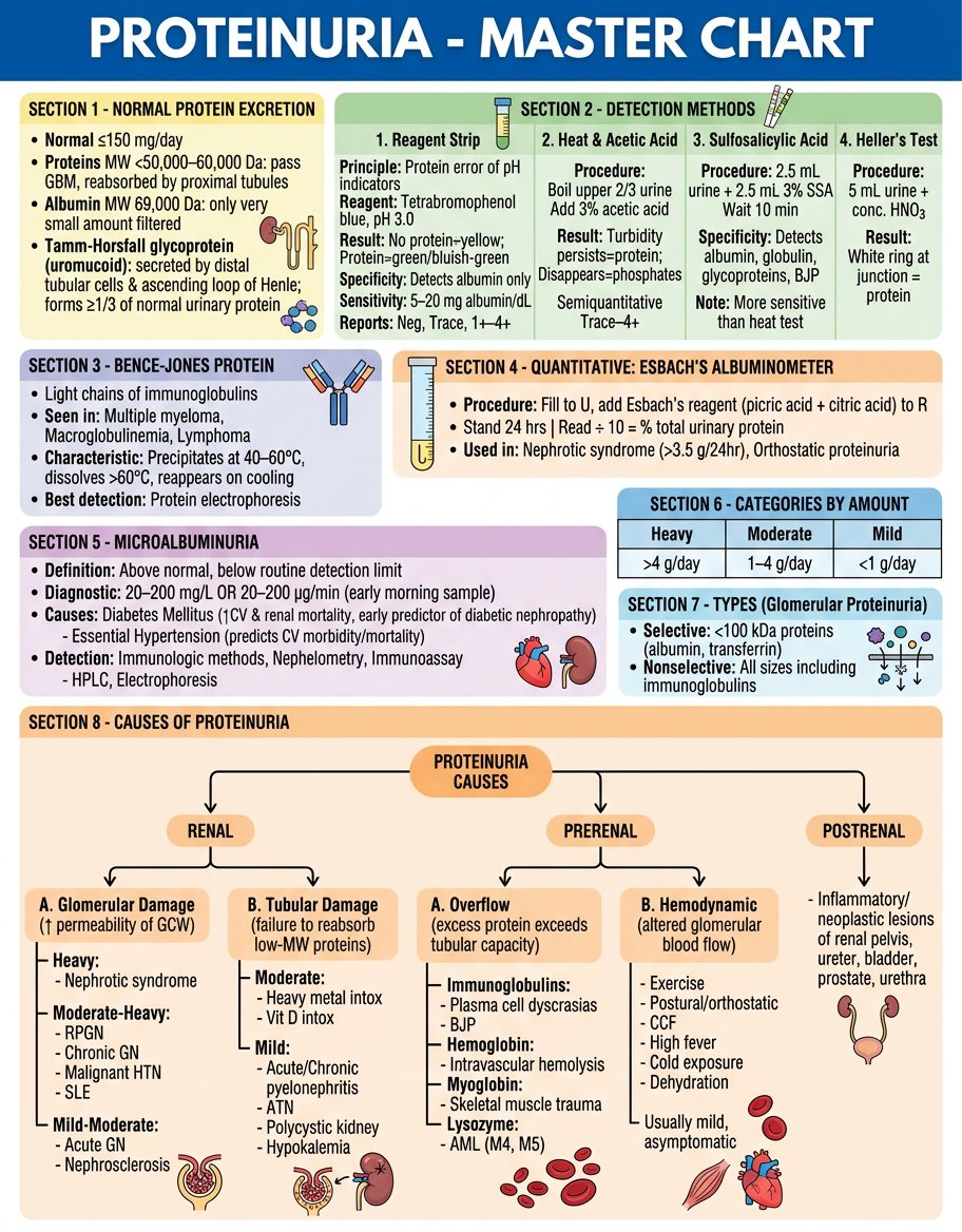
- Normal excretion values and Tamm-Horsfall protein details (top-left)
- 4 Detection methods side by side - Reagent Strip, Heat & Acetic Acid, SSA, Heller's - with principles, procedures, and interpretations
- Bence-Jones Protein box with characteristic thermal precipitation behavior
- Esbach's Albuminometer procedure summary
- Microalbuminuria - diagnostic criteria, causes (DM, HTN), and detection methods
- Categories by amount (Heavy/Moderate/Mild)
- Selective vs. Nonselective glomerular proteinuria
- Full causes flowchart - Renal (Glomerular + Tubular), Prerenal (Overflow + Hemodynamic), and Postrenal - all color-coded