
Occupational cancers
"occupational cancer" AND carcinogens
occupational cancer mesothelioma asbestos lung histology
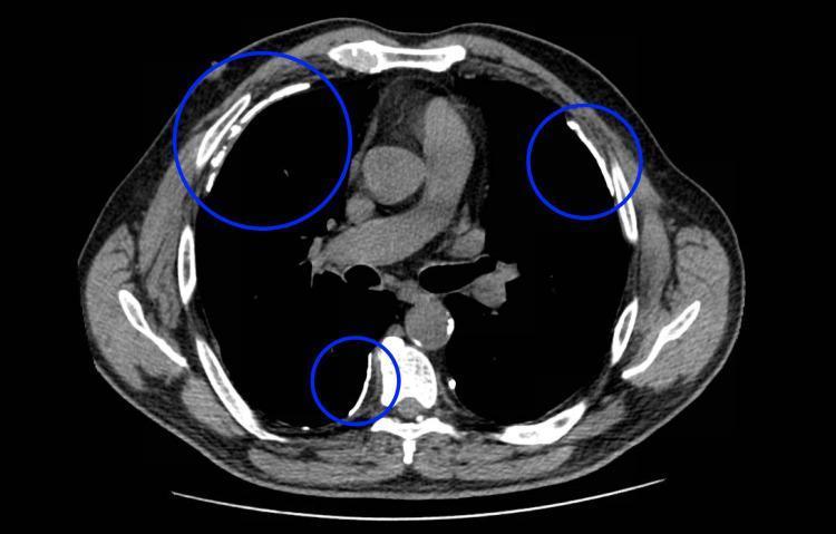
This diagnostic image is an axial (transverse) computed tomography (CT) scan of the thorax at the level of the aortic arch, displayed in a mediastinal window. The image demonstrates hallmark signs of asbestos exposure through the presence of multiple, discrete pleural plaques. These plaques are highlighted with blue circles and appear as well-circumscribed, high-density (hyperattenuating) focal thickenings along the parietal pleura. Key anatomical landmarks include the central mediastinum containing the great vessels, bilateral dark air-filled lungs, and the surrounding thoracic cage with visible ribs and the posterior vertebral column. The plaques are distributed bilaterally along the lateral chest wall and in the paravertebral region, showing a characteristic plateau-like morphology and increased radiodensity compared to adjacent soft tissue, occasionally suggesting focal calcification. This finding is clinically significant for diagnosing asbestos-related pleural disease and serves as a radiological marker for prior occupational exposure, increasing the suspicion for associated conditions like mesothelioma or lung cancer.
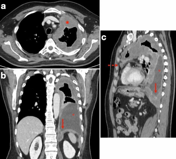
This composite diagnostic image features three contrast-enhanced multidetector CT views (axial, coronal, and sagittal) of a 72-year-old male with a history of asbestos exposure, illustrating the staging of pleural mesothelioma. (a) The axial CT scan at the mid-thoracic level reveals diffuse, irregular, rind-like soft tissue enhancement causing marked pleural thickening that circumferentially encases the left lung. (b) The coronal reconstruction demonstrates a large, loculated pleural effusion in the left hemithorax and clear infiltration of the ipsilateral hemidiaphragm by the enhancing tumor mass. (c) The sagittal view highlights the invasion of the epicardial fat planes, characterized by a lack of distinct demarcation between the tumor and the pericardial layer. Key educational concepts include identifying the 'pleural rind' sign of malignancy, assessing transdiaphragmatic extension, and evaluating the loss of mediastinal fat planes to determine tumor resectability. These findings are consistent with advanced-stage pleural mesothelioma in the context of occupational exposure.
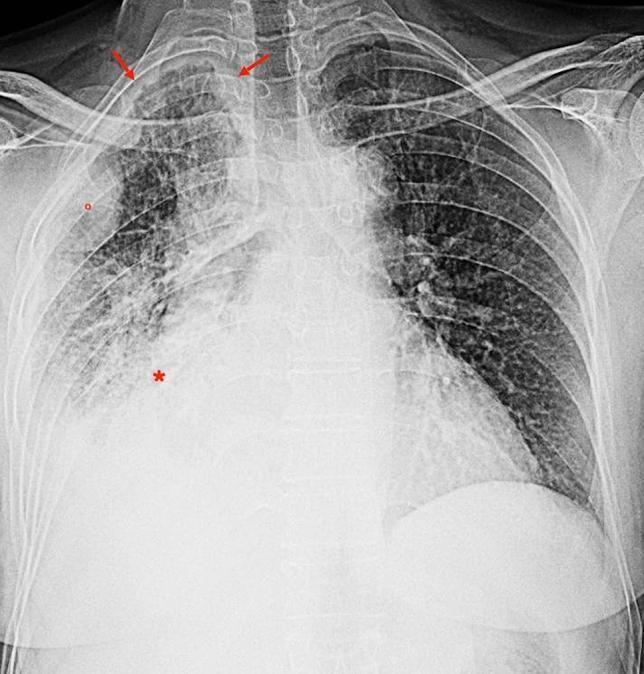
This frontal (posteroanterior) chest radiograph displays significant unilateral pathology of the right hemithorax in a 74-year-old male with a history of occupational asbestos exposure. Primary findings include a large right-sided pleural effusion causing basal opacification and obscuration of the right hemidiaphragm. Adjacent to the effusion, there is consolidation of the lung parenchyma, marked with an asterisk (*). Diffuse, irregular pleural thickening is evident along the right chest wall and apical regions (indicated by red arrows), accompanied by nodular pleural opacities (circled in red). Secondary features include notable right lung volume loss, resulting in a narrowed hemithorax and an ipsilateral shift of the mediastinum towards the right side. In contrast, the left lung appears relatively clear with preserved volume. These radiographic signs are highly suggestive of malignant pleural mesothelioma or advanced pleural disease related to asbestos exposure. This image serves as a clinical example for medical students and residents in radiology and pulmonology to identify signs of pleural malignancy and volume-contracting thoracic processes.
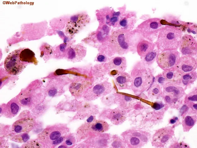
Imaging modality: Light microscopy of a paraffin-embedded, hematoxylin and eosin (H&E) stained lung tissue section. Anatomical context centers on the pulmonary parenchyma, specifically alveolar interstitium and alveolar macrophage-rich regions. Visual features include scattered ferruginous bodies—elongated, golden-brown rod fibers coated with an iron-containing shell—within macrophages and the interstitium, often appearing as dumbbell-like structures. The background may show mild interstitial inflammation and fibrosis consistent with asbestos exposure. Clinically, these histologic findings have high diagnostic significance when correlated with patient history and imaging: ferruginous bodies indicate prior asbestos exposure and raise index for asbestos-related disease, including asbestosis, lung carcinoma, and mesothelioma. This image is particularly relevant for exposure assessment in pathology, occupational health surveillance, and research on asbestos-related pathologies. If available, polarized light can enhance visualization of birefringent asbestos fibers and Prussian blue staining highlights iron in the coating. Overall, the image supports clinical risk stratification and informs management, surveillance strategies, and counseling for at-risk individuals.
Occupational Cancers
Historical Perspective
Characteristics of Occupational Cancers
- They appear only after prolonged exposure
- The latency period between exposure and disease may be 10 to 25 years
- The disease may develop even after cessation of exposure
- The average age of onset is earlier than for cancer in general
- The localization of tumors is remarkably constant for any one occupation
Sites and Carcinogens
1. Lung Cancer (Most Common Occupational Cancer)
| Carcinogen | Occupational Exposures |
|---|---|
| Asbestos | Mining, insulation work, shipyard work, brake lining, textile production |
| Arsenic | Copper/lead/zinc ore smelting, insecticide manufacture, mining |
| Beryllium | Ceramics, electronic/aerospace equipment, mining |
| Cadmium | Battery production, metal plating |
| Chromium (hexavalent) | Chromate production, electroplating, leather tanning, pigment production |
| Nickel | Mining/refining, stainless steel, aluminum production |
| Silica | Mining, quarrying, sandblasting, construction |
| Diesel exhaust | Trucking industry |
| Radon | Underground mining (uranium) |
| Chloromethyl ethers | Chemical manufacturing |
| Polycyclic aromatic hydrocarbons (PAH) | Coal tar, coke oven work, gas industry |
- Asbestos is the single most common occupational cause of lung cancer. Risk is dose-dependent with no clear minimum threshold.
- Cigarette smoking + asbestos: the combination produces a more-than-additive (near-multiplicative) increase in risk - Goldman-Cecil notes risks are "synergistic."
- The U.S. NIOSH estimates ~9,000-10,000 men and ~900-1,900 women per year in the United States develop occupational lung cancer.
2. Mesothelioma
- Smoking is NOT a risk factor for mesothelioma (unlike lung cancer)
- Approximately 3,000 cases occur annually in the United States
- Risk increases with time after first asbestos exposure (latency up to 30-40 years)
- Global asbestos-attributable deaths: ~255,000 annually (30,000 mesothelioma, 184,000 lung cancer)
3. Skin Cancer
- UV radiation (outdoor workers, agriculture, construction, welding): now accounts for ~95% of occupational skin cancers in the UK. OR for SCC = 1.77; OR for BCC = 1.43 in outdoor workers.
- Polycyclic aromatic hydrocarbons (PAH) (tar, pitch, oil fractionation): coke oven workers, gas workers, road makers, tar distillers
- Inorganic arsenic: glass production, copper/zinc/lead smelting, pesticide production
- Ionizing radiation: radiologists, dentists, X-ray technicians, nuclear workers - historically caused radiodermatitis and skin cancers of the hands
- In Australia, UV exposure accounts for 22% and asbestos 21% of compensated occupational cancers
4. Bladder Cancer
- Beta-naphthylamine (2-naphthylamine)
- Benzidine
- Para-aminodiphenyl
- Auramine and magenta (dyes)
5. Leukemia
- Leukemia may appear long after exposure has ceased
- Benzene exposure causes aplastic anemia and acute myeloid leukemia (AML)
6. Head and Neck Cancers
- Group 1 IARC solvents
- Polycyclic aromatic hydrocarbons
- Heavy metals (nickel, chromium - especially sinonasal cancers)
- Wood dust and leather dust: associated with adenocarcinoma of the nasal sinuses in furniture workers and leather workers
7. Other Site-Specific Associations
| Cancer Site | Carcinogen / Industry |
|---|---|
| Sinonasal / nasal sinus | Wood dust (furniture makers), nickel, chromium, leather dust |
| Liver (angiosarcoma) | Vinyl chloride monomer |
| Scrotal cancer | PAHs (chimney sweeps - historical) |
| Pleura (mesothelioma) | Asbestos |
| Skin | UV, PAH, arsenic, ionizing radiation |
| Bone marrow / AML | Benzene, ionizing radiation |
| Nasal cavity | Hardwood dust, isopropyl alcohol production |
IARC Classification
- Group 1: Definite human carcinogens (asbestos, benzene, arsenic, chromium VI, nickel compounds, vinyl chloride, diesel exhaust, radon, formaldehyde, PAHs)
- Group 2A: Probable human carcinogens
- Group 2B: Possible human carcinogens
Prevention and Control
- Elimination or substitution of industrial carcinogens (e.g. replacing benzidine with safer dyes)
- Engineering controls: enclosure of processes, ventilation, automation
- Reduction of exposure below threshold limit values (TLVs) set by ACGIH / national bodies
- Personal protective equipment (PPE): respiratory protection, gloves, coveralls
- Personal hygiene: handwashing, changing clothes before leaving workplace - especially critical
- Biological monitoring: periodic health surveillance of exposed workers
- Medical surveillance with cancer screening in at-risk groups (e.g. CT screening for lung cancer in asbestos workers)
- Pre-employment and periodic examinations to identify early signs
- Legislative controls: national occupational health laws and carcinogen bans
- Worker education and hazard communication (SDS/MSDS)
Global Burden
- Occupational cancers account for approximately 2-8% of all cancers globally, varying by country and industry
- Asbestos causes ~255,000 deaths per year globally (Fishman's)
- The estimated economic loss due to occupational cancers is ~$410 billion globally
- Recent systematic review (2025) confirmed occupational asbestos exposure is associated with gastrointestinal cancers (colorectal, stomach) in addition to lung/pleural malignancies
Housing standard psm