
Cerebral malaria
"cerebral malaria" pathophysiology treatment
cerebral malaria brain histology sequestration
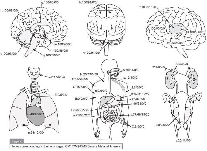
This medical infographic presents a graphical map of parasite sequestration distribution across human organs in four diagnostic categories of malaria: CM1, CM2, CM3 (Cerebral Malaria subtypes), and Severe Malarial Anemia (SMA). The visual consists of anatomical diagrams illustrating the brain (sagittal, coronal, and lateral views), respiratory system, gastrointestinal tract, and renal/reproductive systems. Each organ and tissue site is labeled with a letter key followed by a four-part percentage string (e.g., 100/90/0/0), representing the prevalence of sequestration for CM1, CM2, CM3, and SMA, respectively. Sequestration is defined by high levels of parasitized red blood cells or cytoadherent layers within the microvasculature. Key findings shown include 100% sequestration in all brain regions for CM1, and slightly lower rates (86-93%) for CM2. Significant non-cerebral sequestration is noted in the stomach (75%), large intestine (77%), and small intestine (75%) for CM1. Conversely, CM3 and SMA show minimal to zero sequestration in most organs, except for low levels in the gastrointestinal tract and spleen. This diagram serves as a comparative anatomical study of Plasmodium falciparum pathogenesis and diagnostic classification.
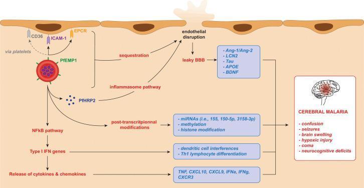
This pathophysiology diagram illustrates the molecular and cellular mechanisms of Plasmodium falciparum-induced cerebral malaria. The diagram depicts an infected red blood cell (iRBC) expressing PfEMP1, which facilitates cytoadherence and sequestration by binding to endothelial receptors CD36 (via platelets), ICAM-1, and EPCR. Simultaneously, the iRBC releases PfHRP2, activating the inflammasome pathway, and triggers the NFkB pathway leading to Type I IFN gene activation and the release of pro-inflammatory cytokines/chemokines (TNF, CXCL10, IFNg). These cascades result in endothelial disruption and a leaky blood-brain barrier (BBB), causing the release of biomarkers such as Ang-1/Ang-2, LCN2, Tau, APOE, and BDNF. Other highlighted processes include post-transcriptional modifications (miRNAs, methylation, histone modification) and immune cell interferences (Th1 differentiation). The culmination of these processes leads to the clinical manifestations of cerebral malaria listed in the final panel: confusion, seizures, brain swelling, hypoxic injury, coma, and neurocognitive deficits.
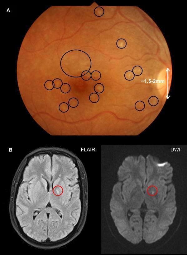
The composite image displays clinical findings in a patient with cerebral malaria, including retinal photography and brain MRI. (A) A fundus retinal photograph showing an orange-hued retina with multiple characteristic lesions of malarial retinopathy. Numerous discrete patches of retinal whitening, ranging from 0.2 to 0.5 mm in diameter, are visible across the macula and fovea (highlighted by black circles). The optic disc is visible at the nasal margin (right) as a reference for scale. (B) Axial brain MRI sections through the level of the basal ganglia. The left image is a T2-FLAIR sequence and the right is a Diffusion-Weighted Imaging (DWI) sequence. Both images demonstrate a focal area of high signal intensity (hyperintensity) in the left globus pallidus (indicated by red circles), representing localized ischemia or edema consistent with cerebral sequestered biomass. These findings illustrate the correlation between microvascular sequestration in the retina and intracranial pathology in severe malaria cases.
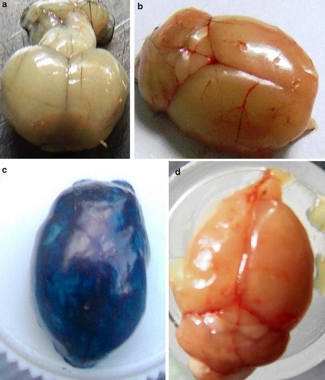
This set of clinical specimen photographs demonstrates an experimental mouse model of cerebral malaria and the evaluation of blood-brain barrier (BBB) integrity using Evans Blue dye. Image (a) shows a brain from an infected, non-stained C57BL/6 mouse exhibiting significant tissue whitening, indicative of cerebral hypoxia and reduced blood flow due to parasitized red blood cell sequestration. Image (b) presents a healthy, uninfected control brain with normal pinkish-tan coloration and visible surface vasculature, reflecting an intact BBB. Image (c) displays an infected brain after intravenous administration of Evans Blue dye; the deep blue staining of the parenchyma indicates substantial leakage of the dye through a compromised BBB, a hallmark of experimental cerebral malaria. Image (d) illustrates a brain from an infected mouse 60 days post-treatment with trioxaquine, showing a return to normal gross morphology, visible vasculature, and an absence of blue staining, confirming clinical recovery and restoration of BBB integrity. This comparison serves as an educational tool for understanding pathophysiology in neuro-parasitology and pharmacological efficacy in treating vascular neurological complications.
"cerebral malaria" management treatment
Cerebral Malaria
Definition
Pathophysiology
1. Cytoadherence and Sequestration
- ICAM-1 (intercellular adhesion molecule-1)
- CD36 (via platelet bridges)
- EPCR (endothelial protein C receptor)
2. Cytokine Storm and Endothelial Injury
- Upregulates endothelial adhesion molecules (ICAM-1), further worsening sequestration
- Disrupts the blood-brain barrier (BBB), causing edema
- Triggers the NF-κB pathway and inflammasome activation (via PfHRP2 release)
3. Reduced Cerebral Blood Flow
Clinical Features
| Feature | Details |
|---|---|
| Impaired consciousness | Delirium progressing to unarousable coma |
| Seizures | Common, especially in children; can be subtle (nystagmus, twitching) |
| Fever | High; may be continuous in P. falciparum unlike the classic paroxysmal pattern |
| Neurological signs | Ataxia, abnormal posturing, opisthotonus, abnormal eye movements |
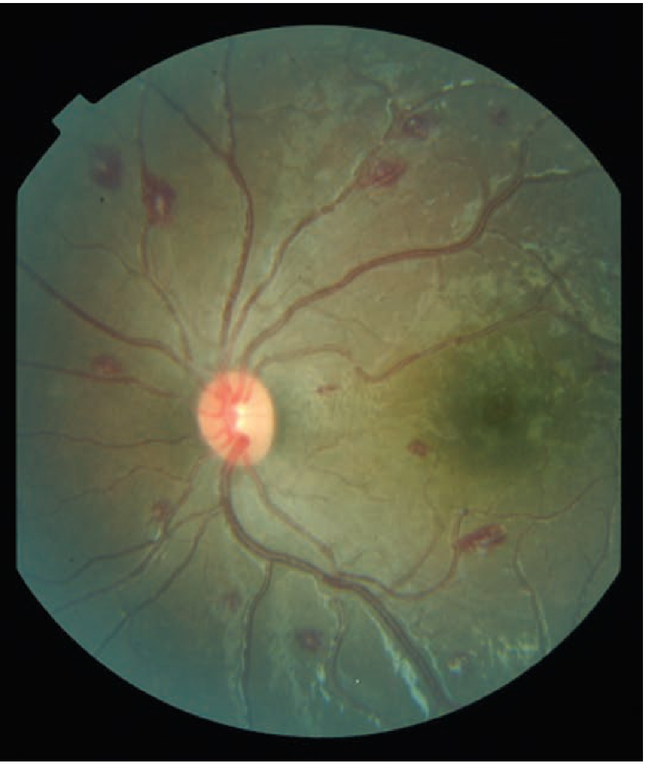
| Malarial retinopathy | Highly specific finding - patches of retinal whitening, white-centered hemorrhages |
| Respiratory | Acute pulmonary edema/ARDS frequently accompanies cerebral malaria |
| Metabolic | Hypoglycemia (worsened by quinine therapy), lactic acidosis |
Malarial Retinopathy - A Key Diagnostic Clue
Diagnosis
- Thick and thin blood films: Diagnostic gold standard. Look for multiple ring forms per RBC, banana-shaped gametocytes
- Rapid antigen tests (RDT): Detect HRP-2 (P. falciparum-specific)
- Blood glucose: Hypoglycemia common and must be corrected immediately
- Lumbar puncture: Opening pressure usually raised in children, normal in adults; CSF is typically clear and normal on routine analysis - but LP is needed to exclude bacterial meningitis
- Brain imaging (CT/MRI): Rule out other causes; may show cerebral edema, petechial hemorrhages
- Parasitemia level: Higher density = greater likelihood malaria is causative in endemic children with many asymptomatic carriers
WHO Criteria for Severe/Complicated Malaria
- Severe anemia (Hb <7 g/dL)
- Acidosis / lactic acidosis
- Hypoglycemia (<2.2 mmol/L)
- Acute kidney injury
- ARDS / pulmonary edema
- Shock ("algid malaria")
- DIC / abnormal bleeding
- Hyperparasitemia (>5% parasitized RBCs)
Treatment
Antimalarial Therapy (Severe/Cerebral Malaria)
| Drug | Adult Dose | Notes |
|---|---|---|
| Artesunate IV (first-line) | 2.4 mg/kg IV at 0, 8, 24 h, then daily | Can give IM if IV not available; WHO-preferred |
| Quinine gluconate IV (alternative) | 6.25 mg base/kg IV load over 2 h, then 0.0125 mg base/kg/min continuous infusion | Requires cardiac monitoring (QTc); always combined with doxycycline or clindamycin |
| + Doxycycline | 100 mg IV q12h x 7 days | Avoid in children <8 years |
| + Clindamycin (children <8 yrs) | 10 mg base/kg IV load, then 5 mg/kg q8h x 7 days | Use instead of doxycycline in children |
- Tintinalli's Emergency Medicine
Supportive Care
- Airway: Intubate if GCS is very low or airway protection is compromised
- Correct hypoglycemia: 50% dextrose IV stat, then dextrose infusion; monitor glucose frequently (quinine stimulates insulin)
- Anticonvulsants: Treat seizures with benzodiazepines; prophylactic phenobarbital is controversial
- Fluids: Cautious IV fluids - avoid fluid overload (risk of pulmonary edema/cerebral edema)
- Fever control: Paracetamol, tepid sponging
- Transfusion: For severe anemia (Hb <7 g/dL) or rapid fall in Hb
- Avoid corticosteroids: Dexamethasone was shown in RCTs to increase complications (gut bleeding, coma prolongation) and is contraindicated
Prognosis and Sequelae
- Mortality: 15-30% even with treatment; up to 80% when acute pulmonary insufficiency is also present
- Neurological sequelae (more common in children than adults):
- Cognitive and language deficits
- Epilepsy
- Behavioral problems
- Deafness, blindness
- Up to 1 in 3 children vs 1 in 20 adults affected
Special Populations
- Children in endemic areas: Most at risk; often already carrying asymptomatic parasitemia, making the diagnosis challenging - retinopathy becomes especially important
- Pregnant women: High risk of severe disease, hypoglycemia, and maternal/fetal death
- Non-immune adults (travelers): Rapid progression; high mortality; always assume P. falciparum until proven otherwise
- Asplenic patients: Extremely high risk; rapidly fatal if untreated
Recent Evidence (PubMed 2024-2025)
- Vathi et al., Ann Intensive Care 2025 - Management and monitoring strategies for severe cerebral malaria in the ICU
- Postels & Katangwe-Chirwa, Semin Pediatr Neurol 2025 - Dedicated review of cerebral malaria (pediatric focus)
- Akafity et al., J Intensive Med 2024 - Diagnosis and ICU management of malaria
Leprosy
leprosy skin lesions lepromatous tuberculoid histology
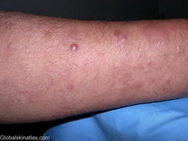
Clinical photograph of skin lesions in lepromatous leprosy. Imaging modality: dermal clinical photography; technique: standard color photograph under ambient lighting; no magnification; no contrast. Anatomical region: forearm skin; laterality not specified; view is a single forearm field. Visual features: generalized reddish-brown, infiltrated papules and nodules with plaques; lesions are densely distributed with some coalescence into larger plaques and surface erythema. The biopsy-proven diagnosis: lepromatous leprosy; histology would reveal diffuse dermal sheets of foamy macrophages (Virchow cells) densely packed with acid-fast bacilli (Mycobacterium leprae). Pathophysiology: infection with M. leprae, an obligate intracellular bacillus that multiplies slowly; prolonged incubation; transmission via close contact; untreated disease causes progressive skin, nerve, limb, and ocular damage. Diagnostic significance: this presentation is characteristic of lepromatous leprosy, a multibacillary form requiring MDT (rifampicin, dapsone, clofazimine). Clinical use cases: educational reference for dermatology, infectious disease, and tropical medicine; aids recognition of diffuse cutaneous involvement, differentiation from tuberculoid forms, and underscores need for early MDT to prevent nerve impairment and disability.
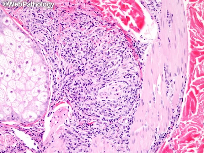
Modality and technique: Light microscopy of a skin punch biopsy from a plaque in borderline lepromatous leprosy was examined with routine hematoxylin and eosin staining and a Wade-Fite acid-fast stain to visualize Mycobacterium leprae. The biopsy demonstrates a dermal and subcutaneous granulomatous infiltrate with nodular architecture and dense lymphohistiocytic crowding. Epithelioid granulomas and foamy (Virchow) macrophages are present, often clustering around small nerves and adnexal structures. Perineural inflammation and thickened nerve trunks are conspicuous, reflecting the clinical finding of palpable thickened nerves. Bacilli are observed inside macrophages, singly or in grape-like globi, on Wade-Fite preparation, confirming infection. The inflammatory pattern is more circumscribed than in classic lepromatous leprosy, yet more numerous lesions and a higher density of lymphocytes distinguish borderline lepromatous disease from tuberculoid forms. Epidermal involvement is minimal; there may be mild interface change without necrosis. The overall histology correlates with a polyclonal, mixed-cell granulomatous response and a propensity to involve cutaneous appendages and nerves. This histopathology supports a Ridley-Jopling borderline lepromatous diagnosis and guides multibacillary treatment decisions, prognosis, and nerve protection strategies. Correlation with clinical features, including symmetrical vs asymmetrical lesions and nerve palsy, improves diagnostic confidence and tailoring of MDT MB regimens. Enhancing educational and research value.
This histopathology image shows a skin biopsy from a patient with borderline lepromatous leprosy. Acquired via light microscopy, tissue sections are stained with Wade-Fite, enabling visualization of acid-fast Mycobacterium leprae bacilli within dermal macrophages and occasionally in association with cutaneous nerves and adnexal structures. The dermal granulomatous infiltrate is nodular and poorly circumscribed with a relatively dense cellular milieu of epithelioid histiocytes, Langhans-type giant cells, and a prominent lymphocytic cuff. Compared with tuberculoid granulomas, BL lesions exhibit increased circumscription of the granulomatous response and greater bacillary load, often forming globi where bacilli cluster within macrophages. The image demonstrates perineural involvement with thickened nerve trunks and bacilli seen singly or in globi within these neural aggregates. Clinically, such histology correlates with numerous skin plaques and nodules, asymmetric distribution, and early sensory impairment. Wade-Fite positivity confirms presence of acid-fast bacilli, distinguishing M. leprae infection from other mycobacterial dermatitis. The diagnostic significance lies in confirming borderline lepromatous leprosy, guiding multidrug therapy, and informing prognosis. Potential educational use includes correlating histology with clinical phenotype, staging within Ridley-Jaworsky spectrum, and emphasizing perineural bacillary invasion. Note: bacillary load and distribution help distinguish BL from LL and are integral to prognosis, disease monitoring, and public health management in endemic regions.
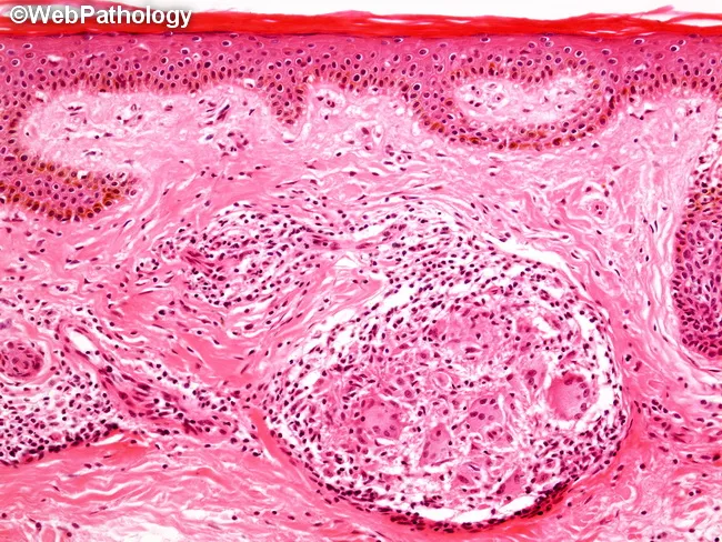
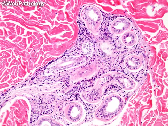
Comprehensive description: Histopathology of a skin biopsy from a cutaneous leprosy lesion prepared with hematoxylin and eosin staining. The dermis and superficial subcutis show non-caseating epithelioid granulomas densely packed around small cutaneous nerve fibers, often forming serpentine contours along nerve courses. Langhans giant cells are present within granulomas, with a predominant lymphocytic infiltrate composed mainly of CD4+ T helper cells, reflecting a robust cell-mediated Th1 immune response. There is no granulomatous zone (Grenz zone) separating the epidermis from the inflammatory infiltrate, and the epidermis itself may show mild involvement. Bacilli are scarce or not detected by routine modified acid-fast staining, consistent with a paucibacillary, tuberculoid form of leprosy. The granulomatous tissue may extend from the dermis into the papillary dermis and occasionally involve the overlying epidermis, though satellitosis around nerve trunks remains a hallmark. Clinically, this pattern correlates with strong protective immunity, limited bacterial burden, and focal nerve destruction or compression by granulomatous inflammation. This image is educational for recognizing neural-centric granulomas in tuberculoid leprosy, differentiating from borderline tuberculoid and lepromatous forms, and understanding how immune status shapes histology. Potential applications include dermatopathology education, differential diagnosis exercises, and correlating histology with therapeutic decision-making in multi-drug therapy regimens for accurate diagnosis.
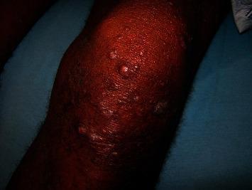
This clinical photograph displays a representative skin lesion characteristic of lepromatous leprosy (LL). The image shows a limb, likely the knee or elbow area, exhibiting diffuse cutaneous thickening. Numerous discrete, erythematous to skin-colored nodules of varying sizes are scattered across the affected surface. These papulonodular lesions demonstrate a tendency toward confluence, forming larger, irregular patches of infiltrated skin. The skin texture appears coarse and indurated, reflecting the high bacillary load and extensive dermal infiltration typical of the polar lepromatous end of the Ridley-Jopling classification. This visual serves as an educational example of the symmetrical, multifocal nodular distribution seen in advanced Mycobacterium leprae infections, highlighting the difference between the paucibacillary tuberculoid form and this multibacillary presentation.
"leprosy" diagnosis treatment multidrug therapy
Leprosy (Hansen Disease)
Definition and Overview
The Organism
- Obligate intracellular pathogen - cannot be cultured in vitro
- Replicates best at 32-34°C (the temperature of human skin and extremities)
- Very long doubling time: ~14 days
- Genome has undergone reductive evolution - highly downsized compared to other mycobacteria
- Key virulence factor: PGL-1 (phenolic glycolipid-1) - critical for invasion of host cells; also inhibits mitochondrial energy metabolism to evade immunity
- BCG vaccination confers some cross-protection (structural similarity to M. tuberculosis)
- Reservoir: humans (primary); wild armadillos (especially in southern USA); some monkey and squirrel species
Transmission
- Mainly via respiratory droplet from the nasal mucosa of untreated multibacillary patients
- Portal of entry: upper respiratory tract (primarily); skin wounds or tattoos (occasionally)
- Long incubation period: 2-10 years (sometimes longer)
- Very low communicability - most exposed people (>95%) never develop disease
- Zoonotic transmission from armadillos is rare but documented
Pathogenesis and Immunology
| Feature | Tuberculoid (TT) | Lepromatous (LL) |
|---|---|---|
| CMI | Strong Th1 | Weak Th1, relative Th2/Treg |
| Antibody | Low | High (hypergammaglobulinemia) |
| Lepromin test | Positive | Negative |
| Bacterial burden | Low (paucibacillary) | High (multibacillary) |
| Tissue granuloma | Well-formed, epithelioid | Poorly formed; foamy macrophages |
| IL-2, IFN-γ | High | Low |
| IL-10, IL-4 | Low | High |
Classification: The Ridley-Jopling Spectrum
TT ←——— BT ——— BB ——— BL ———→ LL
(tuberculoid)  (borderline)  (lepromatous)
- Paucibacillary (PB): 1-5 skin lesions; slit-skin smear negative (TT + BT + some BB)
- Multibacillary (MB): >5 skin lesions; slit-skin smear positive (BB + BL + LL)
Clinical Features
Tuberculoid Leprosy (TT/BT)
- Few (1-5), well-defined, hypopigmented or erythematous skin patches/plaques
- Dry, scaly surface with loss of sensation (anesthesia), anhidrosis, loss of hair
- Asymmetric large peripheral nerve involvement (typically one or two nerve trunks)
- Thickened, palpable nerves (ulnar, lateral popliteal, great auricular, radial cutaneous)
- Paucibacillary - smear negative
Lepromatous Leprosy (LL)
- Innumerable, bilateral, symmetric, diffusely indurated copper-colored/erythematous patches/plaques/nodules
- No initial loss of sensation over lesions (sensation lost late, in "glove and stocking" distribution)
- Smooth, shiny surface; bilateral earlobe thickening; loss of eyebrows (madarosis)
- Coarse facial skin folds = "leonine facies" (lion face)
- Involves eyes, nose, testes, lymph nodes, liver, spleen, bone marrow
- Bacteriologic index 4+ to 6+ with globi on slit-skin smear
Systemic Involvement in LL (multibacillary)
- Eyes: corneal anesthesia, episcleritis, iridocyclitis, lagophthalmos, cataracts, glaucoma, blindness
- Nose/ENT: chronic rhinitis, septal perforation, saddle-nose deformity, epistaxis
- Testes: orchitis → sterility → gynecomastia
- Hands/feet: progressive bone resorption of phalanges, neuropathic arthropathy
- Late: ulceration of palate, tongue, uvula; in advanced disease, palate perforation
Borderline Forms (BT, BB, BL)
- Intermediate features; lesions more variable in size, number, and distribution
- Most clinically unstable - prone to lepra reactions
Special Forms
- Histoid leprosy: Rare variant of LL; waxy, shiny, firm nodules; high bacillary index without globi; spindle-cell granuloma on histology
- Diffuse leprosy of Lucio & Latapi: Non-nodular LL; Mexico/Central America; diffuse shiny "lepra bonita" skin; complicated by Lucio's phenomenon
- Primary neuritic leprosy: Nerve-only involvement, no skin lesions; seen in India/Nepal (2-10% of cases)
Histopathology
- Well-formed epithelioid granulomas with Langhans giant cells in the dermis
- Dense CD4+ Th1 lymphocytic infiltrate
- Granulomas centered on cutaneous nerves (neural-centric) - a hallmark
- No Grenz zone (subepidermal clear zone absent)
- AFB absent or very scarce on Wade-Fite stain
- Diffuse sheets of foamy macrophages (Virchow cells) in the dermis
- Globi (grape-like clusters of bacilli within macrophages) on Wade-Fite stain
- Prominent Grenz zone (clear subepidermal band)
- Many plasma cells; AFB abundant
Diagnosis
-
Clinical: Cardinal signs
- Hypopigmented/erythematous skin patch with loss of sensation
- Thickened peripheral nerve (with or without sensory/motor loss)
- Positive slit-skin smear for AFB
- Any one of the three is sufficient for diagnosis in endemic areas
-
Slit-skin smear: Scraped from earlobes, lesion edges; Ziehl-Neelsen or Wade-Fite stain for AFB; Bacteriologic Index (BI) scored 0-6
-
Lepromin test (Mitsuda reaction): Intradermal injection of heat-killed M. leprae suspension; positive (firm nodule at 4 weeks) in tuberculoid; negative in lepromatous. Not diagnostic - reflects immune status
-
Skin biopsy: Full-thickness biopsy from the raised edge of a skin lesion; provides histologic classification
-
Nerve conduction studies: Detect subclinical neuropathy
-
PCR: Detection of M. leprae DNA in skin/nerve biopsy; useful in smear-negative paucibacillary cases
-
Anti-PGL-1 antibody: Seropositive in 60-90% of multibacillary patients; useful in endemic areas
There is currently no single laboratory test that definitively diagnoses leprosy - diagnosis is primarily clinical.
Leprosy Reactions
Type 1 Reaction (T1R) - Reversal Reaction
- Mechanism: Delayed-type hypersensitivity (Type IV) - sudden upgrade of CMI
- Seen in: Borderline spectrum (BT, BB, BL)
- Skin: Acute redness, swelling, warmth of existing lesions
- Nerve: Painful, tender nerve trunks; neuritis → sensory/motor loss
- "Silent neuritis": Nerve damage without overt inflammation - major cause of disability
- Rarely: fever, malaise, tenosynovitis, edema of hands/feet, arthritis
- Treatment: Prednisolone 40-60 mg/day, tapered over 3-6 months; MDT continued
Type 2 Reaction (T2R) - Erythema Nodosum Leprosum (ENL)
- Mechanism: Immune complex-mediated (Type III hypersensitivity / Arthus phenomenon)
- Seen in: Lepromatous (LL) and borderline lepromatous (BL) - multibacillary forms
- Skin: Evanescent pink-to-red papulonodular lesions on thighs, legs, face; tender, warm; last days
- Systemic: Fever, malaise, lymphadenopathy, arthritis, orchitis, iridocyclitis, nephritis, hepatosplenomegaly
- Severe form: Vesicular, bullous, necrotic ulcerating ENL (erythema nodosum necroticans)
- Long-term complication: amyloidosis
- Treatment: Thalidomide (drug of choice, 100-400 mg/day) - men and post-menopausal women; Prednisolone for women of childbearing age and for neuritis; Clofazimine has anti-inflammatory effect
Lucio's Phenomenon
- Variant of ENL occurring in diffuse LL (Lucio & Latapi type)
- Marked vasculitis + thrombosis → skin hemorrhage and infarction → ulceration
- Can be life-threatening
Treatment: WHO Multidrug Therapy (MDT)
WHO MDT Regimen
| Drug | Paucibacillary (PB) | Multibacillary (MB) |
|---|---|---|
| Dapsone | 100 mg/day | 100 mg/day |
| Rifampin | 600 mg/month (supervised) | 600 mg/month (supervised) |
| Clofazimine | - (not in standard PB regimen) | 50 mg/day + 300 mg/month |
| Duration | 6 months | 12 months |
Drug Mechanisms
| Drug | Action | Notes |
|---|---|---|
| Rifampin | Bactericidal (most potent) | Can worsen reactions by antigen release - avoid during active reversal reaction/ENL |
| Dapsone | Bacteriostatic (folate synthesis inhibitor) | Monitor for hemolysis, methemoglobinemia; G6PD screen first |
| Clofazimine | Bacteriostatic + anti-inflammatory | Skin discoloration (red-brown); useful for managing ENL |
- Harrison's Principles of Internal Medicine 22e; Goodman & Gilman's Pharmacological Basis of Therapeutics
Disability Grading (WHO)
| Grade | Eyes | Hands/Feet |
|---|---|---|
| 0 | No problems | No anesthesia, no impairment |
| 1 | Eye problems without severe visual loss | Anesthesia but no visible impairment |
| 2 | Severe visual impairment (vision <6/60), lagophthalmos, iridocyclitis, corneal opacity | Visible impairment (claw hand, foot drop, ulcers) |
Complications and Sequelae
- Peripheral neuropathy: The single greatest cause of disability
- Clawed hands (ulnar nerve), foot drop (lateral popliteal/common peroneal nerve), wrist drop (radial nerve), facial palsy (facial nerve), lagophthalmos (facial nerve)
- Neuropathic (Charcot) arthropathy: Subluxation, dissolution of foot arches, "rocker-bottom" deformity; in extreme cases, separation of forefoot and hindfoot with tibia becoming weight-bearing
- Trophic ulcers: From anesthesia → painless trauma → non-healing ulcers (plantar surface of feet)
- Saddle-nose deformity: Nasal septal bone destruction in lepromatous leprosy
- Blindness: From corneal anesthesia, lagophthalmos, iridocyclitis
- Amyloidosis: Complication of recurrent ENL; leads to renal failure
- Gynecomastia/infertility: Testicular infiltration in LL
Prevention
- BCG vaccination: Provides 20-80% protection against leprosy (highly variable by region)
- Chemoprophylaxis: Single dose rifampin (SDR) to close contacts of newly diagnosed leprosy cases - reduces incidence by ~57% at 2 years (evidence-based)
- Early detection and treatment: Interrupts transmission; prevents disabilities
- WHO target: elimination as a public health problem (<1 case/10,000 population)
Recent Evidence (PubMed 2024-2026)
- Grijsen et al., Nature Reviews Disease Primers 2024 - Comprehensive updated review of leprosy (highly recommended)
- Marques, Curr Opin Neurol 2024 (PMID 39051539) - Leprous neuropathy: current understanding
- Stuetzle et al., PLoS Negl Trop Dis 2024 (PMID 39264863) - Systematic review on self-healing in leprosy
- Shiozaki et al., Brain Nerve 2026 (PMID 42156030) - 2026 review: Hansen's disease (neurology focus)
- Leprosy is a spectrum disease - the host immune response, not the organism, determines the clinical form
- Nerve damage is the defining morbidity - early diagnosis and treatment of reactions prevent disability
- MDT (rifampin + dapsone ± clofazimine) is curative - provided free globally by WHO
- Leprosy reactions (T1R reversal, T2R/ENL) are immunologic emergencies requiring prompt steroid or thalidomide therapy
- There is still a significant hidden endemic - mathematical models estimate ~4 million undiagnosed cases worldwide