
Peritoneal dialysis
peritoneal dialysis catheter peritoneal membrane diagram
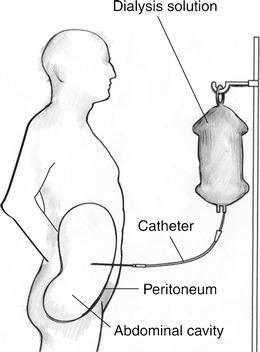
This medical anatomical diagram illustrates the standard setup for Continuous Ambulatory Peritoneal Dialysis (CAPD). The illustration features a lateral profile of a human figure positioned next to a dialysis solution bag suspended on an IV pole. A catheter is shown extending from the solution bag, penetrating the anterior abdominal wall, and passing through the parietal peritoneum. Within the abdominal cavity, the catheter is depicted with its distal tip correctly positioned in the pelvic region. Key anatomical structures and components are labeled, including the 'Dialysis solution,' 'Catheter,' 'Peritoneum,' and 'Abdominal cavity.' This educational visual serves to demonstrate the trans-abdominal pathway and the specific intra-abdominal placement required for effective peritoneal exchange of electrolytes and waste products. The diagram highlights the clinical importance of distal tip placement in the pelvis, which holds the greatest volume of dialysate, to ensure optimal treatment efficacy.
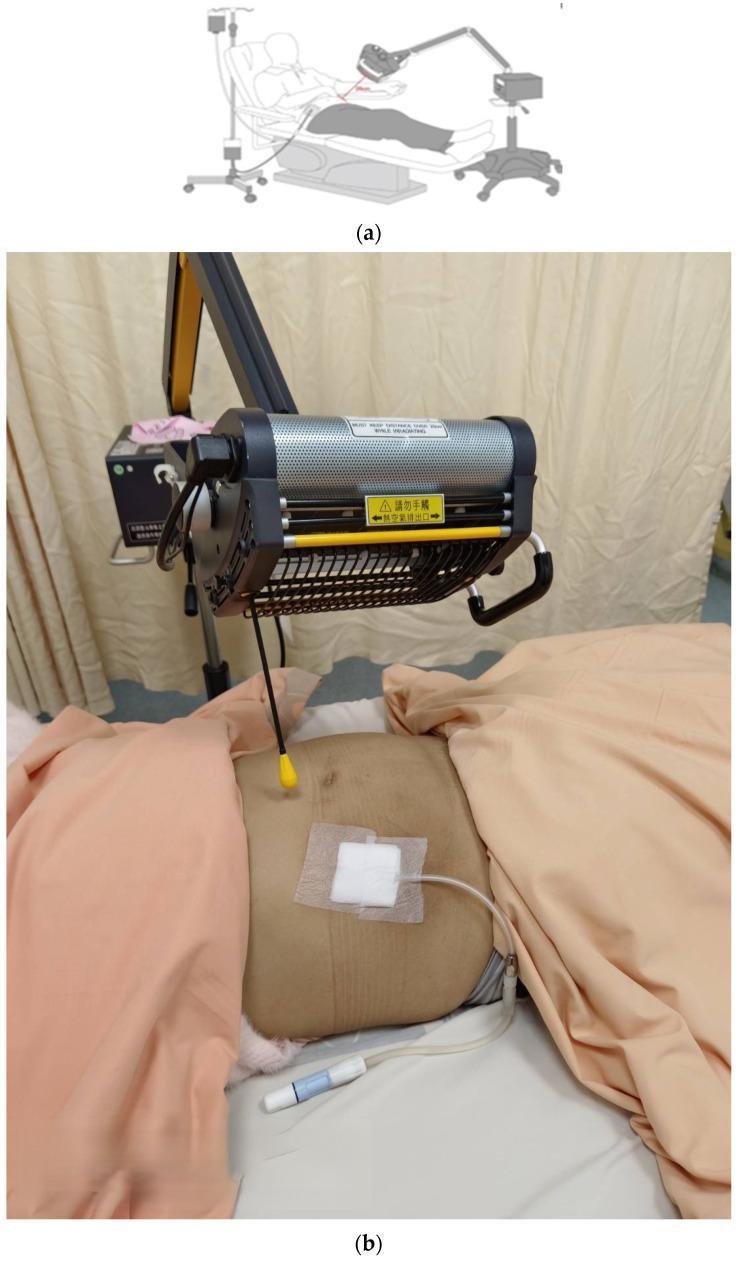
Educational panel illustrating far-infrared (FIR) therapy in a patient undergoing peritoneal dialysis (PD). Part (a) is a schematic diagram showing a patient in a reclined position with a PD bag system and an FIR emitter positioned over the abdomen on an adjustable arm. Part (b) is a clinical photograph showing the real-world application. A patient is supine with the abdomen exposed, featuring a PD catheter exit site secured with a square sterile bandage. The catheter tube connects to a blue-and-white lure lock connector on the bed sheet. Suspended approximately 25 cm above the umbilical region is a WS TY301 FIR emitter, a cylindrical device with a perforated metallic surface for heat dissipation and a yellow safety warning label. The setup demonstrates a clinical trial protocol aimed at studying the effects of FIR radiation on abdominal cavity inflammation and dialysis efficiency. Key concepts include nephrology, home dialysis support, and adjunctive thermal therapy.
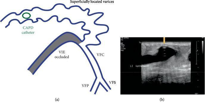
This composite educational resource consists of a schematic diagram and a corresponding diagnostic duplex ultrasound image illustrating a complication of continuous ambulatory peritoneal dialysis (CAPD) catheter placement. Figure (a) is an anatomical diagram showing venous pathology of the lower abdomen and pelvis. It depicts an occluded left external iliac vein (VIE) and a common femoral vein (VFC) branching into the profunda femoris (VFP) and superficial femoral (VFS) veins. A system of tortuous, superficially located abdominal wall varices is shown acting as collateral circulation due to the iliac occlusion. A CAPD catheter is schematically represented transfixing one of these large variceal veins. Figure (b) provides a clinical duplex ultrasound in transverse view, demonstrating a hyperechoic, circular CAPD catheter cross-section (indicated by a yellow arrow) positioned within a dilated, anechoic (dark) lumen of an abdominal wall varicose vein. The image illustrates the anatomical basis for persistent bleeding or anemia in dialysis patients when catheter placement inadvertently involves high-flow collateral venous systems developed secondary to deep venous occlusion.
"peritoneal dialysis"[MeSH Terms] AND outcomes
Peritoneal Dialysis (PD)
1. Overview
2. Peritoneal Membrane: Anatomy and Structure
- A mesothelial cell monolayer covering the abdominal wall and intraabdominal organs, derived from mesenchymal cells, with tight junctions and desmosomes
- Abundant microvilli on the cavity-facing surface, bearing anionic fixed charges -- these increase the total surface area up to 40 m² (the functional peritoneal surface is 1.6-2.0 m²)
- A basement membrane beneath the mesothelium
- A peritoneal interstitium (1-30 μm thick) containing fibroblasts, collagen fibers, and glycosaminoglycans (mainly hyaluronic acid)
- Large pores (>150 Å): transport of large molecules and proteins
- Small pores (40-45 Å): small solute and ion transport
- Ultra-small pores (2-5 Å): aquaporin-1 channels for water transport (free water transport / "transcellular")
3. Transport Physiology
| Mechanism | Description |
|---|---|
| Diffusion | Movement down a concentration gradient. Major pathway for uremic solutes (urea, creatinine, potassium). Most efficient at the start of the dwell when gradient is steepest. |
| Convection | Solute "drag" by bulk water flow (ultrafiltration). Osmotic agent (glucose) creates an osmotic gradient pulling water from plasma into dialysate. |
4. Peritoneal Catheter
Catheter Types
- Material: silicone rubber or polyurethane
- Most common: Tenckhoff catheter (double-cuff, swan-neck, coiled design)
- Intraperitoneal portion options: straight, coiled, Ash (T-fluted), or with silicone disc
- Extraperitoneal: straight or swan-neck design with single or double cuffs
Optimal Design
- Coiled tip: fewer mechanical complications, less outflow obstruction, less pain during infusion, less tip migration
- Swan-neck design: reduces cuff extrusion
- Double cuff: inner cuff in rectus muscle prevents leaks; outer cuff in subcutaneous tissue creates a barrier against exit-site infection tracking inward
Placement Techniques
- Blind (Seldinger)
- Surgical open placement
- Peritoneoscopic
- Laparoscopic
- Moncrief-Popovich (buried technique)
- Fluoroscopic with ultrasound guidance (preferred interventional nephrology technique)
5. PD Solutions
- Osmotic agent: glucose (most common -- 1.5%, 2.5%, or 4.25% dextrose)
- Buffer: lactate (bicarbonate in newer solutions)
- Electrolytes: sodium, calcium, magnesium, chloride
- pH: ~5.5 in standard solutions (more biocompatible pH-neutral solutions available)
6. PD Modalities
| Modality | Description |
|---|---|
| CAPD (Continuous Ambulatory PD) | 3-5 manual exchanges per day; patient performs exchanges manually; continuous dialysis 24 h/day |
| APD (Automated PD) | Machine (cycler) performs exchanges overnight while patient sleeps; variants include CCPD, NIPD, tidal PD |
| Incremental PD | Start with lower dose (e.g., 1-2 exchanges/day in CAPD), increasing as residual kidney function (RKF) declines |
7. Dialysis Adequacy
- K = clearance, t = time, V = volume of distribution of urea
- Historically, weekly Kt/V ≥2.0 was targeted
- Current guidelines: minimum weekly Kt/V of 1.7 (total: peritoneal + renal clearance) for patients with RKF >100 mL/day
- Better preserved in PD patients compared to hemodialysis
- Provides independent survival benefit
- Measured by 24-hour urine collection (average of renal creatinine and urea clearances)
- Assesses peritoneal membrane transport characteristics
- Classifies patients as high, high-average, low-average, or low transporters
- High transporters: rapid glucose absorption, less ultrafiltration -- better suited to APD (shorter dwell times)
- Low transporters: slower solute equilibration -- benefit from longer CAPD dwells
8. Complications
Infectious Complications
- Clinical features: cloudy dialysate, abdominal pain, fever
- Diagnosis: dialysate cell count >100 cells/mm³ (>50% neutrophils), culture, Gram stain
- Common organisms: S. epidermidis, S. aureus, gram-negatives, fungi
- Treatment: intraperitoneal antibiotics (empiric coverage for gram-positive + gram-negative organisms); catheter removal if refractory or fungal peritonitis
- Rate goal: <1 episode per 18 patient-months
- Caused most often by S. aureus and Pseudomonas aeruginosa
- Risk factors: improper exit-site care, nasal carriage of S. aureus
- Prevention: mupirocin nasal/exit-site application
Non-Infectious Complications
| Complication | Notes |
|---|---|
| Catheter tip migration | Up to 35%; causes outflow obstruction; corrected radiologically or surgically |
| Dialysate leaks | Up to 10%; hernia formation, hydrothorax |
| Hernias | Inguinal, umbilical, incisional - due to increased intraabdominal pressure |
| Ultrafiltration failure | Due to peritoneal membrane changes from long-term PD; high transporter state; managed with icodextrin, APD |
| Encapsulating peritoneal sclerosis (EPS) | Rare but serious; fibrotic thickening of peritoneum; most common after >5 years on PD |
| Dyslipidemia | Glucose absorption drives hypertriglyceridemia; common noninfectious complication |
| Hyperglycemia | From glucose absorption, relevant especially in diabetics |
9. Cardiovascular Disease in PD
- Fluid overload and hypertension
- Dyslipidemia from glucose absorption
- Chronic inflammation
- Loss of residual kidney function
10. Patient Outcomes
- 1-, 3-, and 5-year patient survival rates on PD: approximately 89%, 64%, and 49% (Iran registry data)
- Major causes of death: cardiac events (~46%), stroke (~10%), infection (~8%)
- Technique survival (staying on PD without transfer to HD): influenced by peritonitis rates, catheter complications, and ultrafiltration failure
11. PD vs Hemodialysis: Key Differences
| Feature | Peritoneal Dialysis | Hemodialysis |
|---|---|---|
| Location | Home | Hospital/dialysis center |
| Vascular access | Not required | AV fistula/graft/catheter |
| Frequency | Daily (continuous) | 3x/week |
| Residual kidney function | Better preserved | Faster decline |
| Cardiovascular hemodynamics | Gentler | Larger fluid shifts |
| Peritonitis risk | Yes | No |
| Patient independence | High | Lower |
| Protein loss | Yes (in dialysate) | Minimal |
- Brenner and Rector's The Kidney, 2-Volume Set -- Chapters on Peritoneal Dialysis (Chapter 56), Peritoneal Dialysis Catheter Procedures (Chapter 38)
- Comprehensive Clinical Nephrology, 7th Edition -- Chapter on Dose and Adequacy of Peritoneal Dialysis
- Recent evidence: APD vs CAPD Cochrane review (PMID 39258519) | Incremental PD meta-analysis (PMID 39285336) | Percutaneous vs laparoscopic catheter meta-analysis (PMID 41339808)