I now have comprehensive content from multiple surgical textbooks. Let me compile the full exam prep guide.
Oral Exam Prep: Anal Abscess - Surgery Module
Format note: This is structured exactly like your professor's case-based style - anatomy first, then pathology, then a mock case with workup.
PART 1: ANATOMY & PHYSIOLOGY (Brief)
Anal Canal Anatomy
The anal canal is approximately 4 cm long. It has two key landmarks:
- Dentate line (pectinate line): The junction between columnar mucosa (above) and squamous epithelium (below). This is where the anal crypts and anal glands open - the origin of most anal abscesses.
- Anal verge: The distal margin of the anal canal at the skin.
Sphincter Complex:
- Internal anal sphincter (IAS): Smooth muscle, continuation of the circular muscle of the rectum. Involuntary, high resting tone - responsible for ~80% of resting anal pressure.
- External anal sphincter (EAS): Striated skeletal muscle. Voluntary control. Innervated by the pudendal nerve (S3-S4).
- Intersphincteric space: The potential space between IAS and EAS where the anal glands sit. This is ground zero for anal abscess formation.
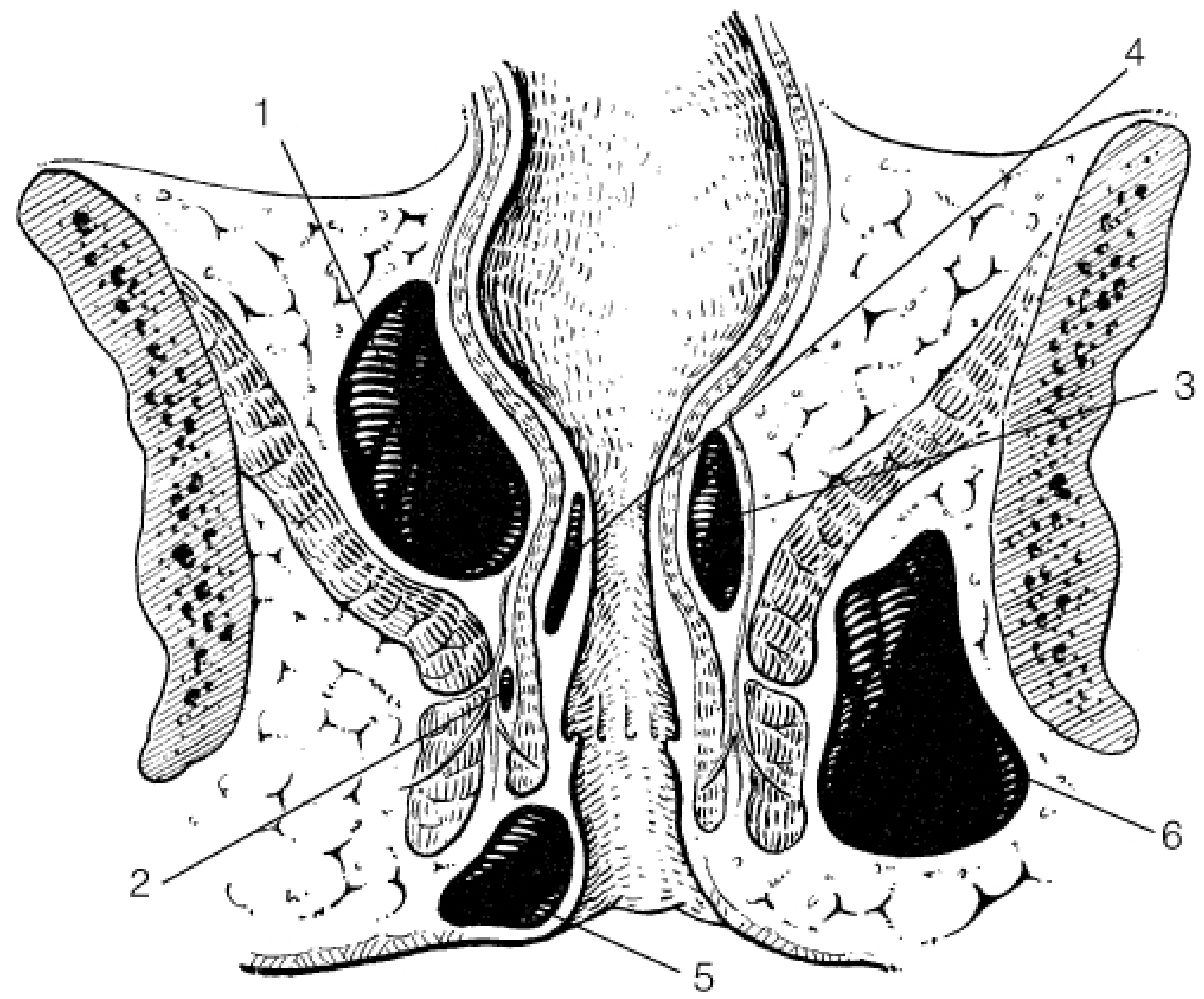
Spaces around the anal canal (clinically important):
| Space | Location | Clinical relevance |
|---|
| Perianal space | Around anal verge | Most common abscess site |
| Ischiorectal fossa | Lateral, below levator ani | Ischiorectal abscess |
| Intersphincteric space | Between sphincters | Primary gland infection site |
| Supralevator space | Above levator ani | Supralevator abscess |
| Submucous space | Between mucosa and IAS | Submucous abscess |
Innervation:
- Above dentate line: Autonomic innervation (visceral pain - poorly localized)
- Below dentate line: Somatic innervation via pudendal nerve (sharp, well-localized pain)
Blood supply: Superior rectal artery (from IMA), middle and inferior rectal arteries (from internal iliac and pudendal arteries respectively).
Lymphatic drainage:
- Above dentate line: Drains upward to pararectal and iliac nodes
- Below dentate line: Drains to inguinal nodes
PART 2: ANAL ABSCESS
Definition & Etiology
An anal/anorectal abscess is a collection of pus in the perianal or perirectal spaces.
>95% are cryptoglandular in origin - infection starts in anal glands at the dentate line, spreads into the intersphincteric space, then tracks in various directions.
Organisms:
- Intestinal origin (cryptoglandular): E. coli, Bacteroides spp., mixed gram-negative + anaerobes
- Skin origin (furuncle/boil): Staphylococcus aureus - these are NOT associated with fistula formation
Key rule: If culture grows intestinal organisms → likely has an underlying fistula (70% of cases). If Staph aureus → no fistula.
Classification by Location (Frequency)
| Type | Frequency | Location | Presentation |
|---|
| Perianal | 60% | Subcutaneous at anal verge | Visible red, tender, fluctuant swelling at anal margin |
| Ischiorectal | 20-25% | Ischiorectal fossa, lateral to EAS | Diffuse buttock swelling, often no visible fluctuance; constitutional symptoms |
| Intersphincteric | 5% | Between IAS and EAS | No visible swelling; pain confirmed only on digital rectal exam |
| Supralevator | 4% | Above levator ani | No visible swelling; fever, malaise; look for pelvic source |
| Submucous | 1% | Between mucosa and IAS | Diagnosed on proctoscopy/EUA |
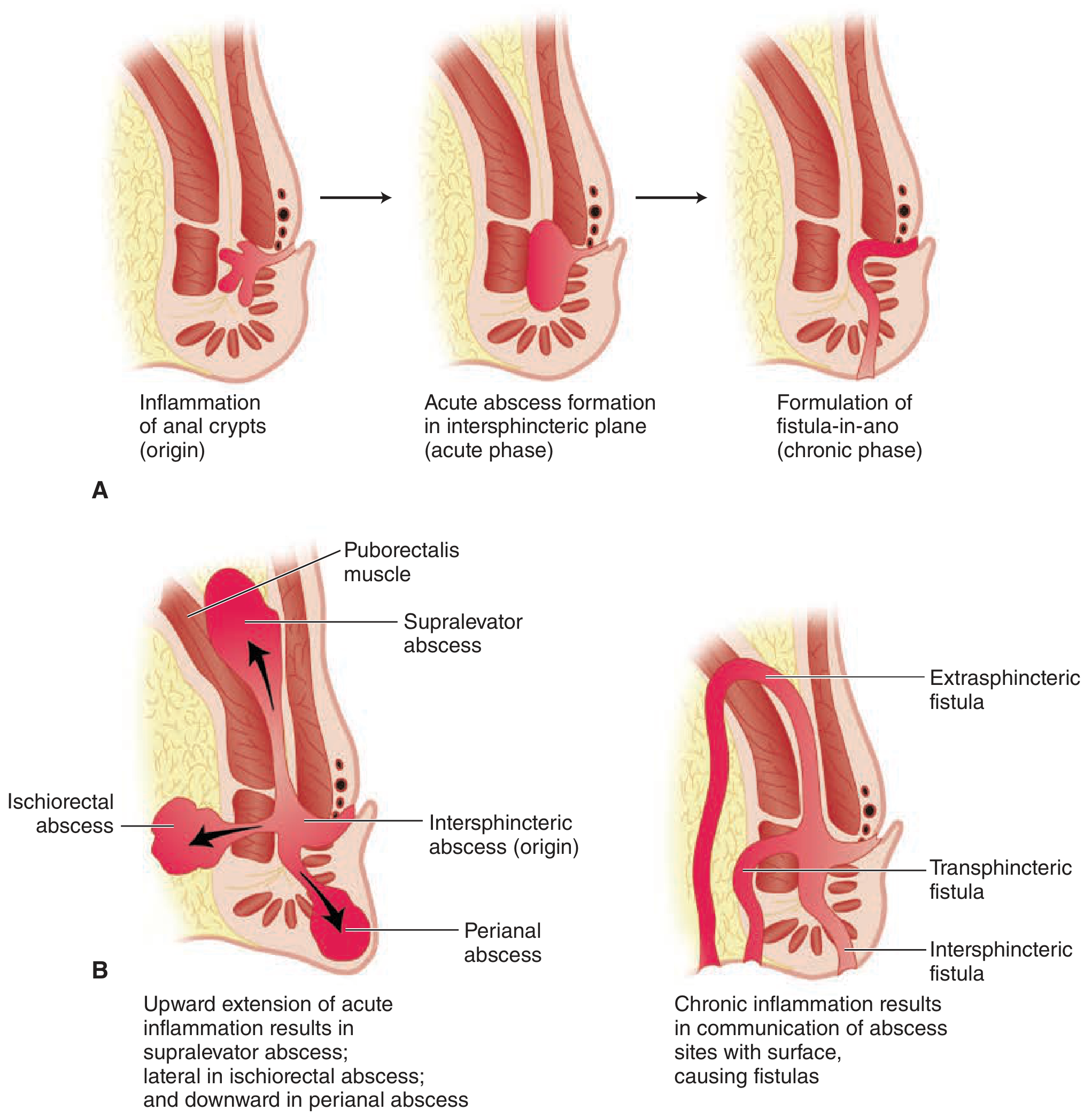
Pathophysiology pathway:
- Anal crypt infection (cryptitis) at dentate line
- Spreads to anal gland in intersphincteric space
- Intersphincteric abscess forms (primary)
- Spreads:
- Downward in intersphincteric plane → Perianal abscess
- Laterally through EAS → Ischiorectal abscess
- Upward in intersphincteric plane → Supralevator abscess
- Inward through IAS → Submucous abscess
Clinical Features
- Severe, constant anal pain (hallmark - worse with sitting, walking, defecation)
- Swelling and erythema around anus (visible in perianal type)
- Fever and constitutional symptoms (especially ischiorectal/supralevator)
- History of previous similar episode that spontaneously drained (30%)
- Secondary causes to ask about: Crohn's disease, HIV/AIDS, malignancy, previous anal surgery, diabetes (immunocompromised)
Diagnosis
- Usually clinical - painful fluctuant swelling near anus
- Digital Rectal Examination (DRE): Essential - identifies intersphincteric/supralevator abscess not visible externally
- Needle aspiration: Confirms pus if not obvious
- Examination Under Anesthesia (EUA): For obscure/complex cases
- MRI: Investigation of choice for complex anorectal sepsis, especially to delineate supralevator/horseshoe extensions
- Anal ultrasound: Can also identify occult abscesses
Labs: CBC (leukocytosis), blood cultures if septic. Culture the pus at the time of I&D.
Treatment
Definitive treatment = Incision and Drainage (I&D)
- Must be done PROMPTLY - do NOT delay waiting for fluctuance
- Antibiotics alone are NOT sufficient and NOT the primary treatment
- Antibiotics are adjuncts ONLY when:
- Extensive surrounding cellulitis
- Immunocompromised (diabetes, HIV, steroids, hematologic disease)
- Valvular heart disease (endocarditis prophylaxis)
Technique by type:
| Abscess type | Drainage technique |
|---|
| Perianal | Cruciate incision or disc excision of skin over abscess, as close to anal canal as possible; break down loculations |
| Ischiorectal | Large incision in perianal skin; cavity explored; mushroom catheter may be placed |
| Intersphincteric | Internal sphincterotomy over the length of abscess to unroof it |
| Submucous | Anal speculum, open mucosa above swelling; often combined with internal sphincterotomy |
| Supralevator (posterior) | Intersphincteric plane opened posteriorly; mushroom catheter sutured in place (failure to do so = recurrence) |
| Supralevator (anterior) | Transverse anal incision anteriorly through intersphincteric plane; mushroom catheter in situ |
PART 3: COMPLICATIONS
Immediate / Short-term
| Complication | Notes |
|---|
| Fistula-in-ano | Most important - occurs in 30-50% after I&D; abscess from intestinal organisms = 70% have a fistula |
| Recurrence | If drainage inadequate; re-evaluate for Crohn's disease |
| Necrotizing fasciitis / Fournier's gangrene | Life-threatening emergency; classic triad: pain + fever + inability to void; requires emergent debridement |
| Perineal sepsis | Extension of infection; medical emergency |
| Bacteremia / Septicemia | Especially in immunocompromised |
Long-term
| Complication | Notes |
|---|
| Anal fistula | Chronic phase of the same disease - tract between internal opening (at dentate line) and external opening (at skin) |
| Fecal incontinence | From sphincter damage (especially if sphincters are divided carelessly) |
| Anal stricture | From scarring |
| Recurrent abscess | Suspect Crohn's disease if recurrent |
PART 4: FISTULA-IN-ANO (the chronic complication you must know)
Goodsall's Rule (predicts fistula tract anatomy):
- External opening anterior to transverse anal line (and within 2 cm): short, straight tract to nearby crypt
- External opening posterior to transverse anal line (or anterior but >2 cm from verge): curved tract to posterior midline crypt
- Exception: Horseshoe fistula - external opening anterior >2 cm, internal opening in posterior midline; requires drainage of postanal space + secondary drainage of anterior extensions
Park's Classification of Fistulas:
- Intersphincteric - most common; tract between sphincters
- Transsphincteric - crosses EAS; low (safe to divide) vs high (risk incontinence)
- Suprasphincteric - above puborectalis; complex
- Extrasphincteric - outside both sphincters; very complex; rare
Surgical treatment:
- Fistulotomy (lay-open): For low, simple intersphincteric/low transsphincteric fistulas
- Seton placement: For high fistulas (to preserve sphincter) - a suture/wire passed through the tract; cutting seton or loose seton
- Division of puborectalis = incontinence - NEVER do this
PART 5: MOCK EXAM CASE (Anal Abscess Style)
A 35/M laborer presents to the ER with severe anal pain for 3 days, aggravated by sitting and defecation. He has fever of 38.5°C and cannot walk properly. On examination, there is a 4 cm erythematous, warm, tender swelling lateral to the anus on the left side. Vital signs: BP 120/80, HR 108, RR 20, T 38.5°C.
Q1: What is your assessment?
Ischiorectal abscess (left-sided, lateral to anus, large, no spontaneous drainage)
- Differential: perianal abscess, pilonidal abscess, Bartholin's abscess (female), hidradenitis suppurativa
Q2: Initial workup at ER
CBC: Leukocytosis (WBC elevated - expect >12,000), possible shift to left
Other labs:
- Blood glucose (screen for diabetes - uncontrolled DM worsens prognosis)
- Blood cultures if febrile/septic
- HIV screening (if high-risk or recurrent)
Imaging:
- MRI pelvis: If supralevator extension or horseshoe spread suspected
- Anal ultrasound: If available
- CT pelvis: If worried about pelvic/intra-abdominal source (especially supralevator abscess)
Workup interpretation (hypothetical values):
- WBC 18 = leukocytosis → supports acute infection/abscess
- Blood glucose elevated → suspect DM, affects healing and risk for necrotizing fasciitis
- Blood cultures → if positive, bacteremia complicating abscess
Q3: Initial management
- IV access, IV fluids if hypotensive/septic
- IV antibiotics as adjunct (if extensive cellulitis, DM, immunocompromised) - e.g., ampicillin-sulbactam or piperacillin-tazobactam + metronidazole
- Analgesia (IV morphine or ketorolac)
- Consent for I&D under anesthesia
- Check bleeding parameters (PT/PTT, platelet count)
Q4: Definitive management
Incision and Drainage (I&D) - the ONLY definitive treatment
- Regional or general anesthesia
- Large cruciate incision over the abscess, as close to anal canal as possible
- Break down all loculations digitally
- Culture the pus
- Pack wound / place mushroom catheter
- Wound care and sitz baths post-op
- Follow-up at 4-6 weeks to evaluate for fistula formation
If fistula found later: Fistulotomy (if low) or seton placement (if high)
Quick Exam Recall Summary
| Point | Key fact |
|---|
| Most common cause | Cryptoglandular (>95%) |
| Most common type | Perianal (60%) |
| Origin | Anal glands at dentate line, intersphincteric space |
| Organisms (cryptoglandular) | E. coli, Bacteroides (intestinal/anaerobes) |
| Organisms (skin/boil) | Staphylococcus aureus - NO fistula |
| Definitive treatment | I&D - NOT antibiotics |
| Antibiotics when? | Cellulitis, DM, immunocompromised, valvular disease |
| Most important complication | Fistula-in-ano (30-50%) |
| Life-threatening complication | Necrotizing fasciitis / Fournier's gangrene |
| Chronic complication | Fistula-in-ano (the chronic phase of the SAME disease) |
| Goodsall's rule posterior | Curved tract to posterior midline |
| High fistula treatment | Seton (NOT fistulotomy - risk of incontinence) |
| MRI use | Investigation of choice for complex/supralevator abscess |
Sources: Maingot's Abdominal Operations, Fischer's Mastery of Surgery 8th ed., Pfenninger and Fowler's Procedures for Primary Care, Sleisenger and Fordtran's GI and Liver Disease
Good luck on your oral exam today! You've got this.