
Anatomy of labyrinth and its surgical importance and its variations related to cochlear implant surgery and Discuss development of the bony and membranous labyrinth for 20 markers question answer each, in detail, in simplified manner, according to standard ENT textbook, along with images that are supposed to be drawn
bony labyrinth anatomy cochlea semicircular canals vestibule diagram
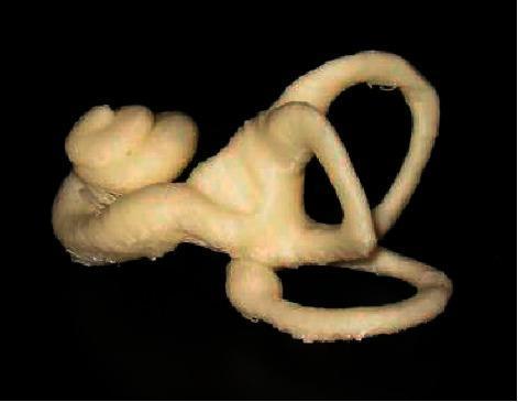
This image presents a 3D computational reconstruction of the human inner ear, derived from a statistical shape model. The anatomical diagram demonstrates the complex three-dimensional relationship between the cochlea, vestibule, and semicircular canals. On the left, the spiral-shaped cochlea is clearly visible with its characteristic bulbous, snail-like winding. This transitions into the central vestibule, from which the three semicircular canals (superior, posterior, and lateral) originate. These canals are depicted as arched, looped structures oriented in nearly orthogonal planes, illustrating their physiological role in sensing rotational head movements. The model features a pale, textured surface, representing the bony labyrinth, set against a high-contrast black background. This visual serves as an educational tool for understanding otolaryngology and vestibular anatomy, particularly for medical students and clinicians studying diagnostic imaging segmentation or the pathophysiology of inner ear disorders like Meniere's disease.
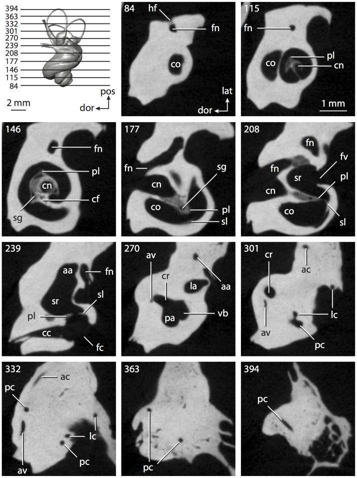
This diagnostic image provides a series of high-resolution CT slices through the petrosal bone, detailing the anatomy of the inner ear. The image set illustrates the complex three-dimensional structure of the bony labyrinth, with specific focus on the cochlea and vestibular apparatus. Slices 84 through 208 show the progressive coiling of the cochlea (co), revealing internal features such as the primary bony lamina (pl), cochlear canal (cn), and secondary bony lamina (sl). The relationship between the cochlea and the facial nerve canal (fn) is visible in the more anterior slices. Progressing posteriorly (slices 239–394), the vestibular system becomes prominent, showcasing the vestibule (vb), spherical recess (sr), and the three semicircular canals: anterior (ac), lateral (lc), and posterior (pc). Key landmarks for anatomical indexing include the fenestra cochleae (fc), fenestra vestibuli (fv), and the common crus (cr). These slices demonstrate the spatial orientation and morphology of the hearing and balance organs within the temporal bone, serving as a critical educational resource for understanding inner ear anatomy and radiological identification of its components.
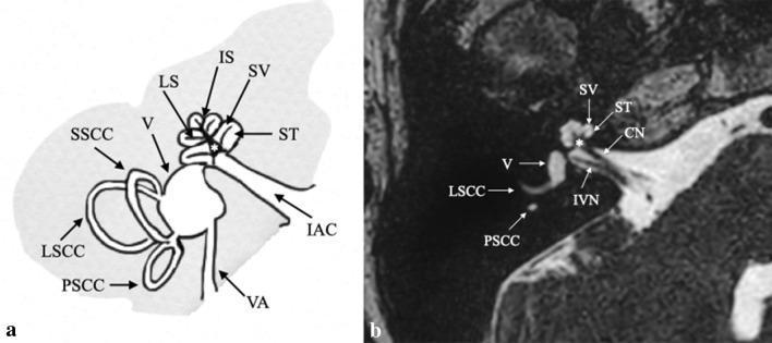
This educational composite depicts the normal anatomy of the human inner ear through an anatomical diagram (a) and a corresponding high-resolution axial MRI (b), specifically a 3D FIESTA sequence. The primary focus is the complex architecture of the bony and membranous labyrinth. Key structures identified include the cochlea, which is shown spiraling around the central modiolus (asterisk), with clear labeling of the scala vestibuli (SV), scala tympani (ST), and the dividing interscalar septum (IS) and osseous spiral lamina (LS). The vestibular system is represented by the vestibule (V), the lateral (LSCC), posterior (PSCC), and superior (SSCC) semicircular canals, and the vestibular aqueduct (VA). The diagnostic MRI correlates these illustrations with clinical findings, showing the cochlear nerve (CN) and inferior vestibular nerve (IVN) within the internal acoustic canal (IAC). This comparison is designed to teach medical students and radiology residents the spatial relationships and internal architecture of the temporal bone's auditory and vestibular apparatus.
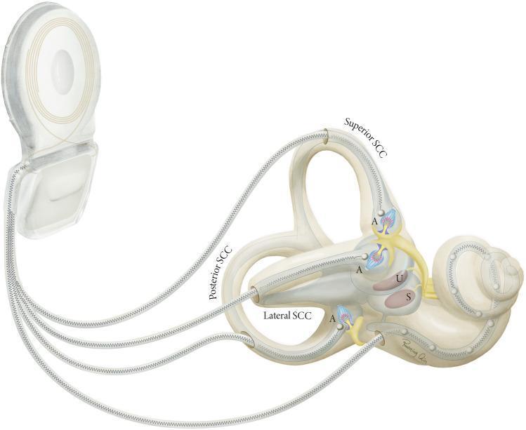
A medical anatomical diagram illustrating a vestibulocochlear implant system integrated with the human inner ear labyrinth. The visual highlights the relationship between the external processor/internal receiver unit and the terminal electrode arrays. The inner ear structures are clearly labeled, showing the three semicircular canals (Superior, Posterior, and Lateral SCC) in their orthogonal orientation, alongside the utricle (U) and saccule (S) within the vestibule. Each semicircular canal is depicted with an electrode lead entering through a bony fenestration to reach the ampulla (A), where it targets the terminal afferent nerve fibers of the crista ampullaris. A separate cochlear lead is shown entering the base of the cochlea. This illustration demonstrates the surgical placement and anatomical targets of vestibular implants designed to restore balance by electrically stimulating the vestibular system's sensory organs.
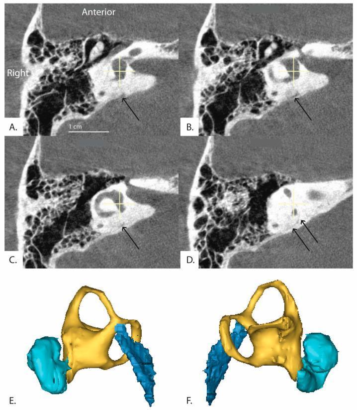
This composite educational resource features high-resolution imaging of the human inner ear anatomy. Panels A-D display sequential axial CT images reformatted in the plane of the horizontal semicircular canal, progressing from an inferior to superior perspective. Black arrows highlight the distal vestibular aqueduct as it traverses the petrous temporal bone. The images demonstrate the spatial relationship between the bony labyrinth, including the vestibule and semicircular canals, and the vestibular aqueduct. Panels E and F provide 3D anatomical reconstructions (right and left views, respectively) illustrating the complex spatial morphology of these structures. The color-coding in the 3D models differentiates key components: the cochlea is shown in cyan, the vestibule and semicircular canals in yellow, and the vestibular aqueduct in dark blue. This visualization is intended to assist in the clinical assessment of inner ear pathologies, such as Meniere's disease or superior canal dehiscence syndrome, by mapping the trajectory and angle of the vestibular aqueduct relative to the labyrinthine structures.
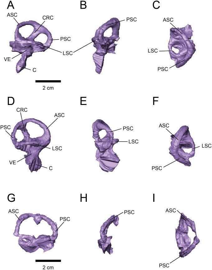
Anatomical diagram presenting virtual 3D endocasts of the endosseous labyrinth (inner ear). The image displays left and right specimens (FPDM-V-9775 and FPDM-V-9677) across nine panels in lateral (A, D, G), posterior (B, E, H), and dorsal (C, F, I) views. Key anatomical structures labeled include the anterior semicircular canal (ASC), posterior semicircular canal (PSC), lateral semicircular canal (LSC), cochlea (C), vestibule (VE), and crus commune (CRC). The ASC is characterized by a generally round, low-arc morphology. The PSC is slightly lower dorsoventrally than the ASC, and both are joined by the crus commune. The LSC is the shortest of the three canals. Ventral to the vestibular apparatus, the elongated cochlear duct (C) is visible in the lateral views. The arrangement illustrates the spatial orientation and comparative morphology of the vestibular and auditory systems, relevant for understanding balance and auditory function within comparative anatomy and diagnostic imaging studies.
membranous labyrinth utricle saccule endolymph perilymph cochlear duct
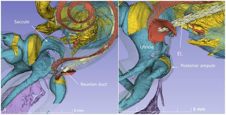
This medical visual comprises two panels (A and B) featuring high-magnification 3D reconstructions of the human inner ear's membranous labyrinth, derived from micro-CT imaging. Panel A highlights the surgical anatomy of the cochlear base, demonstrating the saccule (yellow pouch-like structure) and its connection via the reunion duct (RD, white tubular structure) to the cochlea (reddish spiral). The spatial relationship emphasizes the proximity of the saccule to the surgical pathway. Panel B illustrates a virtual cochlear implant surgery using a round window (RW) approach. A cochlear implant electrode (El, white segmented structure) is shown inserted into the cochlea, passing near the utricle (red) and the posterior ampulla (yellow). The supporting bony and soft tissues are rendered in blue and purple hues. The images serve as educational models to visualize anatomical risks during electrode insertion, specifically the close distances between the round window and vestibular receptors like the saccule and posterior ampulla nerves.
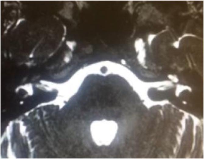
This diagnostic image is an axial T2-weighted MRI focusing on the inner ear and posterior fossa. The scan demonstrates a comparative view of the bilateral cochlear structures. On the left side, the cochlea exhibits a normal high-signal (bright) intensity, indicating the presence of perilymph and endolymph within the membranous labyrinth. In contrast, the right cochlea shows a significant loss of the normal fluid signal, replaced by a darker, hypointense signal intensity and altered morphology characteristic of labyrinthitis ossificans. This finding indicates ossification of the cochlear lumen, a common sequela of bacterial meningitis. The internal auditory canals are visible bilaterally as high-signal intensity fluid-filled channels containing the vestibulocochlear nerve complexes. The pons and cerebellum are visible in the midline and posterior regions, surrounded by bright cerebrospinal fluid. This image is clinically significant for evaluating candidacy for cochlear implantation, as ossification can obstruct electrode insertion.
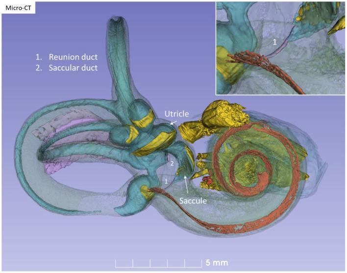
This diagnostic image features a high-resolution 3D reconstruction of the human right internal ear, derived from micro-CT data in Stenver's view. The bony capsule is rendered as a semi-transparent grey-lilac shell to expose the underlying membranous labyrinth and associated neurovascular structures. The cochlea is highlighted by a red-colored basilar membrane spiraling through its turns. The vestibular system, including the three semicircular canals, utricle, and saccule, is depicted in teal/light blue. Key neural components, specifically the vestibular neuro-epithelium and vestibulocochlear nerves, are rendered in bright yellow. Labels identify the 'Reunion duct' (1) and 'Saccular duct' (2), illustrating the fluid pathways connecting the saccule to the cochlear duct (ductus reuniens) and the endolymphatic system. An upper-right inset provides a magnified perspective of the reunion duct (1), emphasizing its anatomical position between the vestibular organ and the cochlear base. This model serves as an educational tool for otolaryngology and neuroanatomy, demonstrating the complex spatial relationships within the temporal bone.
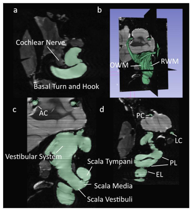
This diagnostic imaging composite displays high-resolution segmented micro-MRI (µMRI) scans of the inner ear anatomy. The images use a green overlay to highlight the lymphatic fluid spaces within the membranous labyrinth. Panel (a) shows a transverse plane depicting the cochlear nerve and the basal turn and hook of the cochlea. Panel (b) provides a 3D volumetric reconstruction, illustrating the spatial relationship between the oval window membrane (OWM) and the round window membrane (RWM). Panel (c) is a sagittal section highlighting the vestibular system and the anterior canal (AC). Panel (d) presents a coronal plane view that clearly differentiates the cochlear duct compartments: the scala tympani, scala media, and scala vestibuli. Additional vestibular structures such as the posterior canal (PC), lateral canal (LC), perilymph (PL), and endolymph (EL) are labeled. This set of images serves as a detailed anatomical reference for the complex fluid-filled chambers and neural structures of the auditory and vestibular systems, relevant for otolaryngology and audiology education.
cochlear implant surgery electrode insertion round window anatomy
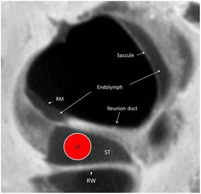
This diagnostic image is a high-resolution micro-computed tomography (micro-CT) cross-section of the human cochlear base, specifically focused on the round window (RW) region. The image illustrates the complex spatial anatomy relevant to cochlear implant (CI) surgery. At the inferior aspect, the round window (RW) leads into the scala tympani (ST). A virtual cochlear implant electrode (labeled 'el' within a red circle) is visualized within the lumen of the scala tympani. Directly superior to the scala tympani is the saccule, containing the endolymphatic space. The reunion duct (RD) is identified on the superior surface of the osseous spiral lamina, connecting the scala media and saccule. Reissner’s membrane (RM) is clearly labeled, separating the fluid compartments. The image highlights the close anatomical proximity between the surgical insertion site (ST/RW) and vestibular structures like the saccule, demonstrating potential risk areas for mechanical trauma during electrode placement. This visualization is intended for advanced otolaryngology education and surgical planning.
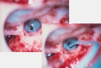
This clinical surgical photograph, captured via endoscopy, shows the intraoperative procedure of a cochlear implant electrode insertion through the round window. The image is split into two frames demonstrating a progression of the 'soft-surgery' technique. The left frame shows the preparation of the round window niche with a small incision or cochleostomy made in the round window membrane, appearing as a dark opening against the bluish-white surface of the membrane. The right frame illustrates the active insertion of the cochlear implant electrode carrier. The electrode is a thin, flexible, translucent cylindrical structure with visible dark internal components (contacts). It is being guided carefully through the opening in the round window to reach the scala tympani of the cochlea. The surrounding surgical field includes vascularized middle ear mucosa with characteristic reddish hue and anatomical landmarks of the tympanic cavity. This visual demonstrates the atraumatic insertion method intended to preserve residual hearing by minimizing mechanical damage to inner ear structures.
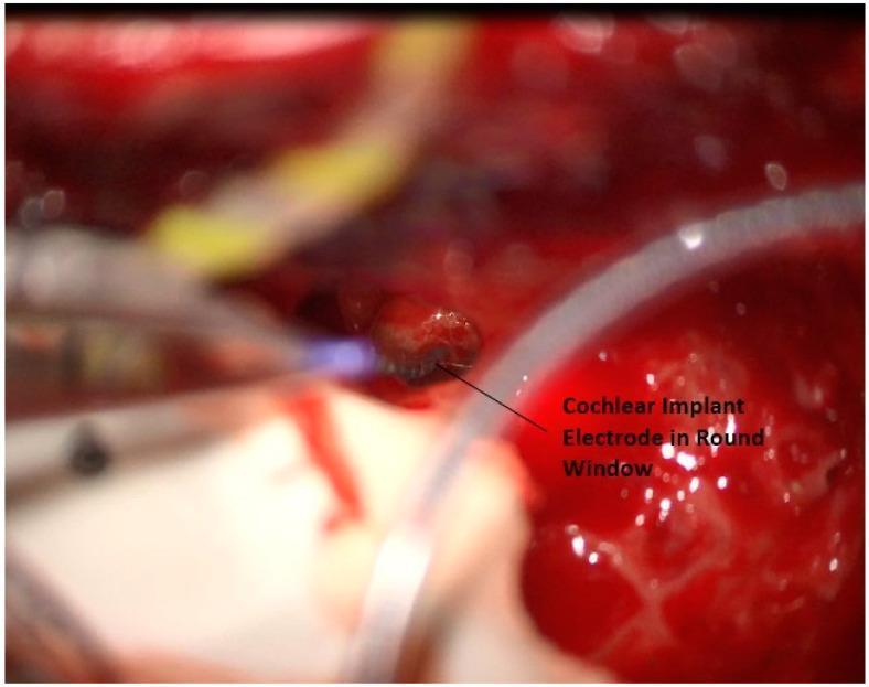
This clinical photograph provides an intraoperative view of a cochlear implant surgery, specifically demonstrating the placement of the electrode array into the round window. The image shows a focused surgical field within the mastoid cavity, characterized by erythematous, moist soft tissue and exposed bone. The primary feature is a dark, segmented cochlear implant electrode array being inserted through the round window niche. A distinctive blue light from a surgical instrument or laser is visible near the insertion point, aiding in precise localization. Surrounding the insertion site, metallic surgical instruments are visible, including a suction tip or dissector, alongside a translucent drainage or suction tube. The surgical field contains evidence of minor bleeding, consistent with active otological surgery. This visual illustrates a critical step in cochlear implantation—accessing the scala tympani via the round window to restore auditory function.
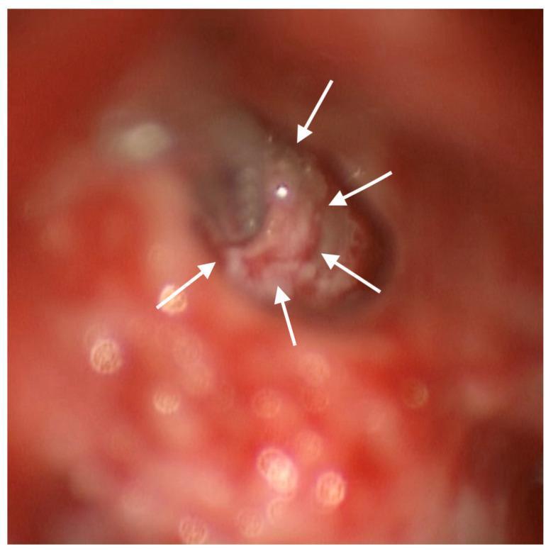
This intraoperative clinical photograph shows a posterior tympanotomy surgical approach to the middle ear during a cochlear reimplantation procedure. The focus is on the round window (RW) region, which has been surgically enlarged to facilitate electrode insertion. A cochlear implant electrode array is visible entering the cochlea through the modified RW opening. Five white arrows indicate a dense mass of light-colored, whitish connective tissue that has been intentionally placed to envelop the electrode array at the entry site. This connective tissue serves as a biological barrier between the implant and the bony margins of the enlarged round window, intended to prevent future osteoneogenesis or fibro-ossification that could damage the device. The surrounding field shows typical erythematous surgical margins of the middle ear mucosa and temporal bone drilling site. This image demonstrates soft-tissue surgical techniques used in revision otologic surgery to manage and prevent implant failure due to reactive tissue formation.
development otic vesicle otocyst embryology inner ear labyrinth
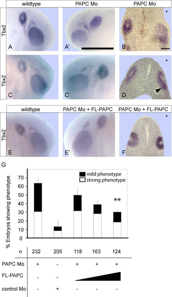
This composite figure illustrates the role of Paraxial Protocadherin (PAPC) in inner ear development using a Xenopus embryo model. (A-F) Micrographs show in situ hybridization (ISH) for Tbx2, an early marker of otic placode and vesicle formation. In wildtype embryos (A, C, E), the otocyst exhibits a rounded morphology with a distinct inner cavity (unstained center). In PAPC antisense morpholino (PAPC Mo) injected embryos, severe phenotypes (A', B) show complete loss of the otocyst cavity and flattened morphology, while mild phenotypes (C', D) show irregular epithelial infolding (arrowhead) and diffuse Tbx2 signal. Transversal sections (B, D) confirm the structural disruption of the otic epithelium on the injected side (asterisks). (E-F) Rescue experiments demonstrate that coinjection of full-length PAPC (FL-PAPC) RNA restores normal otocyst morphology and cavity formation. (G) A stacked bar chart provides statistical quantification of the phenotypes, showing a dose-dependent reduction in strong and mild phenotypes upon increasing FL-PAPC RNA rescue compared to the PAPC Mo group. This research identifies PAPC as a critical regulator of apical-basal cell alignment and morphogenetic movements during otocyst development.
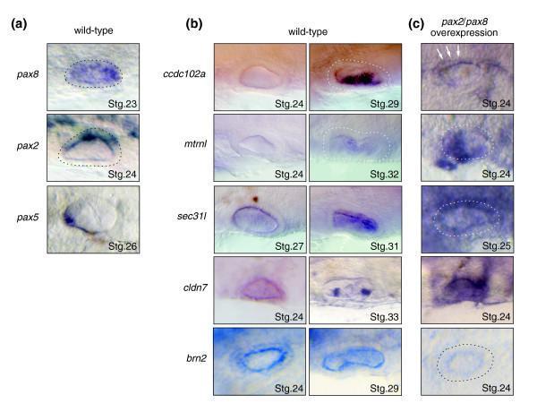
This composite of clinical diagnostic images depicts gene expression patterns in medaka embryos during otic vesicle development, serving as a model for vertebrate inner ear morphogenesis. (a) In wild-type embryos, pax8 (stage 23), pax2 (stage 24), and pax5 (stage 26) show localized purple staining in the otic vesicle epithelium, highlighting their role as upstream regulators. (b) Wild-type expression of downstream candidate genes is shown across stages 24 to 33. ccdc102a transitions from absent to medioventral expression by stage 29; metrnl appears weakly at stage 32; sec31l shifts from general epithelium (stage 27) to medial localization (stage 31); cldn7 is broadly expressed at stage 24 and restricted to medial cristae by stage 33; and brn2 remains restricted to the medial part throughout. (c) Experimental over-expression of pax2/pax8 demonstrates regulatory relationships, where target genes like ccdc102a, mtrnl, sec31l, and cldn7 show ectopic, intensified staining, while brn2 exhibits transcriptional repression (decreased staining) at stage 24. These images illustrate molecular pathways in developmental biology and auditory system embryology.
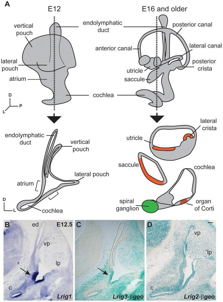
Educational panel illustrating mouse inner ear development and Lrig family gene expression at embryonic stages. (A) Anatomical diagrams compare the immature otic vesicle at E12.5 (left) with the mature labyrinth at E16 (right). Developmental transitions show vertical and lateral pouches evolving into anterior, posterior, and lateral semicircular canals. Cross-sections highlight the differentiation of sensory epithelia (red), including the organ of Corti and cristae, and neurons (green) within the spiral ganglion by E16. (B-D) Microscopic images show mRNA and reporter protein expression at E12.5 using in situ hybridization and X-gal staining. Lrig1 (B) and Lrig3-βgeo (C) exhibit highly restricted, overlapping expression patterns localized to the atrium and the non-sensory domain of the cochlea (indicated by arrows). In contrast, Lrig2-βgeo (D) shows broad, ubiquitous expression throughout the otic epithelium, including the pouches and cochlear duct. This figure demonstrates the unique and overlapping roles of Lrig proteins in regulating inner ear morphogenesis and sensory tissue patterning.
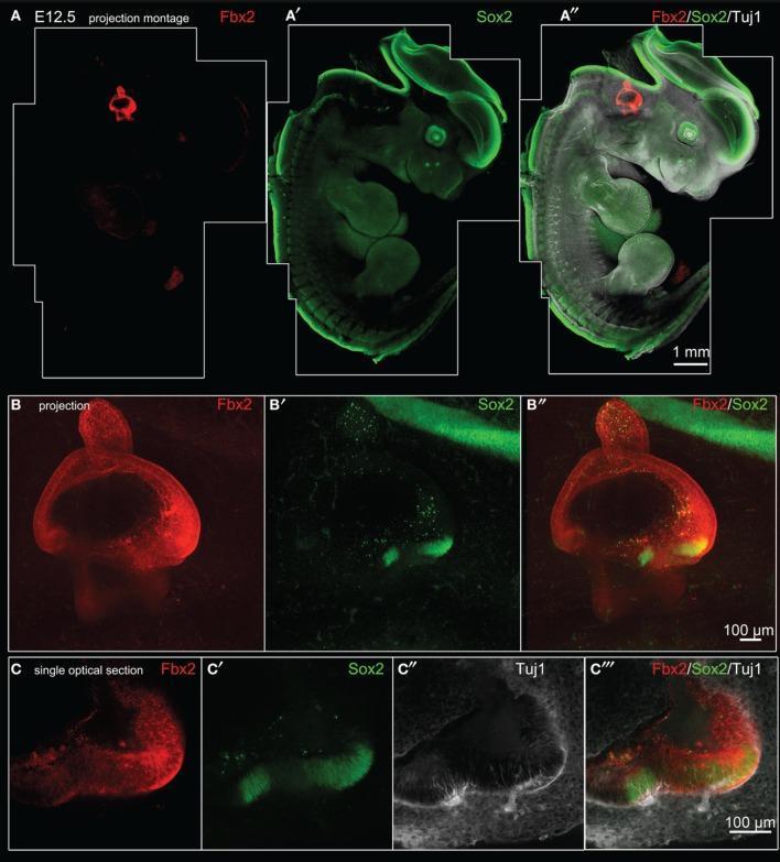
This composite figure presents confocal microscopy images of a mouse embryo at embryonic day 12.5 (E12.5), demonstrating protein expression patterns relevant to inner ear development. Panels A–A″ show merged projection montages of the whole embryo. Fbx2 (red) exhibits highly specific localization restricted to the otic region, while Sox2 (green) shows broad expression across the neural tube and central nervous system. Panels B–B″ provide a high-magnification projection of the otocyst, where Fbx2 is expressed throughout the membranous labyrinth, and Sox2 identifies two distinct bright vestibular prosensory patches in the anterior vestibule. Panels C–C′″ feature single optical sections through the anterior vestibular prosensory patches, revealing the co-localization of Fbx2 (red), Sox2 (green), and Tuj1-labeled neurites (white). The images demonstrate that Fbx2 serves as a robust and highly specific marker for the otic sensory lineage during midgestation, identifying the epithelium that gives rise to the hair cells and supporting cells of the vestibular and cochlear systems.
cochlear cross section scala tympani vestibuli organ of Corti basilar membrane
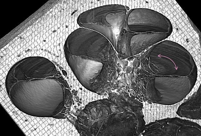
This diagnostic image is a virtual mid-modiolar cross-section of a human cochlea obtained via synchrotron radiation phase contrast imaging (SR-PCI). The image displays the characteristic spiraling architecture of the cochlea, showcasing multiple turns of the bony labyrinth. Within these turns, the three primary fluid-filled chambers—scala vestibuli, scala media (cochlear duct), and scala tympani—are clearly visible as distinct, rounded compartments separated by thin membranes. The osseous spiral lamina is evident as a bony projection extending into the lumen. A magenta arrow specifically identifies the location and orientation of the organ of Corti sitting upon the basilar membrane. The grayscale intensities reflect varying tissue densities, with the surrounding bone and connective tissue exhibiting a porous, mesh-like texture. This advanced imaging modality provides high-contrast visualization of the internal sensory epithelium and membranous partitions, facilitating the study of inner ear anatomy and pathologies related to sensorineural hearing loss (SNHL).
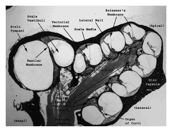
This medical micrograph illustrates a plastic cross-section of a guinea pig cochlea, viewed in a near midmodiolar plane to reveal the anatomy of the inner ear. The cochlea is presented as a coiled labyrinth within the bony otic capsule, showing approximately 2.5 turns from the basal to the apical region. Each turn exhibits three distinct fluid-filled chambers: the scala vestibuli (superior), the scala media (middle), and the scala tympani (inferior). Key anatomical landmarks are labeled, including Reissner's membrane, which serves as the boundary between the scala vestibuli and scala media, and the basilar membrane, which separates the scala media from the scala tympani. The organ of Corti is visible sitting upon the basilar membrane, with the overlying tectorial membrane. Central neural structures are clearly identifiable, specifically the spiral ganglion within Rosenthal's canal and the cochlear nerve fibers. This image serves as a fundamental anatomical reference for auditory physiology and the study of mechanical-to-electrical transduction within the mammalian hearing system.
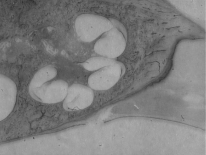
This diagnostic image is a low-magnification (3X) histological cross-section of a mammalian cochlea, specifically showing the spiral structure within the temporal bone. The image clearly demonstrates the classic anatomical partitioning of the cochlear duct into three distinct fluid-filled chambers: the superior scala vestibuli, the middle scala media, and the inferior scala tympani. These chambers are arranged in a triangular configuration within each turn of the cochlear spiral, separated by the vestibular membrane and the basilar membrane. Although shown at a low magnification, the central modiolus around which the cochlea coils is visible, along with outlines of essential auditory structures including the organ of Corti, tectorial membrane, and the stria vascularis located on the lateral wall. The surrounding dense bone provides the structural framework for these sensory pathways. This specimen serves as an educational model for understanding the macro-architecture of the inner ear and the spatial relationship between perilymphatic and endolymphatic spaces essential for auditory transduction.
QUESTION 1: Anatomy of the Labyrinth, Its Surgical Importance, and Variations Related to Cochlear Implant Surgery
INTRODUCTION
PART A: ANATOMY OF THE LABYRINTH
1. Bony Labyrinth
(a) Vestibule
- The central part of the bony labyrinth, roughly oval in shape, measuring about 5 mm in length
- Located medial to the middle ear, lateral to the internal acoustic meatus
- Lateral wall: contains the oval window (fenestra vestibuli), into which the footplate of the stapes fits
- Medial wall: contains two recesses:
- Elliptical recess (posterosuperior) - lodges the utricle
- Spherical recess (anteroinferior) - lodges the saccule
- Posterior wall: opens into the semicircular canals (5 openings)
- Anterior wall: opens into the cochlea
- A narrow canal - the vestibular aqueduct - exits the medial wall and traverses the petrous bone to open at the posterior surface, transmitting the endolymphatic duct
[DRAW]: Diagram of the bony labyrinth showing the vestibule, cochlea, three semicircular canals, oval window, round window, common crus, and their interconnections - lateral view
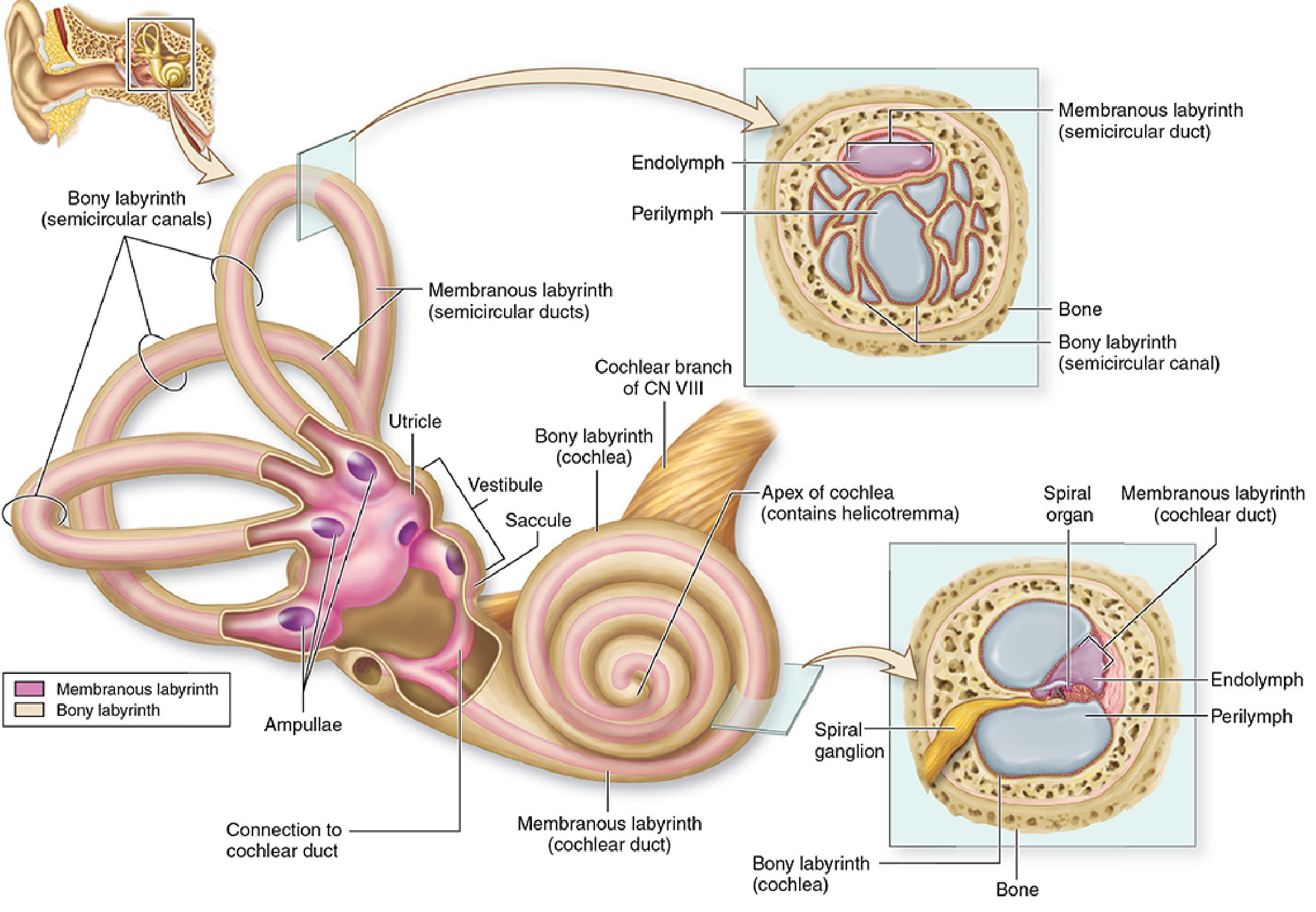
(b) Semicircular Canals
| Canal | Plane | Relation |
|---|---|---|
| Anterior (superior) | Sagittal | Arcuate eminence on petrous bone |
| Posterior | Frontal | Parallel to posterior petrous surface |
| Lateral (horizontal) | Horizontal | Farthest lateral; shortest |
- Each canal has a dilated end (ampulla) containing the crista ampullaris (balance organ)
- The anterior and posterior canals share a common crus (non-ampullary ends join) - so 3 canals open via 5 orifices into the vestibule
- The lateral canal's ampulla opens separately
- Diameter of bony canal: ~1.5-2 mm; membranous duct is much smaller within it
(c) Cochlea
- Makes 2.5 to 2.75 turns around a central bony pillar called the modiolus
- Length of uncoiled cochlea: ~35 mm
- Base (basal turn) faces the middle ear and contains:
- The round window (fenestra cochleae) - closed by the secondary tympanic membrane
- The oval window is just superior to it
- Apex (cupula) points anterolaterally
- The modiolus contains the spiral ganglion (cell bodies of cochlear nerve neurons) in Rosenthal's canal
- The osseous spiral lamina projects from the modiolus like a shelf, partially dividing the cochlear lumen
| Compartment | Contents | Communication |
|---|---|---|
| Scala vestibuli | Perilymph | Opens into vestibule |
| Scala media (cochlear duct) | Endolymph | Closed at apex (caecum cupulare) |
| Scala tympani | Perilymph | Ends at round window |
- Scala vestibuli and scala tympani communicate at the helicotrema at the apex
- Scala media is bounded:
- Superiorly: Reissner's membrane
- Inferiorly: basilar membrane (on which the organ of Corti sits)
- Laterally: stria vascularis (produces endolymph; high K+)
[DRAW]: Cross-section of one cochlear turn showing: Scala vestibuli, Scala media, Scala tympani, Reissner's membrane, basilar membrane, organ of Corti with hair cells, tectorial membrane, stria vascularis, osseous spiral lamina, and spiral ganglion
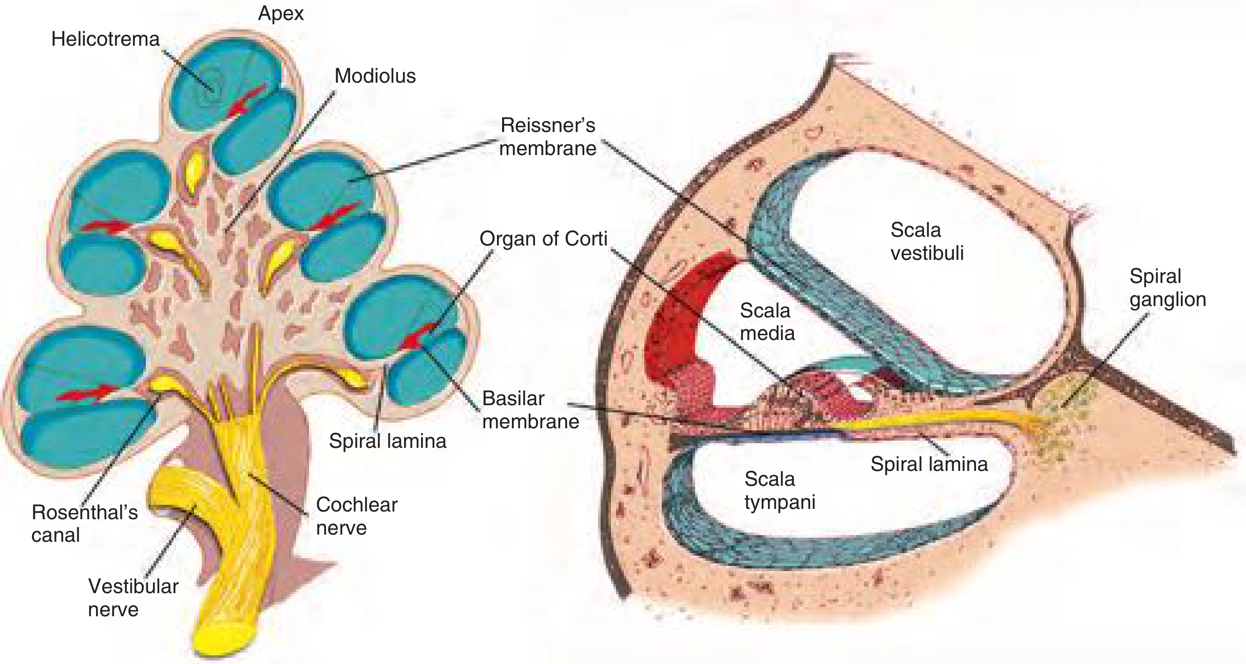
2. Membranous Labyrinth
| Component | Location within bony labyrinth | Function |
|---|---|---|
| Utricle | Elliptical recess of vestibule | Linear horizontal acceleration; head tilt |
| Saccule | Spherical recess of vestibule | Linear vertical acceleration |
| 3 Semicircular ducts | Within semicircular canals | Rotational acceleration |
| Cochlear duct (scala media) | Within cochlea | Hearing (organ of Corti) |
| Endolymphatic duct and sac | Vestibular aqueduct - posterior fossa | Resorption of endolymph |
| Ductus reuniens | Connects saccule to cochlear duct | Communication |
- The utriculosaccular duct joins the utricle and saccule and gives off the endolymphatic duct
- The endolymphatic sac lies extradurally on the posterior petrous surface - it resorbs endolymph
- The macula of the utricle and saccule contains otolith organs (calcium carbonate crystals on hair cells)
- The crista ampullaris in each ampulla detects rotational flow of endolymph
[DRAW]: Membranous labyrinth diagram showing utricle, saccule, 3 semicircular ducts with ampullae, cochlear duct, endolymphatic duct and sac, ductus reuniens, and utriculosaccular duct - with labeling of macula and crista positions
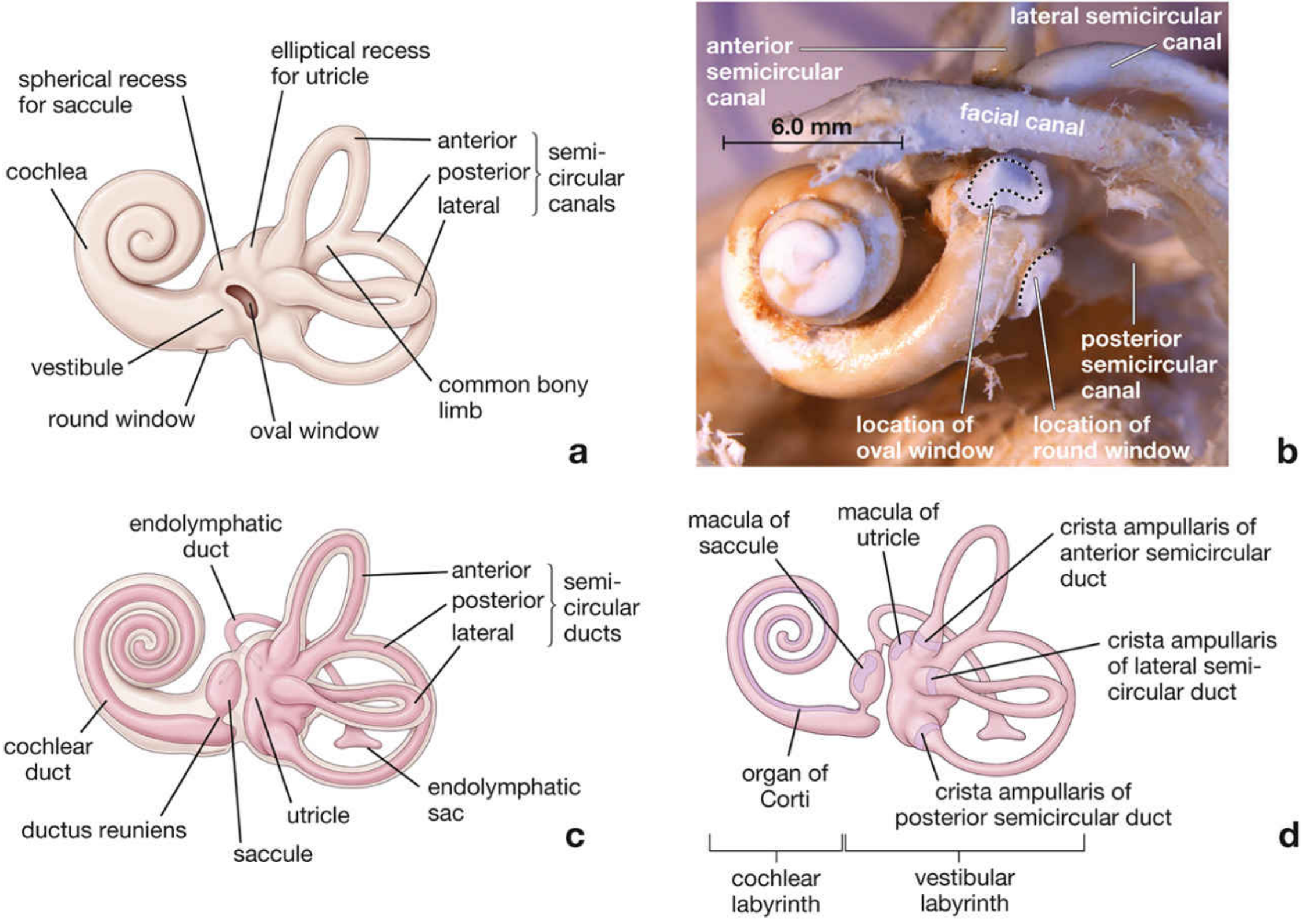
3. Blood Supply of the Labyrinth
- Labyrinthine (internal auditory) artery - a branch of the anterior inferior cerebellar artery (AICA), occasionally from the basilar artery
- Enters the internal acoustic meatus alongside CN VIII
- Divides into:
- Cochlear artery (supplies cochlea)
- Vestibular artery (supplies vestibular apparatus)
- The labyrinthine artery is an end artery - no collateral circulation
- Surgical importance: Any interruption during surgery (e.g., during lateral skull base surgery, translabyrinthine approaches) causes permanent sensorineural hearing loss
4. Nerve Supply
- Cochlear nerve: bipolar cells in spiral ganglion (Rosenthal's canal) - hearing
- Vestibular nerve: bipolar cells in Scarpa's ganglion (within IAC) - balance
PART B: SURGICAL IMPORTANCE OF THE LABYRINTH
1. Relationship to the Facial Nerve
- The facial nerve (CN VII) runs in a bony canal - the Fallopian canal - passing just above the oval window (tympanic segment) and looping behind the lateral semicircular canal (mastoid segment)
- Surgical significance: During mastoidectomy, the lateral semicircular canal is the most reliable landmark to identify the facial nerve. Drilling too close to the canal or oval window risks facial nerve injury and labyrinthine fistula
2. Posterior Tympanotomy (Facial Recess) Approach - for Cochlear Implants
- The triangular facial recess bounded by:
- Anteriorly: chorda tympani nerve
- Posteriorly: facial nerve
- Laterally: fossa incudis
- This approach provides safe access to the round window niche for cochlear implant electrode insertion without entering the external canal
3. Landmarks During Cochlear Implant Surgery
- The round window is the standard insertion point into the scala tympani
- The round window niche faces posteroinferiorly - it must be identified after drilling the facial recess
- The round window membrane is orientated at ~45 degrees and may be partially or fully covered by an overhanging round window niche bone - requiring careful drilling (cochleostomy may then be needed just anteroinferiorly to the round window)
4. Oval Window and Stapedectomy
- Stapes surgery accesses scala vestibuli via the oval window
- Damage to the membranous labyrinth during drilling or prosthesis insertion causes perilymph gusher or SNHL
5. Jugular Bulb
- A high-riding jugular bulb can approach or even dehisce into the hypotympanum
- It may obscure the round window, making cochlear implant surgery hazardous
- Preoperative CT scan is mandatory to assess jugular bulb position
6. Endolymphatic Sac Surgery
- Located on the posterior petrous surface, between the sigmoid sinus and the posterior semicircular canal
- During endolymphatic sac decompression/shunt (for Meniere's disease), careful dissection is needed to avoid entering the semicircular canals posteriorly or injuring the sigmoid sinus
7. The Otic Capsule - Resistance to Resorption
- The otic capsule does NOT undergo endochondral remodeling after birth
- This makes it uniquely resistant to osteoclastic resorption - important in otosclerosis (abnormal remodeling at the fissula ante fenestram)
- This property also means the cochlea is preserved even in advanced chronic ear disease
PART C: ANATOMICAL VARIATIONS RELEVANT TO COCHLEAR IMPLANT SURGERY
1. Round Window Anatomy Variations
- Normal: Round window faces posteriorly and is fully visible after posterior tympanotomy
- Overhanging niche: Bony overhang covers the round window membrane - requires drilling of the niche to visualize the membrane and insert the electrode atraumatically
- False membrane (fibrous tissue over the round window): Occurs in post-inflammatory ears; must be incised carefully
- Position and orientation: The round window membrane orientation varies; it may face more inferiorly or anteriorly
2. Cochlear Ossification (Labyrinthitis Ossificans)
- Post-meningitic or post-inflammatory ossification of the scala tympani is a major surgical challenge in cochlear implantation
- Ossification starts at the base and may partially or completely fill the scala tympani
- Grading (Jackler classification):
- Grade I: Partial (basal turn only)
- Grade II: Complete ossification
- Management: Drill-out of the basal turn; if complete, consider a split electrode or double array
- Preoperative MRI (T2-weighted) is key to assess the extent of ossification
3. Cochlear Malformations (Jackler Classification, 1987)
| Malformation | Embryological Stage | Imaging Features | Surgical Implication |
|---|---|---|---|
| Michel deformity | 3rd week failure | Complete absence of cochlea and vestibule | Cochlear implant contraindicated; ABI considered |
| Common cavity | 4th week | Cochlea and vestibule form a single oval cavity without internal architecture | CI possible; risk of perilymph gusher |
| Cochlear aplasia | 5th week | No cochlea; vestibule present | CI contraindicated |
| IP-I (Mondini-like / incomplete partition type I) | 6th week | Cyst-like cochlea without modiolus or spiral lamina | Risk of gusher; poor CI outcomes |
| IP-II (Classic Mondini) | 7th week | Only 1.5 turns; missing interscalar septum between middle and apical turns; dilated vestibular aqueduct | CI possible; good outcomes |
| IP-III (X-linked) | Various | Absent modiolus; dilated IAC; cochlea present but modiolus deficient | High risk of perilymph/CSF gusher |
| Cochlear hypoplasia | Various | Smaller cochlea (<1.5 turns) | CI possible with shorter electrode |
[DRAW]: Table or diagram of Jackler's classification of cochlear malformations with a sketch of each - common cavity, Mondini (IP-II), Michel, hypoplasia - alongside a normal cochlea for comparison
4. Wide/Large Vestibular Aqueduct (LVAS)
- Defined as diameter >1.5 mm at midpoint on axial CT
- Associated with IP-II (Mondini) and SLC26A4 (Pendrin) gene mutations
- At risk of "third window" effect; sudden SNHL with minor head trauma
- CI is possible; outcomes are generally good
- Intraoperatively: risk of copious endolymph leak; use fascia to seal the round window after insertion
5. Abnormal Facial Nerve Course
- The facial nerve may be dehiscent (no bony covering) over the oval window or may descend more anteriorly than usual
- Can overlie or obstruct the round window
- Preoperative CT is essential; drilling near a dehiscent nerve risks permanent paralysis
6. High Jugular Bulb
- A jugular bulb that extends superior to the floor of the hypotympanum may cover the round window
- May limit posterior tympanotomy or make cochleostomy dangerous due to risk of venous hemorrhage
- May require alternative surgical access
7. Cochlear Nerve Deficiency / Aplasia
- The cochlear nerve may be hypoplastic or absent (seen on MRI in IAC)
- Even if the cochlea is normal, absence of the cochlear nerve means CI will fail
- These patients are candidates for auditory brainstem implant (ABI)
8. Common Cavity with Absent Modiolus
- No modiolus = no spiral ganglion targets for electrode
- Electrode must be positioned against the wall to stimulate ganglion cells broadly
- Risk of high impedances and poor frequency discrimination
[DRAW]: Schematic of the right cochlea from the surgeon's view (looking through posterior tympanotomy), showing: facial nerve, chorda tympani, round window niche, round window membrane, with annotations of the facial recess approach and electrode insertion pathway
SUMMARY TABLE - Surgical Importance
| Anatomical Feature | Surgical Significance |
|---|---|
| Lateral SCC | Landmark for facial nerve in mastoidectomy |
| Round window | Standard entry for CI electrode |
| Oval window | Stapes surgery; perilymph entry |
| Modiolus | Target for CI; absent = poor outcome |
| Endolymphatic sac | Sac decompression in Meniere's disease |
| Labyrinthine artery | End artery; sacrifice = permanent SNHL |
| Vestibular aqueduct | Wide = gusher risk during CI |
| Jugular bulb | High = obscures RW; hemorrhage risk |
| Facial nerve | Dehiscent = injury risk |
QUESTION 2: Development of the Bony and Membranous Labyrinth
INTRODUCTION
PART A: PHYLOGENETIC BACKGROUND (Brief)
- The mammalian inner ear is evolutionarily derived from the lateral line system of fish - a water-motion detection system with hair cells, epidermal placode origin, and innervation by CN VII, IX, X
- As organisms moved to land, the lateral line was enclosed and filled with endolymph (replacing seawater), forming the first true labyrinth
- In ascending vertebrates:
- First: Utricle + 2 semicircular canals (superior and posterior) appear
- Then: Lateral semicircular canal + endolymphatic duct + saccule
- Finally: Lagena (outgrowth of saccule) evolves into the cochlea
- This phylogenetic sequence is recapitulated in human embryology - the pars superior (utricle + superior and posterior SCCs) develops before the pars inferior (saccule + cochlear duct) - explaining why pars superior is relatively more resistant to developmental malformations
PART B: DEVELOPMENT OF THE MEMBRANOUS LABYRINTH
Stage 1: Otic Placode Formation (End of Week 3 - Day 22)
- A plate-like thickening of surface ectoderm appears dorsal to the first branchial groove - the otic placode
- This is induced by signals from the underlying notochord and rhombencephalon (hindbrain, especially rhombomere 5/6)
- Key signals: FGF3, FGF10, Wnt, Pax2, Pax8 transcription factors
[DRAW]: Dorsal view of early embryo at ~22 days showing: surface ectoderm, otic placode (bilateral thickening), notochord, neural tube
Stage 2: Otic Pit Formation (Week 4 - Day 25-28)
- The otic placode invaginates into the underlying mesenchyme, forming the auditory (otic) pit
- The endolymphatic appendage (future endolymphatic duct) appears at this early stage - it is phylogenetically the oldest structure
- The pit deepens rapidly
Stage 3: Otocyst (Otic Vesicle) Formation (End of Week 4 - Day 28-30)
- The otic pit deepens further and pinches off from the surface ectoderm to form a closed epithelial sphere - the otocyst or otic vesicle
- This is entirely ectodermal in origin
- The surrounding mesenchyme begins to condense around the otocyst - this will become the otic capsule (bony labyrinth)
- The vestibulocochlear ganglion (CN VIII precursor neurons) begins to delaminate from the ventromedial wall of the otocyst
[DRAW]: Cross-section at week 4 showing: surface ectoderm, invaginating pit, closed otocyst, surrounding mesenchyme, early endolymphatic appendage on medial wall
Stage 4: Early Differentiation - Folds and Outgrowths (Weeks 5-6)
| Fold | Structure Formed |
|---|---|
| Fold I | Demarcates utricle with 2 early semicircular duct flanges |
| Fold II | Demarcates endolymphatic duct and sac |
| Fold III | Demarcates saccule; gives rise to utriculoendolymphatic valve (Valve of Bast) |
- The endolymphatic duct arises from fold II as a dorsomedial outgrowth
- Two flanges arise from the dorsal part of the otocyst - these are the primordia of the semicircular ducts
- By week 6: The semicircular duct lumina have formed; the macula communis (medial wall sensory patch) has divided into superior and inferior segments
Stage 5: Semicircular Ducts (Weeks 6-8)
- Semicircular ducts form by a plate mechanism: a flat epithelial outgrowth pushes into the mesenchyme and its central portion fuses and is then resorbed, leaving the peripheral rim as the tube
- Order of formation:
- Superior (anterior) semicircular duct - first
- Posterior duct - second
- Lateral duct - last (most phylogenetically recent; most susceptible to malformation)
- Ampullae form at one end of each duct
- The crista ampullaris differentiates within each ampulla from the superior macula segment
Stage 6: Utricle and Saccule (Weeks 5-8)
- The macula communis (from the medial wall of the otocyst) divides:
- Superior segment → macula utriculi, crista of superior SCD, crista of lateral SCD
- Inferior segment → macula sacculi, crista of posterior SCD
- Utricle: forms in posterosuperior otocyst
- Saccule: forms in anteroinferior otocyst
- The utriculosaccular duct connecting them gives off the endolymphatic duct (valve of Bast separates it from utricle)
Stage 7: Cochlear Duct (Weeks 6-8 onwards)
- An outgrowth from the ventral part of the saccule begins to form the cochlear duct around week 6
- This outgrowth coils around the modiolus of the developing bony cochlea:
- 1 turn by week 8
- 1.5 turns by week 10
- Full 2.5 turns achieved by week 25
- The connection between saccule and cochlear duct becomes the ductus reuniens (of Hensen)
- The organ of Corti begins to differentiate from the cochlear duct floor:
- Week 8-10: Specialized hair cells begin to differentiate from the flat epithelium
- Week 12-14: The tunnel of Corti begins to open
- Week 20-25: Organ of Corti approaches mature form; hair cells are present
- Myelination of cochlear nerve completes postnatally
[DRAW]: Sequential diagrams showing: (a) Otocyst with three folds, (b) Elongating vesicle with cochlear outgrowth beginning, (c) 1 turn coil, (d) 2.5 turns achieved - with pars superior and pars inferior labeled at each stage
Stage 8: Endolymphatic Duct and Sac
- Endolymphatic duct arises early (week 4) as the first appendage of the otocyst
- Grows dorsomedially to emerge at the posterior petrous surface
- Expands into the endolymphatic sac by week 8-10
- The sac lies in a groove (vestibular aqueduct) on the posterior petrous surface, extradurally
- Functions: resorption of endolymph; maintenance of endolymph volume and composition
- Abnormal endolymphatic sac/wide aqueduct → Meniere's disease, LVAS syndrome
PART C: DEVELOPMENT OF THE BONY LABYRINTH (OTIC CAPSULE)
Origin
- The otic capsule derives from mesenchyme (neural crest and paraxial mesoderm) surrounding the membranous labyrinth
- It passes through three stages:
- Mesenchymal stage (precartilage condensation, weeks 4-5)
- Cartilaginous stage (enchondral cartilage ossification, week 6 onwards)
- Ossification stage (enchondral ossification into bone, weeks 16-23)
Stage 1: Mesenchymal Condensation (Weeks 4-5)
- The mesenchyme surrounding the otocyst condenses into a precartilage mass that closely follows the shape of the developing membranous labyrinth
Stage 2: Chondrification (Weeks 6-7)
- The precartilage converts into a single piece of hyaline cartilage (the cartilaginous otic capsule) by week 7
- The cartilaginous capsule faithfully molds to the shape of the membranous labyrinth - this is why malformations of the membranous labyrinth are reflected in the shape of the bony capsule on CT scan
Stage 3: Development of Perilymphatic Spaces (Week 8 onwards)
- Critical concept: Perilymphatic spaces form by retrogressive dedifferentiation of the inner layer of the cartilaginous capsule
- By week 8: The innermost mesenchyme/precartilage around the ampullae and vestibule begins to dedifferentiate into a loose, vascular reticulum - the primordial perilymphatic labyrinth
- This reticulum becomes progressively vacuolated and the spaces coalesce
- Sequence of perilymphatic space formation:
- Perilymphatic cistern (of the vestibule, adjacent to the oval window) - appears at end of week 12 - this is the first recognizable space
- Scala tympani - appears soon after week 12, starting as rarefaction under the round window
- Scala vestibuli - appears later as a diverticulum of the perilymphatic cistern, growing toward the cochlear apex
- Helicotrema - the apical communication between scala vestibuli and tympani forms last
Stage 4: Ossification of the Otic Capsule (Weeks 16-23)
- The cartilaginous capsule ossifies by endochondral ossification - one of the most rapid and complete ossification processes in the body
- Ossification proceeds from 14 ossification centers (Anson and Donaldson) that appear between weeks 16-23 and rapidly coalesce
- The entire otic capsule is ossified by week 23 - making the labyrinth the first and only structure to reach adult size in utero (at 23 weeks)
- This early completion has surgical implications: any arrested development before week 23 will be reflected in a malformed (but fully ossified) bony labyrinth visible on CT
[DRAW]: Diagram of the cartilaginous otic capsule showing the 14 ossification centers, the three stages (mesenchyme → cartilage → bone), and the developing perilymphatic spaces alongside the membranous labyrinth
Unique Features of the Otic Capsule Bone
- After ossification, the otic capsule undergoes no further Haversian remodeling - it is permanently endochondral (enchondral) bone
- This makes it the hardest, densest bone in the body
- It does NOT respond to osteoclasts under normal circumstances
- Surgical implication: This bone must be drilled away mechanically - it does not soften or resorb. However, in otosclerosis, abnormal enchondral remodeling occurs at specific sites (mainly fissula ante fenestram, anterior to the oval window)
Special Structures in Bony Labyrinth Development
| Structure | Development |
|---|---|
| Fissula ante fenestram | A normal connective tissue track anterior to the oval window; site of otosclerotic foci |
| Fossula post fenestram | Posterior to oval window; smaller; also involved in otosclerosis |
| Cochlear aqueduct | Bony channel connecting scala tympani to subarachnoid space; transmits perilymph drainage |
| Vestibular aqueduct | Bony channel for endolymphatic duct; wide aqueduct = LVAS |
| Modiolus | Central bony pillar; develops with cartilaginous ossification; contains Rosenthal's canal with spiral ganglion |
PART D: TIMELINE SUMMARY
| Week | Event |
|---|---|
| 3 | Otic placode forms |
| 4 | Otic pit forms; otocyst closes; endolymphatic appendage appears |
| 5 | Mesenchyme condenses; three folds appear; saccule and utricle begin to differentiate |
| 6 | Semicircular duct lumina form; cochlear outgrowth begins; macula communis divides; cartilage starts |
| 7-8 | Perilymphatic reticulum begins; semicircular ducts complete; cochlea has 1 turn |
| 10 | Cochlea has 1.5 turns |
| 12 | Perilymphatic cistern of vestibule forms |
| 14 | Scala tympani appears |
| 16 | Ossification centers begin |
| 23 | Otic capsule reaches adult size and is fully ossified - labyrinth is adult-sized |
| 25 | Cochlear duct completes 2.5 turns; organ of Corti approaches maturity |
[DRAW]: A vertical timeline diagram (3 weeks to 25 weeks) showing each developmental milestone on a single axis, with membranous events on the left and bony/cartilaginous events on the right
PART E: CLINICAL CORRELATIONS (Malformations)
| Arrested Week | Resulting Malformation | Clinical Feature |
|---|---|---|
| Week 3 | Michel deformity (no labyrinth) | Complete SNHL; CI contraindicated |
| Week 4 | Common cavity | SNHL; CSF gusher risk with CI |
| Week 5 | Cochlear aplasia | No cochlea; vestibule present |
| Week 6 | IP-I (absent modiolus + septa) | SNHL; gusher risk |
| Week 7 | Mondini (IP-II) - 1.5 turns | Progressive SNHL; LVAS; CI possible |
| Week 8 | Cochlear hypoplasia | Small cochlea; shorter electrode needed |
| Wide vestibular aqueduct | LVAS + Pendred syndrome | Progressive SNHL; Meniere-like episodes |
KEY POINTS FOR EXAM (20 Marks)
- Otic placode (ectodermal) → otic pit → otocyst (week 3-4) = origin of membranous labyrinth
- Membranous labyrinth = entirely ectodermal; bony labyrinth = from surrounding mesenchyme
- Pars superior (utricle + SCCs) develops before pars inferior (saccule + cochlear duct) - phylogenetically older
- Endolymphatic duct appears earliest (week 4) - before SCCs or cochlea
- Cochlear duct coils to full 2.5 turns by week 25; adult size achieved at week 23
- Otic capsule ossifies from 14 centers (weeks 16-23); no further remodeling - hardest bone in body
- Perilymphatic spaces form by retrogressive dedifferentiation from week 8; perilymphatic cistern is first (week 12)
- Valve of Bast = utriculoendolymphatic valve separating utricle from endolymphatic sac
- Arrested development at specific weeks produces predictable cochlear malformations (Jackler classification)
- Round window variations, wide vestibular aqueduct, cochlear ossification, and aberrant facial nerve are key anatomical variations for cochlear implant planning