
Course of vagus nerve with branches
Reading File
The vagus nerve (CN X) is the longest cranial nerve, originating in the medulla and traveling through the neck, thorax, and abdomen. It carries parasympathetic, sensory, and motor fibers. Let me get detailed textbook content on its full course and branches.
Searching sources...
Finding Sources
Finding Sources
Good results. Let me now read the most comprehensive anatomy sources - Gray's Anatomy for Students and Scott-Brown's, plus the Fischer's Surgery block for surgical anatomy.
Reading File
Reading File
Reading File
Excellent - now let me read the Scott-Brown content on the recurrent laryngeal nerve and the Gray's anatomy section on origin/cranial course, plus the Fischer's surgical anatomy block.
Reading File
Reading File
Finding Sources
Let me now get the Gray's Anatomy cranial origin section and the thoracic/abdominal course content.
Reading File
Reading File
Reading File
I now have comprehensive information. Let me also fetch the key diagram images to enrich the answer.
Loading Image
Loading Image
Excellent - I have all the detailed content needed. Here is the full, well-organized answer on the vagus nerve:
Vagus Nerve (CN X) - Course and Branches
The vagus nerve takes its name from the Latin vagus ("wandering") due to its extensive course from the brainstem to the abdomen. It is the longest cranial nerve and the nerve of the 4th branchial arch.
Fiber Components
The vagus carries five types of fibers:
- GSA (General Somatic Afferent): sensation from larynx, laryngopharynx, auricle, external acoustic meatus, posterior cranial fossa dura
- GVA (General Visceral Afferent): from aortic chemoreceptors/baroreceptors, esophagus, bronchi, lungs, heart, foregut and midgut viscera (reflex sensations, not pain)
- SA (Special Afferent): taste near epiglottis and pharynx
- GVE (General Visceral Efferent): parasympathetic to smooth muscle/glands of pharynx, larynx, and thoraco-abdominal viscera (foregut and midgut)
- BE (Branchial/Special Visceral Efferent): motor to palatoglossus, soft palate muscles (except tensor veli palatini), pharyngeal constrictors (except stylopharyngeus), and all laryngeal muscles
Origin
- Arises as 6-8 rootlets from the posterolateral (anterolateral) surface of the medulla oblongata, just inferior to the glossopharyngeal nerve (CN IX) rootlets, dorsal to the inferior olive
- The cranial root of CN XI joins the vagus at the jugular foramen and contributes motor fibers to the pharyngeal and laryngeal muscles innervated by the vagus
Two Sensory Ganglia
At/near the jugular foramen lie two enlargements:
- Superior (jugular) ganglion - located in the jugular foramen; contains cell bodies for somatic afferent fibers (meningeal and auricular branches)
- Inferior (nodose) ganglion - just distal to the foramen; contains cell bodies for visceral afferent fibers of all other branches
Course and Branches by Region
1. Cranial/Intracranial Portion
The rootlets cross the posterior cranial fossa and converge into the vagus nerve as they enter the jugular foramen. CN IX, CN X, and CN XI all pass through the middle compartment of the jugular foramen together.
Branches given off here:
- Meningeal branch: re-enters the cranium to supply dura mater of the posterior cranial fossa
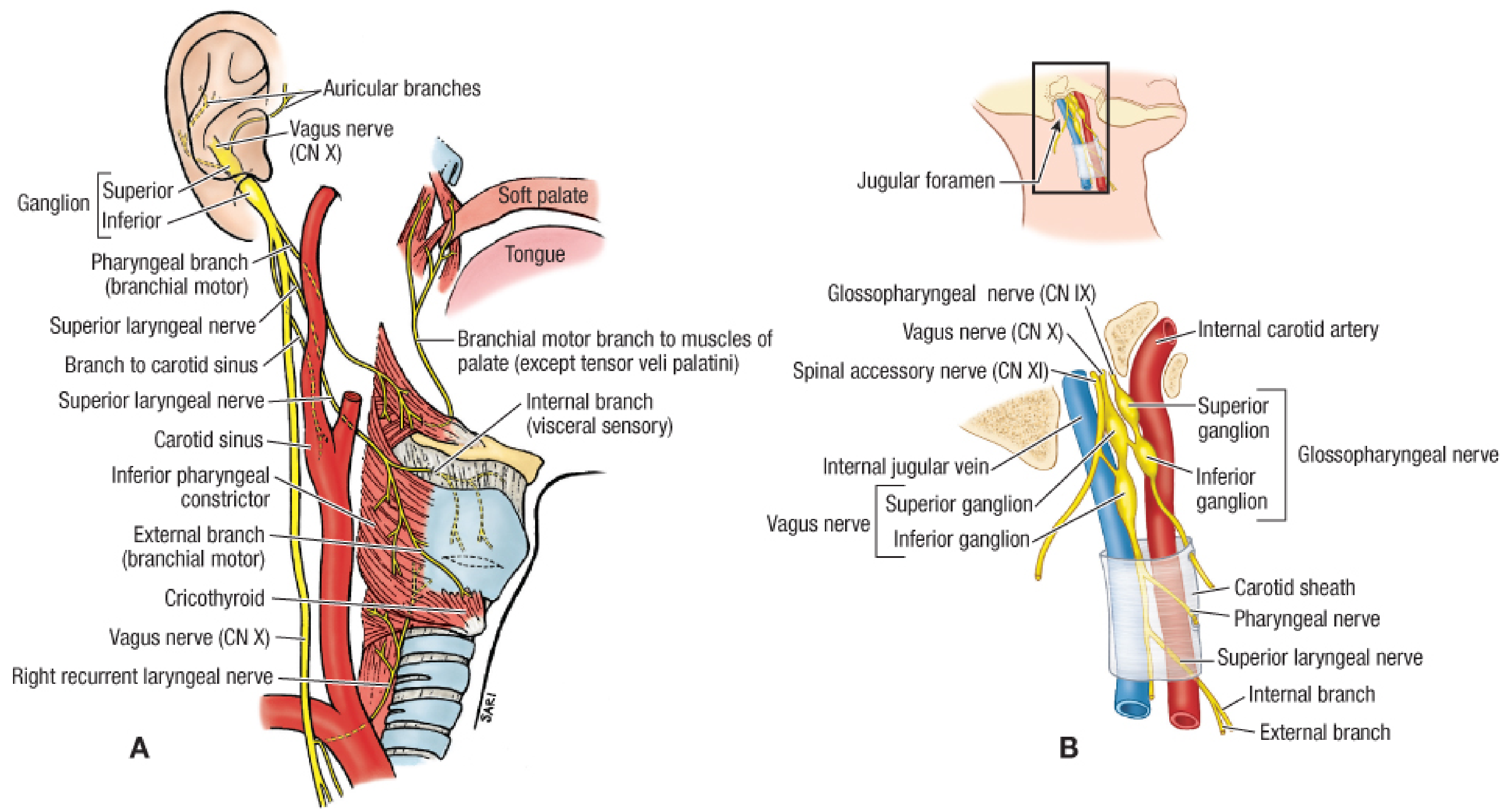
- Auricular branch (Arnold's nerve): passes between the mastoid process and tympanic plate; supplies the tympanic membrane, skin of the posterior pinna, and the posterior external acoustic meatus. Clinically responsible for referred otalgia and the ear-cough reflex (coughing on ear syringing).
2. Cervical Course
After exiting the skull, the vagus descends in the carotid sheath, initially medial to the internal jugular vein (IJV) and internal carotid artery (ICA), then - as it descends - lying posteriorly between the IJV (lateral) and the common carotid artery (medial).
Branches in the neck:
| Branch | Origin | Course & Supply |
|---|---|---|
| Auricular branch | Superior ganglion | Arnold's nerve - see above |
| Pharyngeal branch | Inferior ganglion | Passes between internal and external carotid arteries to join the pharyngeal plexus (with CN IX + sympathetics); supplies all pharyngeal constrictors and soft palate muscles except tensor veli palatini and stylopharyngeus |
| Carotid body branch | Supplements CN IX branches to the carotid body chemoreceptors | |
| Superior laryngeal nerve | Inferior ganglion | Descends medial to ICA and ECA; at the level of the hyoid divides into: |
| - Internal laryngeal nerve | Pierces thyrohyoid membrane with superior laryngeal artery; sensory to laryngeal mucosa above the vocal folds, piriform fossa, cervical esophagus and trachea | |
| - External laryngeal nerve | Runs with the superior thyroid artery on the inferior pharyngeal constrictor; motor to cricothyroid (the only intrinsic laryngeal muscle NOT supplied by the recurrent laryngeal nerve) | |
| Cervical cardiac branches (x2) | Superior branch: arises just below superior laryngeal nerve, passes posteromedial to carotid sheath; Inferior branch: arises at root of neck just above 1st rib; both right cardiac branches + superior left cardiac branch join the deep cardiac plexus; left inferior cardiac branch joins the superficial cardiac plexus | |
| Right recurrent laryngeal nerve | Right vagus as it crosses anterior to right subclavian artery | Hooks posterior to the subclavian artery and ascends in the tracheo-esophageal groove; gives tracheal, esophageal, and pharyngeal branches; terminates as inferior laryngeal nerve - motor to all intrinsic laryngeal muscles except cricothyroid, sensory to subglottic mucosa |
Note on the right RLN: In ~1% of individuals it takes a non-recurrent course (associated with an aberrant right subclavian artery/arteria lusoria).
3. Thoracic Course
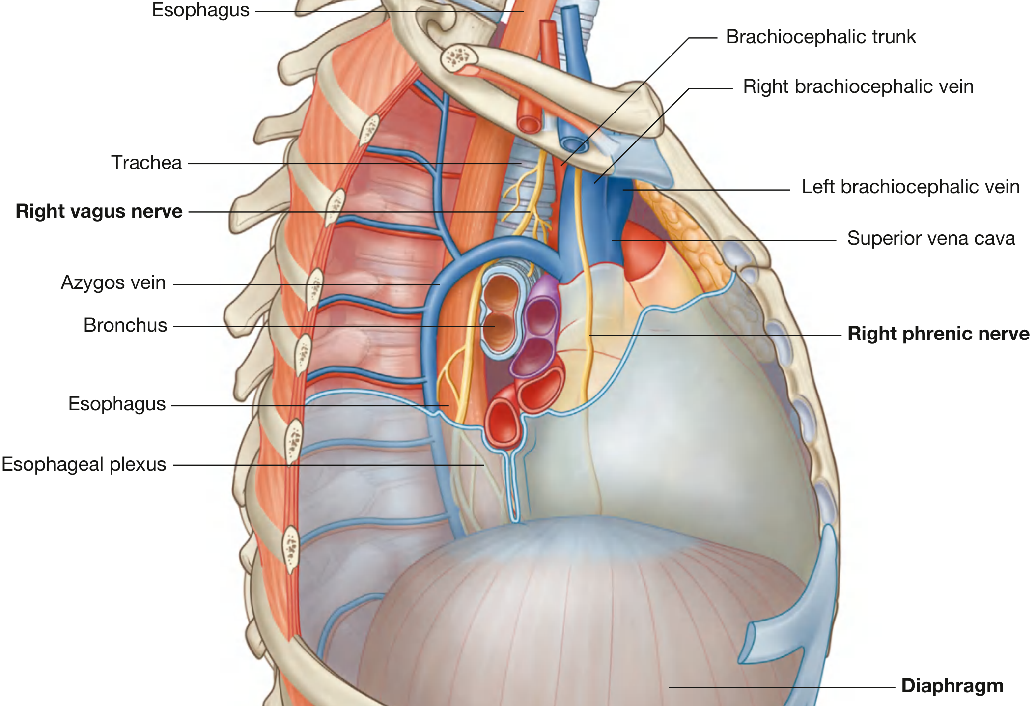
Right vagus nerve:
- Enters the superior mediastinum lying between the right brachiocephalic vein and the brachiocephalic trunk
- Descends toward the trachea, crosses its lateral surface
- Is crossed by the arch of the azygos vein before reaching the esophagus
- Passes posterior to the root of the right lung
- Descends on the posterior surface of the esophagus, contributing to the esophageal plexus - forming the posterior vagal trunk
Left vagus nerve:
- Enters the superior mediastinum posterior to the left brachiocephalic vein, between the left common carotid and left subclavian arteries
- Lies deep to the mediastinal parietal pleura
- Crosses the left side of the arch of the aorta
- At the inferior margin of the arch, just lateral to the ligamentum arteriosum, gives off the left recurrent laryngeal nerve
- The left RLN loops under the arch of the aorta, ascends in the tracheo-esophageal groove, and enters the neck to terminate in the larynx
- The left vagus then passes posterior to the root of the left lung and reaches the esophagus, contributing to the anterior vagal trunk
Thoracic branches:
- Thoracic cardiac branches: from both vagi, join the deep/superficial cardiac plexuses
- Pulmonary branches: form the pulmonary plexuses at the roots of the lungs; supply the bronchi and lungs
- Esophageal branches: form the esophageal plexus around the lower esophagus (left vagus = anterior surface; right vagus = posterior surface)
The plexus around the esophagus reconstitutes as:
- Anterior vagal trunk (mainly left vagus fibers)
- Posterior vagal trunk (mainly right vagus fibers)
Both trunks enter the abdomen through the esophageal hiatus of the diaphragm at the level of T10.
4. Abdominal Course and Branches
Anterior vagal trunk (mainly left vagus):
- Runs on the anterior surface of the esophagus/cardia
- Gives off the hepatic branch(es) - travel in the hepatogastric ligament to the porta hepatis; descending branches supply the pylorus, pyloric sphincter, pancreas, and 1st part of duodenum
- Divides into gastric, pyloric, and celiac branches
- Anterior gastric branches: supply cardia, fundus, and anterior stomach wall
- Principal anterior nerve of the lesser curvature (Latarjet's nerve): the longest gastric branch running along the lesser curvature with the left gastric artery to the pylorus, terminating as "crow's foot" branches (3-4 terminal branches)
- Pyloric branches (usually two): one travels in the lesser omentum, the other from the nerve of Latarjet to the pyloric antrum
Posterior vagal trunk (mainly right vagus):
- Runs on the posterior surface of the esophagus
- Posterior gastric branches: supply the posterior fundus, body, and pyloric antrum (fewer and terminates more proximally than the anterior nerve)
- Celiac branches: the main branch of the posterior trunk; passes to the celiac plexus and through it supplies the small intestine up to the left colic flexure (splenic flexure), plus pancreas, kidneys, and adrenals via the renal plexus
- Right vagus also gives a branch directly to the celiac plexus continuing posteriorly along the lesser curvature
Summary Table
| Region | Key Branches |
|---|---|
| Cranial (intracranial) | Meningeal branch, Auricular branch (Arnold's nerve) |
| Cervical | Pharyngeal branch (pharyngeal plexus), Carotid body branch, Superior laryngeal nerve (internal + external), Cervical cardiac branches (x2), Right recurrent laryngeal nerve |
| Thoracic | Left recurrent laryngeal nerve, Thoracic cardiac branches, Pulmonary branches, Esophageal plexus (forms anterior + posterior vagal trunks) |
| Abdominal | Esophageal branches, Gastric branches, Hepatic branches, Pyloric branches, Celiac branches, Renal branches, Intestinal branches (to left colic flexure) |
Clinically Important Points
- Left RLN loops around the arch of the aorta at the ligamentum arteriosum - vulnerable in mediastinal masses, aortic aneurysm, bronchial/esophageal carcinoma
- Right RLN loops around the right subclavian artery - vulnerable in thyroid/parathyroid surgery; non-recurrent course in ~1% (associated with aberrant subclavian)
- External laryngeal nerve runs with the superior thyroid artery - at risk during thyroid ligation, causing loss of cricothyroid function (voice pitch changes)
- Arnold's nerve (auricular branch) stimulation explains: referred otalgia from laryngeal/esophageal pathology, and cough reflex triggered by ear syringing (Arnold's ear-cough reflex)
- Beahr's triangle (landmark for RLN): bounded by common carotid artery laterally, inferior thyroid vessels superiorly, and RLN medially
- Vagotomy (truncal/selective/highly selective) targets different levels of vagal supply to reduce gastric acid secretion
- The vagus provides parasympathetic supply to gut up to the left (splenic) colic flexure - beyond this, the pelvic splanchnic nerves (S2-S4) take over
Sources: Gray's Anatomy for Students, Fischer's Mastery of Surgery 8th ed., Scott-Brown's Otorhinolaryngology Head & Neck Surgery
This is a shared conversation. Sign in to Orris to start your own chat.