
Details about allergy, pathogenesis,cause treatment and management
"allergy" AND "pathogenesis" AND "treatment"
allergy hypersensitivity IgE mast cell degranulation pathogenesis diagram
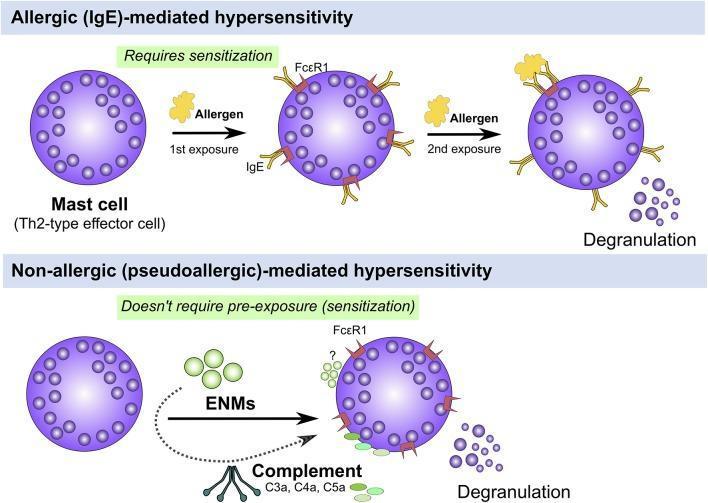
A comparative pathophysiology diagram illustrating two mechanisms of mast cell activation and degranulation in hypersensitivity. The top panel depicts 'Allergic (IgE)-mediated hypersensitivity,' showing a multi-step process: initial sensitization where an allergen exposure leads to IgE antibodies binding to Fć̵R1 receptors on a Th2-type effector mast cell, followed by a second exposure where allergen cross-linking of the IgE-Fć̵R1 complex triggers degranulation. The bottom panel depicts 'Non-allergic (pseudoallergic)-mediated hypersensitivity,' highlighting that this pathway does not require prior sensitization. Instead, activation is triggered by direct interaction with Engineered Nanomaterials (ENMs) or anaphylatoxins from the complement system (C3a, C4a, C5a), leading directly to the release of intracellular granules. Visually, mast cells are shown as spherical purple cells with dense internal granules that are expelled into the extracellular space upon activation. The diagram serves as an educational tool to distinguish classic Type I hypersensitivity from direct mast cell triggers relevant to toxicology and immunology.
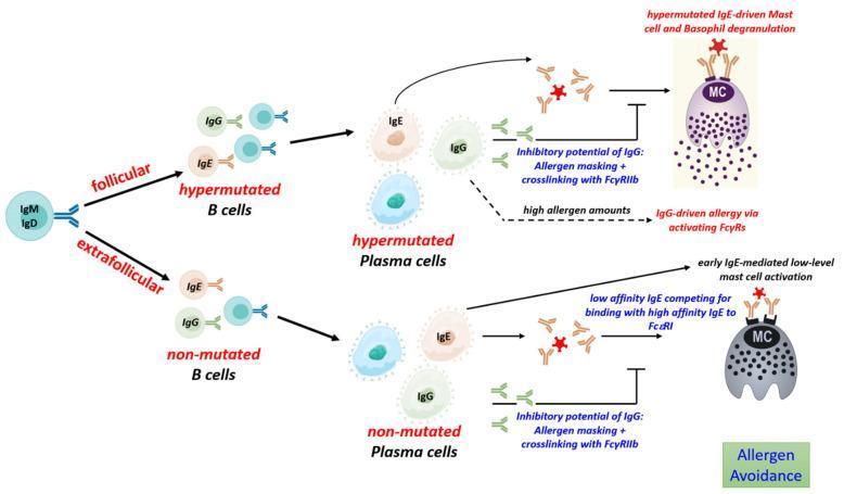
A detailed immunology flow diagram illustrating the B cell response to allergens via follicular and extrafollicular pathways. The process begins with naive IgM/IgD B cells. The 'follicular' pathway leads to somatic hypermutation, producing hypermutated B cells and subsequent hypermutated plasma cells. These secrete high-affinity IgE and IgG. High-affinity IgE binds to mast cells (MC) leading to robust degranulation. The 'extrafollicular' pathway results in non-mutated B cells and plasma cells secreting low-affinity IgE and IgG. This results in competition for FcεRI receptors and low-level mast cell activation. The diagram highlights the regulatory role of IgG, showing its inhibitory potential through allergen masking and crosslinking with FcγRIIb to suppress mast cell activation. Additionally, it notes that high allergen amounts can lead to IgG-driven allergy via activating FcγRs. Key molecular targets include FcεRI and FcγRIIb, while clinical outcomes focus on mast cell and basophil degranulation versus allergen avoidance.
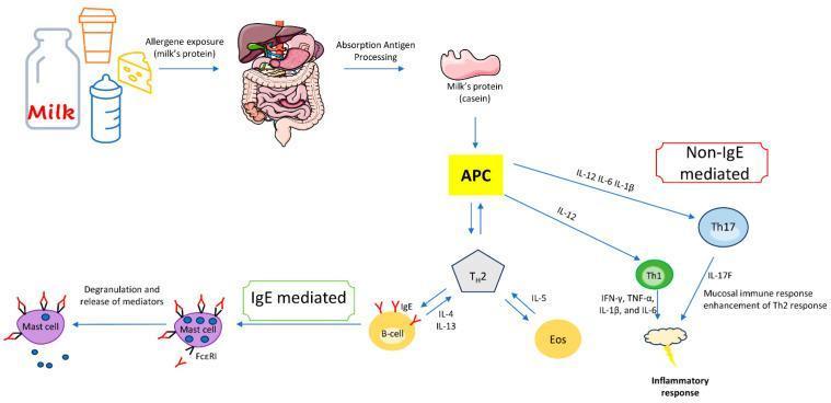
A pathophysiology diagram illustrating the immunological pathways of IgE-mediated and non-IgE-mediated cow's milk protein allergy (CMPA). The process begins with allergen exposure (milk proteins like casein) and subsequent absorption/antigen processing in the gastrointestinal tract. Central to both pathways is the Antigen-Presenting Cell (APC). In the IgE-mediated pathway, the APC activates Th2 cells, which release IL-4 and IL-13 to stimulate B-cells to produce IgE antibodies. These antibodies bind to Fć̑RI receptors on mast cells, leading to degranulation and mediator release. Additionally, Th2 cells release IL-5 to activate eosinophils. In the non-IgE-mediated pathway, the APC releases cytokines (IL-12, IL-6, IL-1̣̂) to activate Th17 and Th1 cells. Th17 cells release IL-17F, contributing to mucosal immune responses, while Th1 cells produce pro-inflammatory cytokines including IFN-̱́, TNF-̱, IL-1̣̂, and IL-6, collectively resulting in a systemic inflammatory response. This diagram provides a comparative overview of Type I hypersensitivity versus cell-mediated allergic responses in pediatric and adult nutrition immunology.
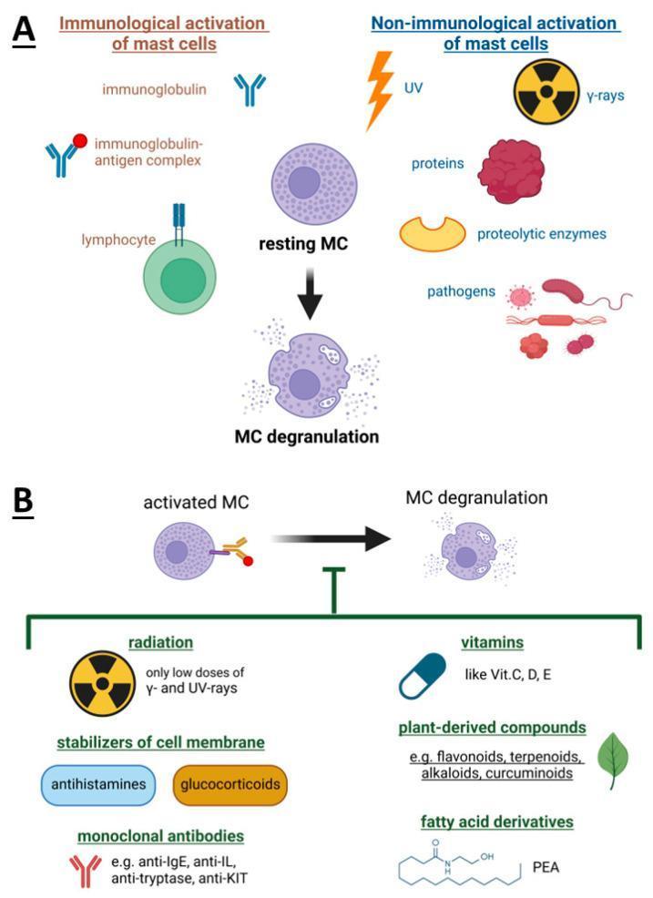
Educational medical diagram illustrating the activation and inhibition of mast cell (MC) degranulation. Section A depicts activation pathways. Immunological activation involves immunoglobulins (IgE), immunoglobulin-antigen complexes, and lymphocyte interactions. Non-immunological triggers include UV/gamma radiation, proteins, proteolytic enzymes, and pathogens. The central visual shows a transition from a 'resting MC' to 'MC degranulation,' characterized by the release of intracellular granules. Section B illustrates inhibitors of the degranulation process. These include pharmacological agents like membrane stabilizers (antihistamines, glucocorticoids) and monoclonal antibodies (anti-IgE, anti-IL, anti-tryptase, anti-KIT). Other listed inhibitory factors include low-dose radiation (UV and gamma), vitamins (C, D, E), plant-derived compounds (flavonoids, curcuminoids), and fatty acid derivatives like Palmitoylethanolamide (PEA). The diagram is designed for cellular immunology education, focusing on therapeutic targets for hypersensitivity reactions and anaphylaxis management.
Allergy: Pathogenesis, Causes, Treatment & Management
1. Definition
2. Types of Hypersensitivity (Gell-Coombs Classification)
| Type | Mechanism | Mediators | Examples |
|---|---|---|---|
| I - Immediate (Allergy) | IgE + Mast cells | Histamine, leukotrienes, prostaglandins | Anaphylaxis, asthma, hay fever, urticaria |
| II - Cytotoxic | IgG/IgM + Complement | Complement, FcR | Hemolytic anemia, Goodpasture's |
| III - Immune Complex | Immune complex deposition | Complement | SLE, serum sickness, post-streptococcal GN |
| IV - Delayed (Cell-mediated) | CD4+ T cells (Th1/Th17), CD8+ CTLs | IFN-γ, IL-17 | Contact dermatitis, TB, graft rejection |
- Robbins & Kumar Basic Pathology, p. 2728-2754
3. Causes and Allergens
- Inhalants: pollens (grass, tree, weed), house dust mites, animal dander, mold spores
- Foods: peanuts, tree nuts, milk, eggs, wheat, shellfish, soy
- Insect venoms: bee, wasp, ant stings
- Drugs: penicillin, aspirin, NSAIDs, contrast media
- Latex and chemicals
- Genetic predisposition (atopy) - strongest single risk factor; multiple susceptibility genes disrupt tolerance pathways
- Environmental factors: early-life microbial exposure, pollution, dietary patterns, urbanization
- Hygiene hypothesis: reduced early microbial exposures skew immunity toward Th2 responses
4. Pathogenesis
Phase 1: Sensitization
- Allergen presentation: Dendritic cells process the allergen and present it to naive CD4+ T helper cells
- Th2 differentiation: In genetically susceptible individuals, T cells differentiate into Th2 cells driven by IL-4. Th2 cells and group 2 innate lymphoid cells (ILC2s) produce IL-4, IL-5, and IL-13 (type 2 cytokines)
- IgE class switching: IL-4 and IL-13 drive B cells to undergo class-switch recombination, producing IgE antibodies specific for the allergen
- Mast cell sensitization: IgE binds with high affinity to FcεRI receptors on mast cells and basophils throughout connective tissues, especially under epithelial barriers. This "loads" mast cells, awaiting re-exposure
- Cellular and Molecular Immunology, p. 1301-1303
Phase 2: Activation (Re-exposure)
- Cross-linking: Multivalent allergen cross-links two or more IgE molecules on the mast cell surface, clustering FcεRI receptors
- Signal transduction: LYN tyrosine kinase phosphorylates ITAMs in the FcεRI β and γ chains → recruits SYK kinase → activates PLCγ → IP3 and DAG → calcium mobilization
- Mast cell degranulation and mediator release (three types):
- Preformed granule contents (released within seconds-minutes): Histamine, tryptase, chymase, heparin, TNF
- Newly synthesized lipid mediators (minutes): Prostaglandin D2, leukotrienes C4, D4, E4 (LTC4/D4/E4 = cysteinyl leukotrienes), PAF
- Cytokines (hours): IL-4, IL-5, IL-13, TNF, GM-CSF
- Cellular and Molecular Immunology, p. 3598-3602
Phase 3: The Immediate Reaction (Minutes)
- Histamine causes vasodilation, increased vascular permeability, smooth muscle contraction, mucus secretion, pruritus
- Leukotrienes (LTC4, LTD4, LTE4) cause sustained bronchospasm (1000x more potent than histamine in airways), mucus secretion
- Prostaglandin D2 causes bronchoconstriction and vasodilation
- Classic skin manifestation: The wheal-and-flare reaction - redness (flare) from vasodilation, soft swelling (wheal) from increased permeability
Phase 4: The Late-Phase Reaction (Hours)
- Cytokines (TNF, IL-4, IL-5) upregulate endothelial adhesion molecules (E-selectin, VCAM-1)
- Eosinophils are recruited via eotaxin (CCL11)/CCR3, IL-5, and complement C5a
- Neutrophils also accumulate
- Eosinophils release major basic protein (MBP), eosinophil cationic protein (ECP), and lipid mediators, causing tissue damage
- Late-phase reactions account for chronic allergic symptoms (nasal congestion, persistent wheezing)
- Charcot-Leyden crystals (galectin-10 aggregates from dying eosinophils) found in chronic type 2 inflammation sites (e.g., bronchi in asthma)
5. Clinical Manifestations
| Organ System | Manifestation |
|---|---|
| Respiratory | Allergic rhinitis (hay fever), bronchial asthma, sinusitis |
| Eyes | Allergic conjunctivitis |
| Skin | Urticaria (hives), angioedema, atopic dermatitis (eczema) |
| GI tract | Food allergy, abdominal pain, vomiting, diarrhea |
| Systemic | Anaphylaxis - cardiovascular collapse (tachycardia, hypotension), severe bronchospasm, urticaria, potentially fatal |
Anaphylaxis is the most severe form: mast cell mediators restrict airways to near asphyxiation and cause cardiovascular collapse. It requires immediate intervention. - Cellular and Molecular Immunology, p. 1302-1303
6. Diagnosis
| Test | Purpose |
|---|---|
| Serum Total IgE | Elevated in atopic individuals |
| Allergen-specific IgE (RAST/ImmunoCAP) | Identifies specific allergen sensitivity (foods, pollens, animals, insects, molds) |
| Skin prick test (SPT) | Gold standard for identifying IgE-mediated sensitization - wheal-and-flare response |
| Intradermal test | More sensitive than SPT; for drug and venom allergy |
| Basophil activation test | Flow cytometry-based; useful when skin testing is not possible |
| Oral food challenge | Definitive diagnosis of food allergy |
| CBC with differential | Eosinophilia suggests atopy/allergy |
- Henry's Clinical Diagnosis and Management by Laboratory Methods, p. 2760-2762
7. Treatment & Management
A. Avoidance (Primary Prevention)
- Identify and eliminate the allergen (most effective when feasible)
- Dietary elimination for food allergies; HEPA filters for dust mites/mold; hypoallergenic products for latex
B. Pharmacological Treatment
1. Antihistamines (H1 Blockers)
- Mechanism: Competitively block H1 receptors, preventing histamine effects
- First-generation (sedating): Diphenhydramine, chlorphenamine - used for acute reactions, pruritus
- Second-generation (non-sedating): Cetirizine, loratadine, fexofenadine, levocetirizine - preferred for chronic management (allergic rhinitis, urticaria)
- Role: Most effective against histamine-driven symptoms; less effective against leukotriene-driven bronchospasm
2. Corticosteroids
- Mechanism: Broad anti-inflammatory; reduce cytokine production, inhibit late-phase reaction, reduce eosinophil recruitment
- Intranasal (fluticasone, budesonide, mometasone): First-line for allergic rhinitis
- Inhaled (fluticasone, budesonide): First-line for allergic asthma
- Topical (hydrocortisone, triamcinolone): Atopic dermatitis
- Systemic (prednisolone, methylprednisolone): Severe/refractory allergic reactions, status asthmaticus
3. Epinephrine (Adrenaline) - First-line for Anaphylaxis
-
Route: Intramuscular (anterolateral thigh), 0.3-0.5 mg (1:1000)
-
Mechanism: α1-agonist (vasoconstriction, reduces angioedema); β2-agonist (bronchodilation); β1-agonist (increases cardiac output)
-
Early treatment with epinephrine is associated with lower hospitalization risk
-
IV epinephrine carries higher cardiovascular complication risk vs IM route
-
Patients at risk should carry an epinephrine auto-injector (EpiPen)
-
Goldman-Cecil Medicine, Treatment and Prevention, p. 503-505
4. Leukotriene Receptor Antagonists (LTRAs)
- Montelukast, zafirlukast: Block CysLT1 receptors for leukotrienes LTC4/D4/E4
- Used adjunctively for allergic asthma and rhinitis
5. Mast Cell Stabilizers
- Sodium cromoglicate, nedocromil: Prevent mast cell degranulation
- Used prophylactically for allergic asthma and conjunctivitis
6. Bronchodilators (for Asthma)
- Short-acting β2-agonists (SABAs): Salbutamol/albuterol - rescue therapy
- Long-acting β2-agonists (LABAs): Salmeterol, formoterol - used with ICS
7. Biologics (Targeted Therapy)
- Omalizumab (anti-IgE): Binds free IgE, prevents mast cell sensitization; for moderate-severe allergic asthma, chronic urticaria
- Dupilumab (anti-IL-4Rα): Blocks IL-4 and IL-13 signaling; approved for atopic dermatitis, asthma, chronic rhinosinusitis with nasal polyps
- Mepolizumab, benralizumab (anti-IL-5/IL-5Rα): Target eosinophilic asthma
- Tezepelumab (anti-TSLP): Blocks upstream innate alarmin, broadest asthma biologic
C. Allergen Immunotherapy (Desensitization)
| Route | Details |
|---|---|
| Subcutaneous Immunotherapy (SCIT) | Injections with increasing doses over 3-5 years; most evidence base |
| Sublingual Immunotherapy (SLIT) | Drops or tablets under the tongue; similar efficacy to SCIT, safer profile, can be home-administered |
- Reduces symptom scores by 32% and medication needs by 41% (grass pollen SCIT)
- Long-lasting benefit even after treatment stops
- Reduces risk of developing asthma in children with rhinitis
- May prevent sensitization to new allergens
- Shifts Th2 → Th1/Treg balance
- Induces IL-10 and TGF-β producing regulatory T cells
- Increases blocking IgG4 antibodies
- Reduces mast cell and basophil reactivity
D. Anaphylaxis Emergency Management
| Step | Action |
|---|---|
| 1 | Remove/stop the allergen trigger |
| 2 | Epinephrine IM (anterolateral thigh), 0.3-0.5 mg - do not delay |
| 3 | Call emergency services; lay patient flat (Trendelenburg) |
| 4 | 100% oxygen via face mask |
| 5 | IV fluid resuscitation (aggressive) for hypotension |
| 6 | Salbutamol/albuterol nebulized for bronchospasm |
| 7 | H1 antihistamine (IV/IM diphenhydramine) - adjunctive |
| 8 | IV corticosteroid (hydrocortisone/methylprednisolone) - reduces late-phase |
| 9 | Monitor for biphasic reaction (4-8 hours) |
- Campbell Walsh Wein Urology, Anaphylaxis section
E. Management of Specific Conditions
| Condition | First-line Management |
|---|---|
| Allergic rhinitis | Intranasal corticosteroids ± 2nd-gen antihistamines; SLIT/SCIT |
| Allergic asthma | ICS ± LABA; SABA rescue; omalizumab/biologics for severe disease |
| Atopic dermatitis | Topical corticosteroids; moisturizers; dupilumab for moderate-severe |
| Food allergy | Strict avoidance; epinephrine auto-injector; oral immunotherapy (emerging) |
| Urticaria | 2nd-gen antihistamines; omalizumab for chronic spontaneous urticaria |
| Drug allergy | Stop drug; desensitization protocols if re-exposure needed; HLA genotyping (e.g., HLA-B57:01 before abacavir) |
8. Summary Diagram
- Allergy = Type I hypersensitivity = IgE + mast cell degranulation
- Two phases: immediate (minutes, histamine-driven) and late-phase (hours, eosinophil-driven)
- Epinephrine IM is the only life-saving treatment for anaphylaxis - use it first, not antihistamines
- Immunotherapy is the only disease-modifying treatment; biologics (especially dupilumab, omalizumab) represent a major advancement for refractory disease
- Drug allergy desensitization is safe and effective when re-exposure to the causative drug is necessary