
How to study the fetal sex determination in USG
fetal sex determination ultrasound male female genitalia
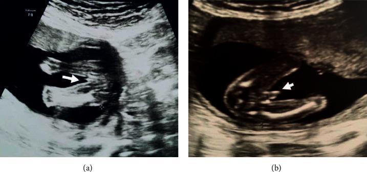
This diagnostic imaging set consists of two grayscale fetal ultrasound images in the transverse plane, focusing on the external genitalia during the second trimester (approximately 18–20 weeks). Image (a) displays the characteristic sonographic appearance of a female fetus. An arrowhead points to a series of parallel, hyperechoic linear structures representing the labia majora and labia minora, often referred to as the 'hamburger sign' in clinical practice. Image (b) displays the sonographic appearance of a male fetus. The arrowhead indicates a distinct dome-shaped structure representing the phallus and scrotum. A central, longitudinal echogenic line is visible at the base of the structure, which is a characteristic finding for male genital development at this gestational age. These images serve as educational examples for fetal sex determination during routine prenatal screening. Key visual indicators highlighted include the presence of parallel lines (female) versus a protruding, dome-shaped morphologic structure (male). The surrounding environment shows typical hypoechoic amniotic fluid and varying echogenicity of fetal soft tissue.
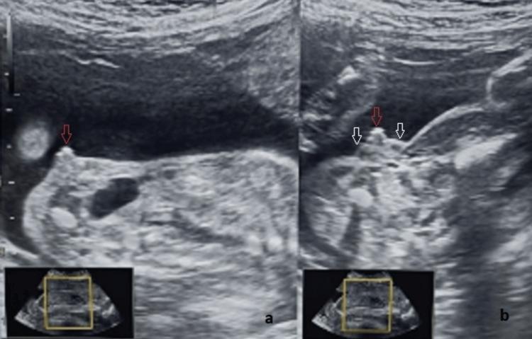
This diagnostic ultrasound illustrates fetal clitoromegaly and labial hypertrophy at 25 weeks of gestation, a key finding in the evaluation of ambiguous genitalia and congenital adrenal hyperplasia. Image (a) provides a sagittal view, highlighting a significantly thickened and prominent clitoris (indicated by a red arrow) protruding from the genital region. Image (b) presents an axial view of the same anatomy, where the enlarged clitoris (red arrow) is flanked by two distinct, hypoechoic, and homogenous labial swellings (white arrows). These findings demonstrate a departure from the normal 'three-line sign' of female genitalia, with the outer labial lines appearing divergent and convex rather than parallel. The imaging modality is B-mode prenatal sonography. This case is educationally significant for distinguishing between female genital virilization and severe male hypospadias during prenatal screening, emphasizing the importance of multi-planar assessment (sagittal and axial) and the identification of internal pelvic organs like the uterus for accurate sex determination.
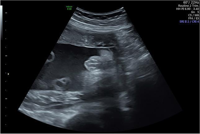
This diagnostic ultrasound image demonstrates the external genitalia of a female fetus at 30 weeks and 4 days of gestation. The primary sonographic finding is the 'multiple parallel linear echo pattern' (also known as the hamburger sign), which is characteristic of the developing labia majora and labia minora. This pattern consists of three or more hyperechoic, parallel lines representing the labial folds, often separated by a thin hypoechoic midline cleft. The image is captured in a transverse perineal plane, clearly showing the absence of a male phallus or scrotum. This visualization is a key educational marker used in prenatal imaging to confirm female fetal sex and assess normal urogenital development in the third trimester. The surrounding structures include the anechoic amniotic fluid and adjacent fetal limbs, providing context for the perineal orientation.
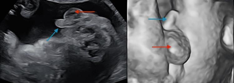
This composite diagnostic image displays a 20-week gestation level 2 ultrasound scan focusing on fetal perineal anatomy. The image is split into two views: a standard 2D grayscale sonogram on the left and a 3D/surface-rendered reconstruction on the right. In both views, the external male genitalia are clearly visualized and annotated with colored arrows. A blue arrow identifies the penis, which appears as an elongated, echogenic projection. A red arrow identifies the scrotum, seen as a rounded, more inferiorly positioned structure. The 3D view provides enhanced spatial perspective and surface topography of the genital phenotype. This imaging is used during routine second-trimester anatomy scans for sex determination and to screen for genital anomalies. In this clinical context, the visualization of a clear male phenotype is used to confirm fetal sex, particularly when investigating discrepancies with non-invasive prenatal testing (NIPT).
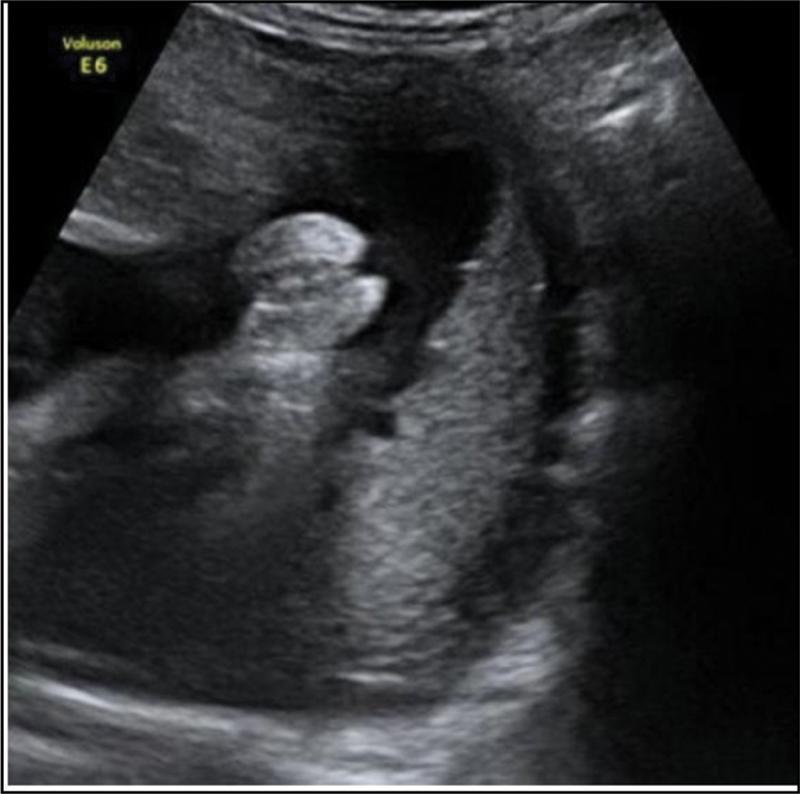
This diagnostic image is a gray-scale prenatal ultrasound from a midtrimester scan, specifically focusing on the fetal perineum to identify external genitalia. The scan displays a transverse view of the pelvic region. Key anatomical features include two parallel hyperechoic (bright) linear structures representing the labia majora. Centrally located between these lines is a smaller, distinct hyperechoic nodule corresponding to the clitoris, forming the characteristic 'hamburger sign' indicative of female fetal sex. The surrounding anechoic (dark) areas represent amniotic fluid, providing the necessary acoustic window for visualization. This image is clinically significant for routine obstetric screening, demonstrating the standard morphological markers used for sex determination and the assessment of external genital development during the second trimester of pregnancy.
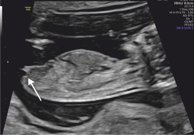
This diagnostic ultrasound image shows a midline sagittal view of a human fetus at 13 weeks of gestation during the first trimester. The fetus is oriented horizontally with its caudal end positioned toward the left of the frame. The primary educational focus is fetal sex determination through the assessment of the genital tubercle. A white arrow points to the genital tubercle located at the caudal extremity of the fetal torso. The tubercle is oriented parallel (at an angle of less than 30 degrees) to the horizontal axis of the fetus, which is a sonographic marker indicative of female gender development. The image clearly demonstrates the relationship between the genital tubercle and the lumbosacral spine/rump contour. This technique is clinically significant in prenatal screening for early gender identification and the evaluation of normal external genitalia development.
first trimester genital tubercle angle sex determination 12 weeks
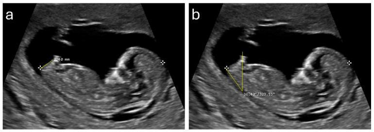
Diagnostic ultrasound images showing two side-by-side midsagittal views (a and b) of a fetus at approximately 12 weeks of gestation. The fetus is positioned in a supine orientation within the amniotic sac, with visible anatomical landmarks including the head, spine, and caudal region. The images demonstrate specific sonographic markers used for fetal sex determination. Panel (a) illustrates the measurement of the anogenital distance (AGD), shown as a linear caliper measurement (7.40 mm) extending from the anterior edge of the anus to the tip of the genital tubercle. Panel (b) illustrates the genital tubercle angle (GTA), showing an angular measurement (36.47 degrees) formed between a horizontal reference line and the longitudinal axis of the genital tubercle. These sonographic markers are clinically relevant for early prenatal screening and gender identification, as larger AGD and higher GTA values are statistically associated with male fetal development during the first trimester.
This diagnostic ultrasound image shows a midline sagittal view of a human fetus at 13 weeks of gestation during the first trimester. The fetus is oriented horizontally with its caudal end positioned toward the left of the frame. The primary educational focus is fetal sex determination through the assessment of the genital tubercle. A white arrow points to the genital tubercle located at the caudal extremity of the fetal torso. The tubercle is oriented parallel (at an angle of less than 30 degrees) to the horizontal axis of the fetus, which is a sonographic marker indicative of female gender development. The image clearly demonstrates the relationship between the genital tubercle and the lumbosacral spine/rump contour. This technique is clinically significant in prenatal screening for early gender identification and the evaluation of normal external genitalia development.
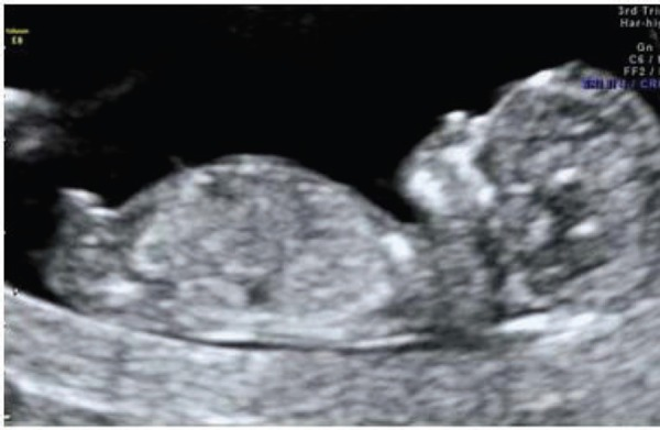
**Imaging Modality:** Obstetric Ultrasound (2D grayscale). **Anatomical Region:** Intrauterine environment showing a single fetus in a mid-sagittal plane. **Clinical Context:** First-trimester screening, approximately 12–13 weeks gestation. **Key Visual Features:** * **Fetal Anatomy:** The image captures the fetus in a profile view, demonstrating the head (cranium), thorax, and abdomen. The fetal spine is visible along the lower aspect of the trunk. * **Cranial Structures:** Early ossification of the skull is evident. The nasal bone and facial profile are distinguishable. * **Nuchal Region:** The nuchal translucency (NT) space—the sonographic appearance of subcutaneous fluid behind the fetal neck—is visible for assessment. * **Genitourinary Markers:** A prominent genital tubercle (phallus) is visualized at the caudal end of the torso. The cranial orientation of the tubercle (angle relative to the lumbosacral spine) is a sonographic marker used for early fetal sex determination, suggesting male development. * **Environment:** The fetus is surrounded by anechoic amniotic fluid, providing clear visualization of the fetal borders. **Diagnostic Significance:** This plane is critical for assessing crown-rump length (CRL), nuchal translucency thickness, and early anatomical surveys including the presence of the nasal bone and genital tubercle orientation.
Fetal Sex Determination on Ultrasonography (USG)
Why It Is Clinically Important
- Detection of fetuses at risk of severe X-linked disorders (e.g., Duchenne muscular dystrophy, haemophilia)
- Diagnosis of disorders of sex development (DSD) / ambiguous genitalia
- Assessment of gonadal abnormalities (e.g., undescended testes)
Note: Sex determination by USG for non-medical (social) reasons is legally prohibited in several countries including India (PCPNDT Act).
1. First Trimester (11-14 Weeks) - Genital Tubercle Method
Principle
Technique
- Obtain a strict mid-sagittal plane of the fetus (same plane as NT measurement)
- Identify the genital tubercle at the caudal end of the trunk
- Measure the Genital Tubercle Angle (GTA) - the angle between the tubercle and the horizontal reference line (lumbosacral spine)
Sonographic Findings
| Parameter | Male | Female |
|---|---|---|
| Tubercle direction | Points cranially (upward, toward head) | Points caudally (downward, parallel to spine) |
| GTA | >30° from horizontal (typically ~36-45°) | <10° from horizontal (nearly parallel) |
| Anogenital Distance (AGD) | Longer | Shorter |
| Appearance | Prominent upward projection | Flat or parallel to dorsal surface |
- (a) Anogenital distance (AGD) measured from anus to tip of tubercle
- (b) GTA of ~36° = male fetus
Accuracy
- ~70-75% accuracy at 11 weeks; improves to ~85-90% by 13-14 weeks
- Requires strict mid-sagittal plane; prone to error with incorrect fetal position
2. Second Trimester (18-22 Weeks) - External Genitalia Visualization
Technique
- Scan in transverse plane through the fetal perineum (between the thighs)
- Adequate amniotic fluid is needed for an acoustic window
- Fetal position must allow perineum visualization (in 30% of cases, position prevents this)
Sonographic Signs
MALE Fetus
| Sign | Description |
|---|---|
| Turtle sign | The penis and scrotum appear as a round dome-shaped structure with a central echogenic line - resembling a turtle head protruding from the shell |
| Direct visualization | Penis as an elongated echogenic structure; scrotum as a rounded inferior structure |
| Testes | Can be seen as hypoechoic oval structures within the scrotum after 22-28 weeks |
FEMALE Fetus
| Sign | Description |
|---|---|
| Hamburger sign (three-line sign) | Three parallel hyperechoic lines - the two outer lines represent the labia majora and the central echogenic dot/line is the clitoris/labia minora, resembling a hamburger in cross-section |
| Absence of male structures | No phallus or scrotum seen |
- (a) Female - arrowhead shows three parallel lines = "hamburger sign" (labia majora + clitoris)
- (b) Male - arrowhead shows dome-shaped phallus + scrotum
Accuracy at 18-22 Weeks
- ~95-99% accuracy when genitalia are clearly visible
- Male sex is slightly easier to confirm than female
- Only 100% certain finding for male: Confirmation of testes in the scrotum (possible at 22-36 weeks)
3. Third Trimester (>28 Weeks)
- Genitalia are larger and easier to visualize
- Scrotal testes are the most definitive sign for male sex
- Can be limited by decreased amniotic fluid (oligohydramnios) and fetal position
- 3D ultrasound can enhance visualization when 2D is inconclusive
Summary Table: Sex Determination by Trimester
| Trimester | Gestational Age | Method | Key Sign | Accuracy |
|---|---|---|---|---|
| 1st | 11-14 weeks | Genital tubercle angle | >30° = male; <10° = female | 75-90% |
| 2nd | 18-22 weeks | Transverse perineal view | Hamburger sign (F) / Turtle sign (M) | 95-99% |
| 3rd | >28 weeks | Direct visualization | Scrotal testes (M) / labia (F) | >99% (if seen) |
Pitfalls and Limitations
- Fetal position - in ~30% of fetuses, the perineum cannot be visualized
- Oligohydramnios - reduced fluid limits acoustic window
- Labial edema may mimic a scrotum (false male)
- Penile agenesis / micropenis - may be missed or misidentified as female
- Hypospadias - may alter appearance of male genitalia
- Clitoromegaly (e.g., in congenital adrenal hyperplasia) may mimic a phallus
- Umbilical cord between the thighs can be mistaken for a penis ("cord between legs" artifact)
- Incorrect plane in first trimester leads to false angle measurement
When Genitalia Appear Ambiguous: DSD
- Congenital Adrenal Hyperplasia (CAH) - virilized XX female (most common cause)
- Ovotesticular DSD - rare, external genitalia always ambiguous
- Androgen insensitivity syndrome
- Turner syndrome (45,XO) or Klinefelter (47,XXY)
Other Methods for Fetal Sex Determination
| Method | Notes |
|---|---|
| Cell-free fetal DNA (cfDNA/NIPT) | Most accurate non-invasive method from 10 weeks; detects Y chromosome in maternal blood |
| Amniocentesis | Karyotype - gold standard but invasive (risk of miscarriage ~0.5%) |
| Chorionic Villus Sampling (CVS) | Karyotype from 10-13 weeks; invasive |
| 3D/4D Ultrasound | Improves visualization in difficult cases; useful in third trimester |
What is L- carnitine
L-carnitine biochemistry function clinical uses deficiency
L-Carnitine
What Is It?
Biosynthesis
- Synthesized primarily in the liver and kidneys from lysine and methionine
- Requires cofactors: vitamin C (ascorbic acid), vitamin B6 (pyridoxine), niacin (B3), and iron - deficiency of any of these can impair synthesis
- Skeletal and cardiac muscle cannot synthesize carnitine - they depend entirely on uptake from blood
- ~97% of total body carnitine is stored in skeletal muscle
Primary Biochemical Role: The Carnitine Shuttle
| Step | Event | Enzyme |
|---|---|---|
| 1 | LCFA activated to acyl-CoA in cytosol | Acyl-CoA synthetase (thiokinase) - uses ATP |
| 2 | Acyl group transferred from CoA to carnitine → forms acylcarnitine | CPT-I (outer mitochondrial membrane) |
| 3 | Acylcarnitine transported into matrix; carnitine recycled back | Carnitine-acylcarnitine translocase |
| 4 | Acyl group transferred back to CoA in matrix | CPT-II (inner mitochondrial membrane) |
| 5 | Free carnitine returns to cytosol via translocase | Completes the cycle |
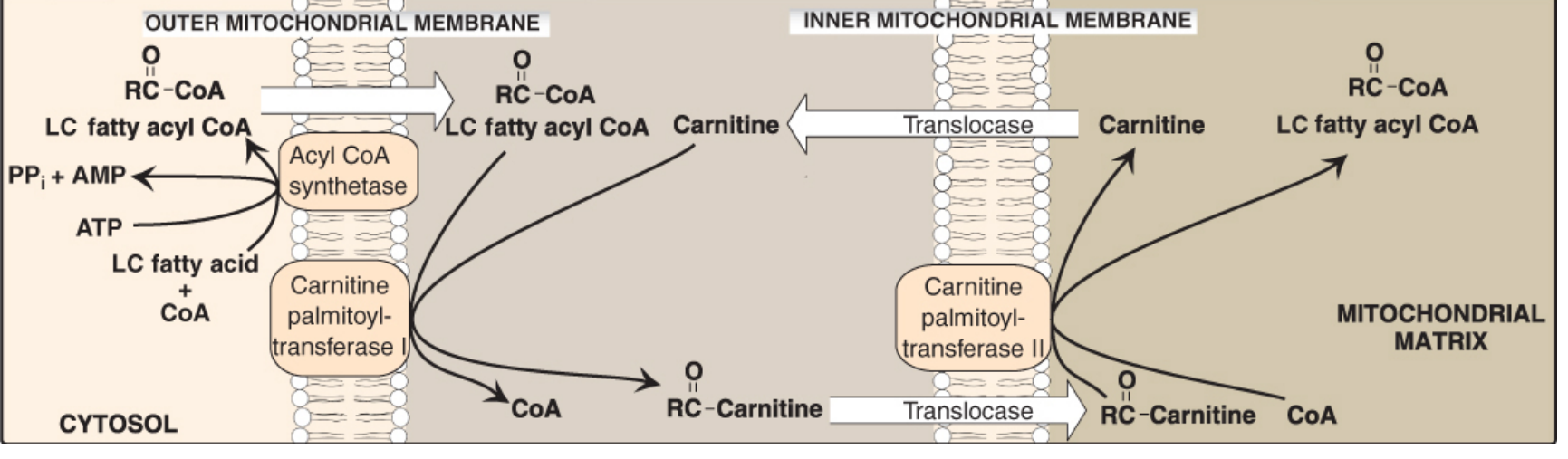
Key point: Fatty acids with ≤12 carbons (short- and medium-chain) do NOT need carnitine and can enter mitochondria directly. Carnitine is essential only for long-chain fatty acids (>12 carbons).
Regulation
- Malonyl-CoA (the first committed intermediate in fatty acid synthesis) is a potent inhibitor of CPT-I, thus preventing simultaneous fatty acid synthesis and beta-oxidation - Lippincott's Illustrated Biochemistry, p. 543
Dietary Sources
| Source | Carnitine Content |
|---|---|
| Red meat (beef, lamb) | Highest (~60-180 mg/100g) |
| Pork | Moderate (~24 mg/100g) |
| Fish | Low-moderate |
| Dairy | Small amounts |
| Plant foods (fruits, vegetables, grains) | Very low or absent |
Clinical Uses / Supplementation
| Condition | Rationale |
|---|---|
| Primary carnitine deficiency | First-line treatment; lifelong L-carnitine supplementation (reverses cardiomyopathy and muscle weakness) |
| Secondary carnitine deficiency | IV/oral replacement (dialysis patients, liver disease, valproate therapy) |
| Hemodialysis patients | Carnitine is removed by dialysis; supplementation improves anemia, muscle cramps, fatigue |
| Valproic acid toxicity | Valproate depletes carnitine by impairing renal reabsorption |
| Organic acidemias (propionic, methylmalonic acidemia) | Carnitine binds toxic acyl-CoA derivatives and promotes urinary excretion |
| Cardiomyopathy | Supports cardiac energy metabolism |
| Male infertility | High concentration in epididymis; sperm maturation and motility support |
| Athletic performance / weight loss | Used as supplement; evidence is modest |
| Alzheimer's disease | Acetyl-L-carnitine (ALC) form improves cognition in some studies |
Dosing (from Harriet Lane Handbook)
| Indication | Route | Dose |
|---|---|---|
| Primary carnitine deficiency - Child | Oral | 50-100 mg/kg/day ÷ every 8-12 hr; max 3 g/day |
| Primary carnitine deficiency - Adult | Oral | 330 mg - 1 g/dose BID-TID; max 3 g/day |
| Secondary deficiency | IV | 50 mg/kg loading dose; then 50 mg/kg/day ÷ every 4-6 hr; up to 300 mg/kg/day |
Carnitine Deficiency
Primary Carnitine Deficiency (PCD)
- Caused by defect in OCTN2 (organic cation transporter novel 2) - the high-affinity carnitine transporter in heart, skeletal muscle, and kidney
- Results in urinary carnitine wasting and cellular carnitine depletion
- Presents with: cardiomyopathy, skeletal muscle weakness, hypoketotic hypoglycemia (especially during fasting)
- Fatal if untreated; responds well to L-carnitine supplementation - Ganong's Review, p. 880
Secondary Carnitine Deficiency (SCD)
- Liver disease (reduced synthesis)
- Renal disease / dialysis (urinary loss)
- Valproic acid, pivampicillin (drug-induced urinary excretion)
- Organic acidemias (toxic acylcarnitines excreted)
- Diabetes, sepsis, heart failure, chronic alcoholism
- Premature infants (limited synthesis + low body stores)
CPT Deficiencies
- CPT-I deficiency - affects liver primarily; severe hypoglycemia during fasting, coma, death
- CPT-II deficiency - most common form affects skeletal muscle: presents as muscle weakness + myoglobinuria after prolonged exercise
Side Effects
- Nausea, vomiting, abdominal cramps, diarrhea
- "Fishy" body odor (due to trimethylamine production by gut bacteria)
- Seizures (reported even in patients without prior history)
- High-dose caution in renal failure / ESRD - accumulation of trimethylamine and trimethylamine-N-oxide (potentially toxic metabolites)
- Serious hypersensitivity reactions (including anaphylaxis) with IV use in dialysis patients
Summary
| Feature | Detail |
|---|---|
| Chemical class | Amino acid derivative (quaternary ammonium) |
| Precursors | Lysine + Methionine |
| Site of synthesis | Liver, kidneys |
| Storage | 97% in skeletal muscle |
| Primary function | Long-chain fatty acid transport into mitochondria (carnitine shuttle) |
| Key enzyme | CPT-I (rate-limiting; inhibited by malonyl-CoA) |
| Active form | L-carnitine (levocarnitine) |
| Acetylated form | Acetyl-L-carnitine (ALC) - crosses blood-brain barrier; used for neurological conditions |