
How to distinguish between gender of fetus in 11 week gestation, with images and simplified techniques on usg
Reading File
Finding Sources
Searching PubMed
"fetal sex determination" AND ultrasound AND first trimester
Searching the Web
first trimester ultrasound fetal sex determination genital tubercle angle 30 degrees accuracy 11 weeks
Finding Sources
Searching Images
male fetus genital tubercle cephalad angle greater than 30 degrees midsagittal 12 weeks
Searching Images
fetal external genitalia ultrasound three lines sign female labia male scrotum penis
query_images_db
query_images_db
Reading Article
PMID: 39798051
Sex determination on ultrasound at 11 weeks
At 11 weeks the external genitalia are still in the indifferent stage. The only usable marker is the angle of the genital tubercle (the "nub") on a midsagittal view. At this gestation it is unreliable, so use it only as a provisional impression.
Why it is hard at 11 weeks
- The genital tubercle looks the same in both sexes until about 14 weeks.
- Tubercle length does not help. It is actually larger in the female in early development, and using length as a criterion has led to mistakes (Langman's Medical Embryology, "External Genitalia in the Female").
- Real anatomy (penis and scrotum, or labia) only becomes visible from about 14 weeks onward.
Simplified technique (the angle method)
- Get a true midsagittal view.
- This is the same view used for crown-rump length and nuchal translucency.
- You should see the nasal bone and tip, the palate, the diencephalon, and the spine in profile.
- The fetus should be horizontal, with the lumbosacral skin line clear.
- Magnify the image. The rump and perineum should fill most of the screen.
- Find the genital tubercle. It is a small echogenic nub between the legs, at the caudal end of the torso.
- Draw a reference line. Use a horizontal line through the lumbosacral skin surface, along the back or rump.
- Compare the tubercle axis with that line.
| Finding | Interpretation |
|---|---|
| Tubercle points cranially, more than 30 degrees above the horizontal | Male |
| Tubercle parallel (about 0 to 10 degrees) or pointing down or convergent | Female |
| Angle 10 to 30 degrees | Indeterminate. Do not assign a sex. |
- If the fetus is poorly positioned, the legs may be crossed, or the fetus may be in an oblique or flexed position. In that case, wait for fetal movement, ask the mother to cough or walk briefly, or rescan later. Transvaginal scanning can help if the fetus is low.
Mnemonic: Up like a flag (more than 30 degrees) is a boy. Flat or down is a girl.
Images
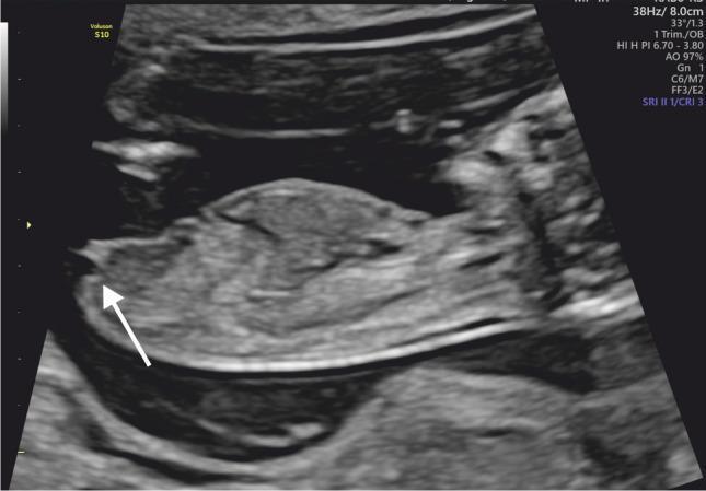
This midsagittal view at 13 weeks shows the female pattern. The tubercle (arrow) is parallel to the horizontal axis, at less than 30 degrees.
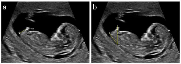
This 12-week image shows how to measure. Panel (a) is the anogenital distance, 7.40 mm. Panel (b) is the genital tubercle angle, 36.47 degrees, which is more than 30 degrees and so consistent with male. A larger anogenital distance and a higher angle are both associated with male fetuses.
Accuracy: why 11 weeks is the weak point
In the original prospective study of 172 fetuses, accuracy was:
- 70.3% at 11 weeks
- 98.7% at 12 weeks
- 100% at 13 weeks
At 11 weeks, 56% of males were wrongly called female. Only 5% of females were wrongly called male. So a "female" call at 11 weeks is least reliable, because many males still have a flat tubercle.
A 2025 systematic review and meta-analysis of 28 studies found pooled sensitivity rising from 69% at 11 weeks to 89% at 12 weeks and 96% at 13 weeks. Mean first-trimester accuracy was 85%. Female sex was detected more easily in the first trimester, and male sex more easily later.
Practical tips
- Take the measurement at a CRL of about 45 mm or more. Accuracy improves with larger CRL, and the male angle widens with CRL.
- A good image and an experienced operator matter more than the equipment.
- Report it as "provisional", or defer it. Say "indeterminate" when the angle is 10 to 30 degrees.
- Confirm at 12 to 14 weeks, and definitively at the 18 to 20 week anomaly scan. There you look for the penis and scrotum in males, and for the labia ("three lines" or "hamburger" sign) in females.
- Where an answer is needed before 14 weeks, cell-free DNA (NIPT) for Y-chromosome sequences or invasive karyotyping is far more accurate.
Legal and ethical note
In India, the PCPNDT Act prohibits disclosing fetal sex by any means. Many other countries have similar restrictions. Check your local law and clinic policy. Sex is documented or disclosed only where there is a medical indication, such as sex-linked disease risk or suspected disorders of sex development.
Sources: Langman's Medical Embryology (embryology of the external genitalia), the 1999 first-trimester gender study above, and the 2025 meta-analysis.
This is a shared conversation. Sign in to Orris to start your own chat.